

The DOI matches its Crossref record; 14 of the 14 references it cites were found in a registry or an index.
A Rare Case of Pulmonary and Gastrointestinal Mucormycosis Due to Rhizopus spp. in a Child with Chronic Granulomatous Disease
Open accessAbstract
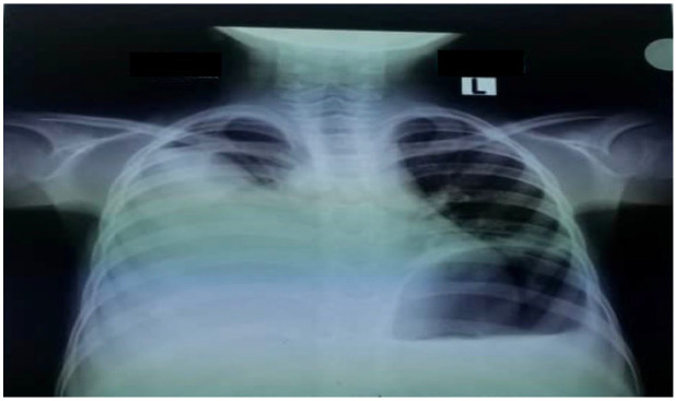
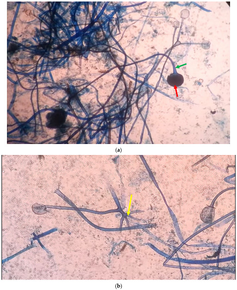
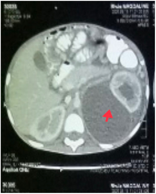
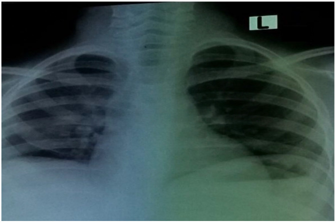
Mucormycosis is a rare but serious fungal infection caused by a mold family known as the Mucorales. These fungi exist throughout the environment, especially in the soil, leaves, compost piles, or decaying woods. Humans contract mucormycosis by coming in contact with the spores from fungus either by inhalation or through cuts on the skin. The population at risk for this life-threatening infection includes diabetes mellitus patients, cancer patients, premature infants, burn patients, and immunocompromised patients. The fungi that most commonly cause mucormycosis are the Rhizopus species, and the least represented are Apophysomyces species. Common clinical manifestations of mucormycosis include pulmonary, cutaneous, rhinocerebral, and gastrointestinal mucormycosis. Cases of lung mucormycosis are often misdiagnosed because of non-specific clinical symptoms and radiological features, and in many cases, have been diagnosed as aspergillosis due to similarities in signs, symptoms, and imaging presentation of the lungs. We present a pediatric case of a 6-year-old from Togo who presented to our hospital in Nigeria with dyspnea, fever, and abdominal pain of five-day duration. The child’s symptoms began 6-months prior, with dry cough, fever, fatigue, and chest pain and abdominal pain. The hospital in Togo where he lived suspected infection with tuberculosis (TB) despite a false-positive Mantoux test and negative chest X-ray. He was initially treated for TB with Isoniazid and vitamin B6 and was discharged home. Six months later, his symptoms have not improved, but became more severe with high grade fever 40 °C (oral reading), anorexia, fatigue, tachypnea, abdominal distention, and cough. The patient was immediately referred to our hospital in Abuja, Nigeria where more specific tests were ordered. He was eventually diagnosed with chronic granulomatous disease induced pulmonary and gastrointestinal (GI) mucormycosis due to Rhizopus spp. In this report, we discuss an unusual clinical presentation of an infection caused by Rhizopus spp., its management, and outcomes in a child with chronic granulomatous disease (CGD).
Overview
The claims and statistics of this paper haven’t been checked yet.
Run the check — free, no account needed. It reads the open-access text and checks the sources it cites.
Integrity signals
Registry
Cites
Statistics
Figures
Verification
Claims the paper makes
The claims and statistics of this paper haven’t been checked yet.
Run the check — free, no account needed. It reads the open-access text and checks the sources it cites.
Claims other papers make about this one
Each is checked against this paper’s own text: does it say what the citing paper says it does?
The source backs part of the claim, not all of it — or backs it for a different population, condition or comparison.
In the citing paper
“The presence of such ARGs and VFs may have clinical relevance, particularly in immunocompromised individuals or those with disrupted gut barriers, where opportunities for opportunistic infection or horizontal gene transfer may be increased.22,29-35”
From Safety and efficacy concerns in Indian probiotics: Insights from whole-genome sequencing and in vitro assessment (2026) · cites this paper as [31]
Experimental — accuracy not yet measured
Self-check
Percentages, sums and reported differences re-computed from the paper’s own numbers.
The self-check needs the paper’s full text.
Citations
Cites
Ribes, J. A., Vanover-Sams, C. L., & Baker, D. J. (2000) · Clinical Microbiology Reviews · doi:10.1128/cmr.13.2.236 · Cited by 1,861
Ribes, J. A., Vanover-Sams, C. L., & Baker, D. J. (2000) · Clinical Microbiology Reviews · doi:10.1128/cmr.13.2.236-301.2000 · Cited by 1,702
A Controlled Trial of Interferon Gamma to Prevent Infection in Chronic Granulomatous Disease
The International Chronic Granulomatous Disease Cooperative Study Group* (1991) · New England Journal of Medicine · doi:10.1056/nejm199102213240801 · Cited by 832
The rising trend of invasive zygomycosis in patients with uncontrolled diabetes mellitus
Chakrabarti, A., Das, A., Mandal, J., Rudramurthy, S. M., George, V. K., Tarai, B., Rao, P., Panda, N., Verma, S. C., & Sakhuja, V. (2006) · Medical Mycology · doi:10.1080/13693780500464930 · Cited by 376
Zygomycosis (mucormycosis): emerging clinical importance and new treatments
Greenberg, R. N., Scott, L. J., Vaughn, H. H., & Ribes, J. A. (2004) · Current Opinion in Infectious Diseases · doi:10.1097/00001432-200412000-00003 · Cited by 257
Zygomycosis in Children: A Systematic Review and Analysis of Reported Cases
Zaoutis, T. E., Roilides, E., Chiou, C. C., Buchanan, W. L., Knudsen, T. A., Sarkisova, T. A., Schaufele, R. L., Sein, M. T., Sein, T., Prasad, P. A., et al. (2007) · The Pediatric Infectious Disease Journal · doi:10.1097/inf.0b013e318062115c · Cited by 225
Slavin, M. A., van Hal, S. J., Sorrell, T. C., Lee, A., Marriott, D. J. E., Daveson, K., Kennedy, K., Hajkowicz, K. M., Halliday, C. L., Athan, E., et al. (2015) · Clinical Microbiology and Infection · doi:10.1016/j.cmi.2014.12.021 · Cited by 189
Beobachtungen über Lungenmycose beim Menschen
Fürbringer, P. (1876) · Archiv für Pathologische Anatomie und Physiologie und für Klinische Medicin · doi:10.1007/bf01878266 · Cited by 76
Mucormycosis of the gastrointestinal tract in children
MOONEY, J. E., & Wanger, A. R. (1993) · The Pediatric Infectious Disease Journal · doi:10.1097/00006454-199310000-00013 · Cited by 58
Hyperbaric Oxygen as an Adjunct in Zygomycosis: Randomized Controlled Trial in a Murine Model
Barratt, D. M., Meter, K. V., Asmar, P., Nolan, T., Trahan, C. G., García-Covarrubias, L., & Metzinger, S. E. (2001) · Antimicrobial Agents and Chemotherapy · doi:10.1128/aac.45.12.3601-3602.2001 · Cited by 48
Cited by
Sorted by citations
What Is New in Pulmonary Mucormycosis?
Danion, F., Coste, A., Hyaric, C. L., Melenotte, C., Lamoth, F., Calandra, T., Garcia‐Hermoso, D., Aimanianda, V., Lanternier, F., & Lortholary, O. (2023) · Journal of Fungi · doi:10.3390/jof9030307 · Cited by 62
Mycoproteins and their health‐promoting properties: Fusarium species and beyond
Lee, D., Pan, J. H., Kim, D., Heo, W., Shin, E., Kim, Y. J., Shim, Y. Y., Reaney, M. J. T., Ko, S., Hong, S., et al. (2024) · Comprehensive Reviews in Food Science and Food Safety · doi:10.1111/1541-4337.13365 · Cited by 39
Rodrigues, M. A., Teiga-Teixeira, P., & Esteves, A. (2025) · Foods · doi:10.3390/foods14081320 · Cited by 9
Three case reports of pulmonary mucormycosis with a review of the literature
Zhang, Z., & Wang, M. (2025) · Frontiers in Medicine · doi:10.3389/fmed.2025.1580912 · Cited by 3
Madhumathi, I., Ramesh, V., Rathakrishnan, B., Abirami, B., Gunasekaran, K., Kumar, T. S., Jeyasankar, P., Raja, G., Selvam, K., Rajendran, S., et al. (2026) · Gut microbes reports · doi:10.1080/29933935.2026.2668863
Metadata
- Authors
- Opara, N. U.
- Affiliations
- Charleston Area Medical Center (US)
- Corresponding author
- Opara, Nnennaya U.
- ORCID
- Opara, Nnennaya U.
- Venue
- Infectious Disease Reports
- Publisher
- MDPI AG
- Published
- 2022-08-08
- Volume
- 14
- Issue
- 4
- Pages
- 579-586
- ISSN
- 2036-7430 (ISSN-L)
- 2036-7449
- PMC
- PMC9408395
- OpenAlex
- W4293105298
- Cited by
- 8
- References
- 13
- Citations by year
- 2026: 1 · 2025: 4 · 2024: 1 · 2023: 1 · 2022: 1
- FWCI
- 0.86
- Type
- journal article
- Language
- en
- Open access
- gold
- doi.org
- Licence
- Locations
- Infectious Disease Reports (publishedVersion, cc-by)
- PubMed Central (publishedVersion, cc-by)
- PubMed (publishedVersion)
- DOAJ (DOAJ: Directory of Open Access Journals) (submittedVersion, cc-by-sa)
- Topics
- Health Sciences › Medicine › Infectious Diseases › Antifungal resistance and susceptibility
- Health Sciences › Medicine › Infectious Diseases › Infectious Diseases and Mycology
- Health Sciences › Medicine › Infectious Diseases › Fungal Infections and Studies
- Keywords
- mucormycosis, Rhizopus, chronic granulomatous disease, pulmonary mucormycosis, gastrointestinal mucormycosis, pediatric fungal infection, misdiagnosis as tuberculosis, immunocompromised patients, Mucorales, antifungal therapy
About these checks
Registry, retraction, Cites and statistics results are computed, not judged, with one exception: a printed reference that no index lists is read once by a model before it counts as a citation error. Claims are extracted by a model and kept only when their quote appears verbatim in the text; each cited source is then checked, with a model’s help, for whether it says what the claim relies on. Those verdicts can be wrong — read the claim’s quote and the cited source before relying on one.
Statistics and arithmetic are recomputed, not judged.
Confidence bands are shown only for fields calibrated on our annotated test set. None is calibrated yet, so no band is shown.
What counts as a claim ›How we measure confidence ›How we check this ›
Are you an author, or do you see a problem with this page? Request a review
