

The DOI matches its Crossref record; 8 of the 8 references it cites were found in a registry or an index.
In Vitro Models of Biological Barriers for Nanomedical Research
Abstract
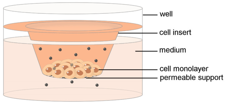
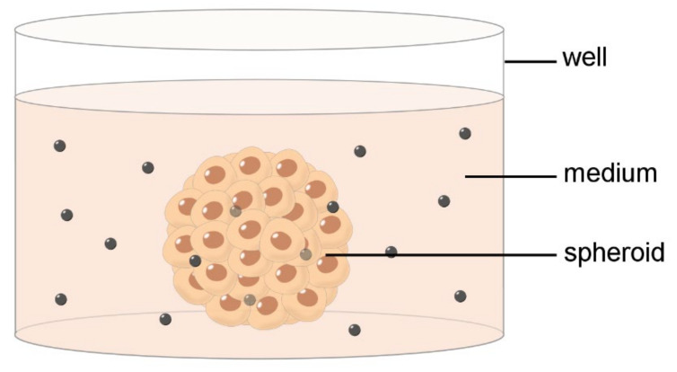
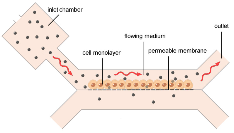
Nanoconstructs developed for biomedical purposes must overcome diverse biological barriers before reaching the target where playing their therapeutic or diagnostic function. In vivo models are very complex and unsuitable to distinguish the roles plaid by the multiple biological barriers on nanoparticle biodistribution and effect; in addition, they are costly, time-consuming and subject to strict ethical regulation. For these reasons, simplified in vitro models are preferred, at least for the earlier phases of the nanoconstruct development. Many in vitro models have therefore been set up. Each model has its own pros and cons: conventional 2D cell cultures are simple and cost-effective, but the information remains limited to single cells; cell monolayers allow the formation of cell–cell junctions and the assessment of nanoparticle translocation across structured barriers but they lack three-dimensionality; 3D cell culture systems are more appropriate to test in vitro nanoparticle biodistribution but they are static; finally, bioreactors and microfluidic devices can mimicking the physiological flow occurring in vivo thus providing in vitro biological barrier models suitable to reliably assess nanoparticles relocation. In this evolving context, the present review provides an overview of the most representative and performing in vitro models of biological barriers set up for nanomedical research.
Overview
The claims and statistics of this paper haven’t been checked yet.
Run the check — free, no account needed. It reads the open-access text and checks the sources it cites.
Integrity signals
Registry
Cites
Statistics
Figures
Verification
Claims the paper makes
The claims and statistics of this paper haven’t been checked yet.
Run the check — free, no account needed. It reads the open-access text and checks the sources it cites.
Claims other papers make about this one
Each is checked against this paper’s own text: does it say what the citing paper says it does?
In the citing paper
“Earlier overviews have typically organized systems by material class or by a single trigger, but they seldom cross-compare how different design logics perform against specific tumor barriers or how deeply they penetrate in three-dimensional models and in vivo [16].”
From Overcoming tumor microenvironment barriers: transformable and bioinspired nanomedicine strategies for deep tumor penetration (2026) · cites this paper as [16]
Self-check
Percentages, sums and reported differences re-computed from the paper’s own numbers.
The self-check needs the paper’s full text.
Citations
Cites
Organs-on-chips at the frontiers of drug discovery
Esch, E. W., Bahinski, A., & Huh, D. (2015) · Nature Reviews Drug Discovery · doi:10.1038/nrd4539 · Cited by 1,185
Optimization of lipid nanoparticles for intramuscular administration of mRNA vaccines
Hassett, K. J., Benenato, K. E., Jacquinet, E., Lee, A., Woods, A., Yuzhakov, O., Himansu, S., Deterling, J., Geilich, B. M., Ketova, T., et al. (2019) · Molecular Therapy — Nucleic Acids · doi:10.1016/j.omtn.2019.01.013 · Cited by 927
Tumour-on-a-chip provides an optical window into nanoparticle tissue transport
Albanese, A., Lam, A. K., Sykes, E. A., Rocheleau, J. V., & Chan, W. C. W. (2013) · Nature Communications · doi:10.1038/ncomms3718 · Cited by 330
Paek, J., Park, S. E., Lu, Q., Park, K.-T., Cho, M., Oh, J. M., Kwon, K. W., Yi, Y.-S., Song, J. W., Edelstein, H. I., et al. (2019) · ACS Nano · doi:10.1021/acsnano.9b00686 · Cited by 238
Colorectal tumor-on-a-chip system: a 3D tool for precision onco-nanomedicine
Carvalho, M. R., Barata, D., Teixeira, L. M., Giselbrecht, S., Reis, R. L., Oliveira, J. M., Truckenmüller, R., & Habibovic, P. (2019) · Science Advances · doi:10.1126/sciadv.aaw1317 · Cited by 230
Ma, H.-L., Jiang, Q., Han, S., Wu, Y., Tomshine, J. C., Wang, D., Gan, Y., Zou, G., & Liang, X.-J. (2012) · Molecular Imaging · doi:10.2310/7290.2012.00012 · Cited by 182
Tumor-Vasculature-on-a-Chip for Investigating Nanoparticle Extravasation and Tumor Accumulation
Wang, H.-F., Ran, R., Liu, Y., Hui, Y., Zeng, B., Chen, D., Weitz, D. A., & Zhao, C.-X. (2018) · ACS Nano · doi:10.1021/acsnano.8b06846 · Cited by 161
Huang, K., Boerhan, R., Liu, C., & Jiang, G. (2017) · Molecular Pharmaceutics · doi:10.1021/acs.molpharmaceut.7b00726 · Cited by 74
Research works printed but not linked: 132
- Weber, 1999, Nanomedicine, Health Forum J., 42, 36
- Codullo, 2019, Imatinib-Loaded Gold Nanoparticles Inhibit Proliferation of Fibroblasts and Macrophages from Systemic Sclerosis Patients and Ameliorate Experimental Bleomycin-Induced Lung Fibrosis, J. Control Release, 310, 198, 10.1016/j.jconrel.2019.08.015 · DOI
- Inchaurraga, 2019, The Effect of Thiamine-Coating Nanoparticles on Their Biodistribution and Fate Following Oral Administration, Eur. J. Pharm. Sci., 128, 81, 10.1016/j.ejps.2018.11.025 · DOI
- Ak, 2021, An Intravenous Application of Magnetic Nanoparticles for Osteomyelitis Treatment: An Efficient Alternative, Int. J. Pharm., 592, 119999, 10.1016/j.ijpharm.2020.119999 · DOI
- Arango, 2021, Fluorine Labeling of Nanoparticles and In Vivo 19F Magnetic Resonance Imaging, ACS Appl. Mater. Interfaces, 13, 12941, 10.1021/acsami.1c01291 · DOI
- Changsan, 2021, Dry Powder Inhalation Formulation of Chitosan Nanoparticles for Co-Administration of Isoniazid and Pyrazinamide, Pharm. Dev. Technol., 26, 181, 10.1080/10837450.2020.1852570 · DOI
- Costanzo, M., Esposito, E., Sguizzato, M., Lacavalla, M.A., Drechsler, M., Valacchi, G., Zancanaro, C., and Malatesta, M. (2021). Formulative Study and Intracellular Fate Evaluation of Ethosomes and Transethosomes for Vitamin D3 Delivery. Int. J. Mol. Sci., 22. · DOI
- Zou, 2021, Accelerating Transdermal Delivery of Insulin by Ginsenoside Nanoparticles with Unique Permeability, Int. J. Pharm., 605, 120784, 10.1016/j.ijpharm.2021.120784 · DOI
- Albanese, 2012, The Effect of Nanoparticle Size, Shape, and Surface Chemistry on Biological Systems, Annu. Rev. Biomed. Eng., 14, 1, 10.1146/annurev-bioeng-071811-150124 · DOI
- Malatesta, M. (2021). Transmission Electron Microscopy as a Powerful Tool to Investigate the Interaction of Nanoparticles with Subcellular Structures. Int. J. Mol. Sci., 22. · DOI
- Gamboa, 2013, In Vitro and in Vivo Models for the Study of Oral Delivery of Nanoparticles, Adv. Drug Deliv. Rev., 65, 800, 10.1016/j.addr.2013.01.003 · DOI
- Trickler, 2014, Porcine Brain Microvessel Endothelial Cells Show Pro-Inflammatory Response to the Size and Composition of Metallic Nanoparticles, Drug Metab. Rev., 46, 224, 10.3109/03602532.2013.873450 · DOI
- Pashaiasl, 2019, Application of Nanomaterials in Three-Dimensional Stem Cell Culture, J. Cell Biochem., 120, 18550, 10.1002/jcb.29133 · DOI
- Van Zundert, I., Fortuni, B., and Rocha, S. (2020). From 2D to 3D Cancer Cell Models-The Enigmas of Drug Delivery Research. Nanomaterials, 10. · DOI
- Giusti, 2014, A Novel Dual-Flow Bioreactor Simulates Increased Fluorescein Permeability in Epithelial Tissue Barriers, Biotechnol. J., 9, 1175, 10.1002/biot.201400004 · DOI
- Carton, 2017, Incubation under Fluid Dynamic Conditions Markedly Improves the Structural Preservation in Vitro of Explanted Skeletal Muscles, Eur. J. Histochem., 61, 2862
- Schmid, 2018, A Perfusion Bioreactor System for Cell Seeding and Oxygen-Controlled Cultivation of Three-Dimensional Cell Cultures, Tissue Eng. Part C Methods, 24, 585, 10.1089/ten.tec.2018.0204 · DOI
- Cappellozza, 2021, Integrated Microscopy and Metabolomics to Test an Innovative Fluid Dynamic System for Skin Explants In Vitro, Microsc. Microanal., 27, 923, 10.1017/S1431927621012010 · DOI
- Whitesides, 2006, The Origins and the Future of Microfluidics, Nature, 442, 368, 10.1038/nature05058 · DOI · Paper page
- Bhatia, 2014, Microfluidic Organs-on-Chips, Nat. Biotechnol., 32, 760, 10.1038/nbt.2989 · DOI · Paper page
- Pardridge, 2003, Blood-Brain Barrier Drug Targeting: The Future of Brain Drug Development, Mol. Interv., 3, 90, 10.1124/mi.3.2.90 · DOI · Paper page
- Abbott, 2010, Structure and Function of the Blood–Brain Barrier, Neurobiol. Dis., 37, 13, 10.1016/j.nbd.2009.07.030 · DOI · Paper page
- Hirrlinger, 2010, The Cytosolic Redox State of Astrocytes: Maintenance, Regulation and Functional Implications for Metabolite Trafficking, Brain Res. Rev., 63, 177, 10.1016/j.brainresrev.2009.10.003 · DOI
- Scheiber, 2013, Astrocyte Functions in the Copper Homeostasis of the Brain, Neurochem. Int., 62, 556, 10.1016/j.neuint.2012.08.017 · DOI
- Zhang, 2021, Development of Polymeric Nanoparticles for Blood-Brain Barrier Transfer-Strategies and Challenges, Adv. Sci., 8, 2003937, 10.1002/advs.202003937 · DOI · Paper page
- Bowman, 1983, Brain Microvessel Endothelial Cells in Tissue Culture: A Model for Study of Blood-Brain Barrier Permeability, Ann. Neurol., 14, 396, 10.1002/ana.410140403 · DOI
- Prades, 2012, Delivery of Gold Nanoparticles to the Brain by Conjugation with a Peptide That Recognizes the Transferrin Receptor, Biomaterials, 33, 7194, 10.1016/j.biomaterials.2012.06.063 · DOI
- Kuo, 2012, Methylmethacrylate–Sulfopropylmethacrylate Nanoparticles with Surface RMP-7 for Targeting Delivery of Antiretroviral Drugs across the Blood–Brain Barrier, Colloids Surf. B Biointerfaces, 90, 75, 10.1016/j.colsurfb.2011.09.048 · DOI
- Kuo, 2013, Solid Lipid Nanoparticles Carrying Chemotherapeutic Drug across the Blood-Brain Barrier through Insulin Receptor-Mediated Pathway, J. Drug Target., 21, 730, 10.3109/1061186X.2013.812094 · DOI
- Lu, 2005, Cationic Albumin Conjugated Pegylated Nanoparticle with Its Transcytosis Ability and Little Toxicity against Blood–Brain Barrier, Int. J. Pharm., 295, 247, 10.1016/j.ijpharm.2005.01.043 · DOI
- Tian, 2012, In Vitro and in Vivo Studies on Gelatin-Siloxane Nanoparticles Conjugated with SynB Peptide to Increase Drug Delivery to the Brain, Int. J. Nanomed., 7, 1031
- Gil, 2005, A Relevant in Vitro Rat Model for the Evaluation of Blood-Brain Barrier Translocation of Nanoparticles, Cell Mol. Life Sci., 62, 1400, 10.1007/s00018-005-5094-3 · DOI
- Grover, 2014, Brain-Targeted Delivery of Docetaxel by Glutathione-Coated Nanoparticles for Brain Cancer, AAPS PharmSciTech, 15, 1562, 10.1208/s12249-014-0165-0 · DOI
- Heggannavar, 2019, Preparation of Transferrin-Conjugated Poly-ε-Caprolactone Nanoparticles and Delivery of Paclitaxel to Treat Glioblastoma across Blood–Brain Barrier, Emergent Mater., 2, 463, 10.1007/s42247-019-00033-9 · DOI
- Kiss, 2018, Niosomes Decorated with Dual Ligands Targeting Brain Endothelial Transporters Increase Cargo Penetration across the Blood-Brain Barrier, Eur. J. Pharm. Sci., 123, 228, 10.1016/j.ejps.2018.07.042 · DOI
- Hanada, 2014, Cell-Based in Vitro Blood–Brain Barrier Model Can Rapidly Evaluate Nanoparticles’ Brain Permeability in Association with Particle Size and Surface Modification, Int. J. Mol. Sci., 15, 1812, 10.3390/ijms15021812 · DOI
- Xu, 2015, Silver Nanoparticles Induce Tight Junction Disruption and Astrocyte Neurotoxicity in a Rat Blood-Brain Barrier Primary Triple Coculture Model, Int. J. Nanomed., 10, 6105
- Bergmann, 2018, Blood–Brain-Barrier Organoids for Investigating the Permeability of CNS Therapeutics, Nat. Protoc., 13, 2827, 10.1038/s41596-018-0066-x · DOI
- Hudecz, 2020, Ultrathin Silicon Membranes for in Situ Optical Analysis of Nanoparticle Translocation across a Human Blood–Brain Barrier Model, ACS Nano, 14, 1111, 10.1021/acsnano.9b08870 · DOI
- Gromnicova, R., Davies, H.A., Sreekanthreddy, P., Romero, I.A., Lund, T., Roitt, I.M., Phillips, J.B., and Male, D.K. (2013). Glucose-Coated Gold Nanoparticles Transfer across Human Brain Endothelium and Enter Astrocytes In Vitro. PLoS ONE, 8. · DOI
- Sokolova, 2020, Transport of Ultrasmall Gold Nanoparticles (2 Nm) across the Blood–Brain Barrier in a Six-Cell Brain Spheroid Model, Sci. Rep., 10, 18033, 10.1038/s41598-020-75125-2 · DOI
- Papademetriou, I., Vedula, E., Charest, J., and Porter, T. (2018). Effect of Flow on Targeting and Penetration of Angiopep-Decorated Nanoparticles in a Microfluidic Model Blood-Brain Barrier. PLoS ONE, 13. · DOI
- Englert, 2016, Crossing the Blood-Brain Barrier: Glutathione-Conjugated Poly(Ethylene Imine) for Gene Delivery, J. Control Release, 241, 1, 10.1016/j.jconrel.2016.08.039 · DOI
- Falanga, 2017, Shuttle-Mediated Nanoparticle Transport across an in Vitro Brain Endothelium under Flow Conditions, Biotechnol. Bioeng., 114, 1087, 10.1002/bit.26221 · DOI
- Nowak, 2020, Size, Shape, and Flexibility Influence Nanoparticle Transport across Brain Endothelium under Flow, Bioeng Transl Med., 5, e10153, 10.1002/btm2.10153 · DOI
- Salman, 2020, Design and Validation of a Human Brain Endothelial Microvessel-on-a-Chip Open Microfluidic Model Enabling Advanced Optical Imaging, Front. Bioeng. Biotechnol., 8, 573775, 10.3389/fbioe.2020.573775 · DOI
- Peng, 2020, In Situ Surface Modification of Microfluidic Blood–Brain-Barriers for Improved Screening of Small Molecules and Nanoparticles, ACS Appl. Mater. Interfaces, 12, 56753, 10.1021/acsami.0c17102 · DOI
- Lee, 2020, Modeling Nanocarrier Transport across a 3D In Vitro Human Blood-Brain–Barrier Microvasculature, Adv. Healthc. Mater., 9, 1901486, 10.1002/adhm.201901486 · DOI
- Tricinci, 2020, A 3D Biohybrid Real-Scale Model of the Brain Cancer Microenvironment for Advanced In Vitro Testing, Adv. Mater. Technol., 5, 2000540, 10.1002/admt.202000540 · DOI
- Anderson, 2020, The Tumor Microenvironment, Curr. Biol., 30, R921, 10.1016/j.cub.2020.06.081 · DOI · Paper page
- Abreu, 2021, In Vitro and In Vivo Tumor Models for the Evaluation of Anticancer Nanoparticles, Adv. Exp. Med. Biol., 1295, 271, 10.1007/978-3-030-58174-9_12 · DOI
- Du, 2015, Core-Cross-Linking Accelerates Antitumor Activities of Paclitaxel–Conjugate Micelles to Prostate Multicellular Tumor Spheroids: A Comparison of 2D and 3D Models, Biomacromolecules, 16, 1470, 10.1021/acs.biomac.5b00282 · DOI
- Rane, T.D., and Armani, A.M. (2016). Two-Photon Microscopy Analysis of Gold Nanoparticle Uptake in 3D Cell Spheroids. PLoS ONE, 11. · DOI
- Lugert, 2019, Cellular Effects of Paclitaxel-Loaded Iron Oxide Nanoparticles on Breast Cancer Using Different 2D and 3D Cell Culture Models, Int. J. Nanomed., 14, 161, 10.2147/IJN.S187886 · DOI
- Kostarelos, 2004, Binding and Interstitial Penetration of Liposomes within Avascular Tumor Spheroids, Int. J. Cancer, 112, 713, 10.1002/ijc.20457 · DOI · Paper page
- Huang, 2012, Size-Dependent Localization and Penetration of Ultrasmall Gold Nanoparticles in Cancer Cells, Multicellular Spheroids, and Tumors In Vivo, ACS Nano, 6, 4483, 10.1021/nn301282m · DOI
- Kim, 2010, The Delivery of Doxorubicin to 3-D Multicellular Spheroids and Tumors in a Murine Xenograft Model Using Tumor-Penetrating Triblock Polymeric Micelles, Biomaterials, 31, 7386, 10.1016/j.biomaterials.2010.06.004 · DOI
- Wang, 2013, Doxorubicin Delivery to 3D Multicellular Spheroids and Tumors Based on Boronic Acid-Rich Chitosan Nanoparticles, Biomaterials, 34, 4667, 10.1016/j.biomaterials.2013.03.008 · DOI
- Wojnilowicz, 2018, Glycogen-Nucleic Acid Constructs for Gene Silencing in Multicellular Tumor Spheroids, Biomaterials, 176, 34, 10.1016/j.biomaterials.2018.05.024 · DOI
- Gibot, 2014, Polymeric Micelles Encapsulating Photosensitizer: Structure/Photodynamic Therapy Efficiency Relation, Biomacromolecules, 15, 1443, 10.1021/bm5000407 · DOI
- Cantisani, 2015, Biocompatible Nanoparticles Sensing the Matrix Metallo-Proteinase 2 for the on-Demand Release of Anticancer Drugs in 3D Tumor Spheroids, Colloids Surf. B Biointerfaces, 135, 707, 10.1016/j.colsurfb.2015.08.016 · DOI
- Moreira, 2017, Thermo- and PH-Responsive Nano-in-Micro Particles for Combinatorial Drug Delivery to Cancer Cells, Eur. J. Pharm. Sci., 104, 42, 10.1016/j.ejps.2017.03.033 · DOI
- Sims, 2017, Distribution of PLGA-Modified Nanoparticles in 3D Cell Culture Models of Hypo-Vascularized Tumor Tissue, J. Nanobiotechnol., 15, 67, 10.1186/s12951-017-0298-x · DOI
- Goodman, 2007, Increased Nanoparticle Penetration in Collagenase-Treated Multicellular Spheroids, Int. J. Nanomed., 2, 265
- Ho, 2012, Penetration of Endothelial Cell Coated Multicellular Tumor Spheroids by Iron Oxide Nanoparticles, Theranostics, 2, 66, 10.7150/thno.3568 · DOI
- Priwitaningrum, 2016, Tumor Stroma-Containing 3D Spheroid Arrays: A Tool to Study Nanoparticle Penetration, J. Control Release, 244, 257, 10.1016/j.jconrel.2016.09.004 · DOI
- Xu, 2014, A Hydrogel-Based Tumor Model for the Evaluation of Nanoparticle-Based Cancer Therapeutics, Biomaterials, 35, 3319, 10.1016/j.biomaterials.2013.12.080 · DOI
- Belli, 2017, Dynamics of Nanoparticle Diffusion and Uptake in Three-Dimensional Cell Cultures, Colloids Surf. B Biointerfaces, 149, 7, 10.1016/j.colsurfb.2016.09.046 · DOI
- Biondi, 2013, Sub-100 Nm Biodegradable Nanoparticles: In Vitro Release Features and Toxicity Testing in 2D and 3D Cell Cultures, Nanotechnology, 24, 045101, 10.1088/0957-4484/24/4/045101 · DOI
- Le, 2016, A Collagen-Based Multicellular Tumor Spheroid Model for Evaluation of the Efficiency of Nanoparticle Drug Delivery, Artif. Cells Nanomed. Biotechnol., 44, 540, 10.3109/21691401.2014.968820 · DOI
- Brancato, 2017, 3D Tumor Microtissues as an in Vitro Testing Platform for Microenvironmentally-Triggered Drug Delivery Systems, Acta Biomater., 57, 47, 10.1016/j.actbio.2017.05.004 · DOI
- Ng, 2008, A Perfusable 3D Cell-Matrix Tissue Culture Chamber for in Situ Evaluation of Nanoparticle Vehicle Penetration and Transport, Biotechnol. Bioeng., 99, 1490, 10.1002/bit.21698 · DOI
- Kwak, 2014, Simulation of Complex Transport of Nanoparticles around a Tumor Using Tumor-Microenvironment-on-Chip, J. Control Release, 194, 157, 10.1016/j.jconrel.2014.08.027 · DOI · Paper page
- Yang, 2015, Evaluation of Photodynamic Therapy Efficiency Using an in Vitro Three-Dimensional Microfluidic Breast Cancer Tissue Model, Lab. Chip, 15, 735, 10.1039/C4LC01065E · DOI
- Tang, 2017, A Biomimetic Microfluidic Tumor Microenvironment Platform Mimicking the EPR Effect for Rapid Screening of Drug Delivery Systems, Sci. Rep., 7, 9359, 10.1038/s41598-017-09815-9 · DOI · Paper page
- Chen, 2018, A Novel 3D Breast-Cancer-on-Chip Platform for Therapeutic Evaluation of Drug Delivery Systems, Anal. Chim. Acta, 1036, 97, 10.1016/j.aca.2018.06.038 · DOI
- Wang, 2020, Tumor-Microenvironment-on-a-Chip for Evaluating Nanoparticle-Loaded Macrophages for Drug Delivery, ACS Biomater. Sci. Eng., 6, 5040, 10.1021/acsbiomaterials.0c00650 · DOI
- McCormick, 2021, Measuring Nanoparticle Penetration Through Bio-Mimetic Gels, Int. J. Nanomed., 16, 2585, 10.2147/IJN.S292131 · DOI
- Kim, 2014, Probing Nanoparticle Translocation across the Permeable Endothelium in Experimental Atherosclerosis, Proc. Natl. Acad. Sci. USA, 111, 1078, 10.1073/pnas.1322725111 · DOI
- Kiew, 2017, Preparation and Characterization of an Amylase-Triggered Dextrin-Linked Graphene Oxide Anticancer Drug Nanocarrier and Its Vascular Permeability, Int. J. Pharm., 534, 297, 10.1016/j.ijpharm.2017.10.045 · DOI
- Ho, 2017, A Facile Method to Probe the Vascular Permeability of Nanoparticles in Nanomedicine Applications, Sci. Rep., 7, 707, 10.1038/s41598-017-00750-3 · DOI
- Moore, 2017, Cellular Shuttles: Monocytes/Macrophages Exhibit Transendothelial Transport of Nanoparticles under Physiological Flow, ACS Appl. Mater. Interfaces, 9, 18501, 10.1021/acsami.7b03479 · DOI
- Brown, 2019, Shape Effect in Active Targeting of Nanoparticles to Inflamed Cerebral Endothelium under Static and Flow Conditions, J. Control Release, 309, 94, 10.1016/j.jconrel.2019.07.026 · DOI
- Schuerle, 2019, Synthetic and Living Micropropellers for Convection-Enhanced Nanoparticle Transport, Sci. Adv., 5, eaav4803, 10.1126/sciadv.aav4803 · DOI
- Geys, 2006, In Vitro Study of the Pulmonary Translocation of Nanoparticles: A Preliminary Study, Toxicol. Lett., 160, 218, 10.1016/j.toxlet.2005.07.005 · DOI
- George, 2015, Development of an in Vitro Model of Human Bronchial Epithelial Barrier to Study Nanoparticle Translocation, Toxicol. Vitr., 29, 51, 10.1016/j.tiv.2014.08.003 · DOI
- Brandenberger, 2010, Effects and Uptake of Gold Nanoparticles Deposited at the Air-Liquid Interface of a Human Epithelial Airway Model, Toxicol. Appl. Pharm., 242, 56, 10.1016/j.taap.2009.09.014 · DOI
- Maarschalkerweerd, 2015, Toxicity Assessment of Aggregated/Agglomerated Cerium Oxide Nanoparticles in an in Vitro 3D Airway Model: The Influence of Mucociliary Clearance, Toxicol. Vitr., 29, 389, 10.1016/j.tiv.2014.10.017 · DOI
- Raemy, 2011, Cerium Oxide Nanoparticle Uptake Kinetics from the Gas-Phase into Lung Cells in Vitro Is Transport Limited, Eur. J. Pharm. Biopharm., 77, 368, 10.1016/j.ejpb.2010.11.017 · DOI
- Falavigna, M., Stein, P.C., Flaten, G.E., and Pio di Cagno, M. (2020). Impact of Mucin on Drug Diffusion: Development of a Straightforward In Vitro Method for the Determination of Drug Diffusivity in the Presence of Mucin. Pharmaceutics, 12. · DOI
- Huh, 2010, Reconstituting Organ-Level Lung Functions on a Chip, Science, 328, 1662, 10.1126/science.1188302 · DOI · Paper page
- Zhang, 2018, A 3D Human Lung-on-a-Chip Model for Nanotoxicity Testing, Toxicol. Res., 7, 1048, 10.1039/C8TX00156A · DOI
- Doryab, 2021, A Bioinspired in Vitro Lung Model to Study Particokinetics of Nano-/Microparticles Under Cyclic Stretch and Air-Liquid Interface Conditions, Front. Bioeng. Biotechnol., 9, 616830, 10.3389/fbioe.2021.616830 · DOI
- Di Tommaso, N., Gasbarrini, A., and Ponziani, F.R. (2021). Intestinal Barrier in Human Health and Disease. Int. J. Env. Res. Public Health, 18. · DOI
- Jin, 2012, Goblet Cell-Targeting Nanoparticles for Oral Insulin Delivery and the Influence of Mucus on Insulin Transport, Biomaterials, 33, 1573, 10.1016/j.biomaterials.2011.10.075 · DOI · Paper page
- Thomsen, 2014, Mucus Barrier-Triggered Disassembly of SiRNA Nanocarriers, Nanoscale, 6, 12547, 10.1039/C4NR01584C · DOI
- Jia, 2021, “Mucus-on-Chip”: A New Tool to Study the Dynamic Penetration of Nanoparticulate Drug Carriers into Mucus, Int. J. Pharm., 598, 120391, 10.1016/j.ijpharm.2021.120391 · DOI
- Ragnarsson, 2005, Transport of Nanoparticles across an in Vitro Model of the Human Intestinal Follicle Associated Epithelium, Eur. J. Pharm. Sci., 25, 455, 10.1016/j.ejps.2005.04.015 · DOI
- Fievez, 2007, An Improved in Vitro Model of Human Intestinal Follicle-Associated Epithelium to Study Nanoparticle Transport by M Cells, Eur. J. Pharm. Sci., 30, 380, 10.1016/j.ejps.2006.12.006 · DOI
- Kadiyala, 2010, Transport of Chitosan-DNA Nanoparticles in Human Intestinal M-Cell Model versus Normal Intestinal Enterocytes, Eur. J. Pharm. Sci., 39, 103, 10.1016/j.ejps.2009.11.002 · DOI
- Schimpel, 2014, Development of an Advanced Intestinal in Vitro Triple Culture Permeability Model to Study Transport of Nanoparticles, Mol. Pharm., 11, 808, 10.1021/mp400507g · DOI
- Jaradat, 2020, Prediction of the Enhanced Insulin Absorption across a Triple Co-Cultured Intestinal Model Using Mucus Penetrating PLGA Nanoparticles, Int. J. Pharm., 585, 119516, 10.1016/j.ijpharm.2020.119516 · DOI
- Brun, 2014, Titanium Dioxide Nanoparticle Impact and Translocation through Ex Vivo, In Vivo and In Vitro Gut Epithelia, Part Fibre Toxicol., 11, 13, 10.1186/1743-8977-11-13 · DOI
- Gullberg, 2006, Identification of Cell Adhesion Molecules in the Human Follicle-Associated Epithelium That Improve Nanoparticle Uptake into the Peyer’s Patches, J. Pharm. Exp., 319, 632, 10.1124/jpet.106.107847 · DOI
- Walczak, 2015, Translocation of Differently Sized and Charged Polystyrene Nanoparticles in in Vitro Intestinal Cell Models of Increasing Complexity, Nanotoxicology, 9, 453, 10.3109/17435390.2014.944599 · DOI
- Walczak, 2015, In Vitro Gastrointestinal Digestion Increases the Translocation of Polystyrene Nanoparticles in an in Vitro Intestinal Co-Culture Model, Nanotoxicology, 9, 886, 10.3109/17435390.2014.988664 · DOI
- Liang, 2022, Advances of Microfluidic Intestine-on-a-Chip for Analyzing Anti-Inflammation of Food, Crit. Rev. Food Sci. Nutr., 62, 4418, 10.1080/10408398.2021.1875395 · DOI
- Olaizola, 2022, Versatile Membrane-Based Microfluidic Platform for in Vitro Drug Diffusion Testing Mimicking in Vivo Environments, Nanomedicine, 39, 102462, 10.1016/j.nano.2021.102462 · DOI
- Wolf, 2010, 3D-Wound Healing Model: Influence of Morphine and Solid Lipid Nanoparticles, J. Biotechnol., 148, 24, 10.1016/j.jbiotec.2010.01.001 · DOI
- Hao, 2017, Epidermal Penetration of Gold Nanoparticles and Its Underlying Mechanism Based on Human Reconstructed 3D Episkin Model, ACS Appl. Mater. Interfaces, 9, 42577, 10.1021/acsami.7b13700 · DOI
- Bengalli, 2021, In Vitro Skin Toxicity of CuO and ZnO Nanoparticles: Application in the Safety Assessment of Antimicrobial Coated Textiles, NanoImpact, 21, 100282, 10.1016/j.impact.2020.100282 · DOI
- Jun, S.-H., Kim, H., Lee, H., Song, J.E., Park, S.G., and Kang, N.-G. (2021). Synthesis of Retinol-Loaded Lipid Nanocarrier via Vacuum Emulsification to Improve Topical Skin Delivery. Polymers, 13. · DOI
- Hou, 2017, Layer-by-Layer 3D Constructs of Fibroblasts in Hydrogel for Examining Transdermal Penetration Capability of Nanoparticles, SLAS Technol., 22, 447, 10.1177/2211068216655753 · DOI
- Han, 2020, Zinc Sulfide Nanoparticles Improve Skin Regeneration, Nanomedicine, 29, 102263, 10.1016/j.nano.2020.102263 · DOI
- McLaughlin, 2016, Sprayable Peptide-Modified Silver Nanoparticles as a Barrier against Bacterial Colonization, Nanoscale, 8, 19200, 10.1039/C6NR07976H · DOI
- Brodwolf, 2020, Faster, Sharper, More Precise: Automated Cluster-FLIM in Preclinical Testing Directly Identifies the Intracellular Fate of Theranostics in Live Cells and Tissue, Theranostics, 10, 6322, 10.7150/thno.42581 · DOI
- Zheng, 2019, Ag Modified Mesoporous Bioactive Glass Nanoparticles for Enhanced Antibacterial Activity in 3D Infected Skin Model, Mater. Sci. Eng. C Mater. Biol. Appl., 103, 109764, 10.1016/j.msec.2019.109764 · DOI
- Paterson, 2020, Multifunctional Copper-Containing Mesoporous Glass Nanoparticles as Antibacterial and Proangiogenic Agents for Chronic Wounds, Front. Bioeng. Biotechnol., 8, 246, 10.3389/fbioe.2020.00246 · DOI
- Wang, 2021, Biofilm-Infected Human Clusteroid Three-Dimensional Coculture Platform to Replace Animal Models in Testing Antimicrobial Nanotechnologies, ACS Appl. Mater. Interfaces, 13, 22182, 10.1021/acsami.1c02679 · DOI
- Carvalho, 2017, Co-Encapsulation of Paclitaxel and C6 Ceramide in Tributyrin-Containing Nanocarriers Improve Co-Localization in the Skin and Potentiate Cytotoxic Effects in 2D and 3D Models, Eur. J. Pharm. Sci., 109, 131, 10.1016/j.ejps.2017.07.023 · DOI
- Giacone, 2020, Effect of Nanoemulsion Modification with Chitosan and Sodium Alginate on the Topical Delivery and Efficacy of the Cytotoxic Agent Piplartine in 2D and 3D Skin Cancer Models, Int. J. Biol. Macromol., 165, 1055, 10.1016/j.ijbiomac.2020.09.167 · DOI
- Poulsen, 2015, Kinetics of silica nanoparticles in the human placenta, Nanotoxicology, 9, 79, 10.3109/17435390.2013.812259 · DOI
- Walker, 2015, The toxicity, transport and uptake of nanoparticles in the in vitro BeWo b30 placental cell barrier model used within NanoTEST, Nanotoxicology, 9, 66, 10.3109/17435390.2013.833317 · DOI
- Tutar, 2021, Modeling of Artificial 3D Human Placenta, Cells Tissues Organs, 10, 1
- Li, 2012, Effects of quantum dots on different renal proximal tubule cell models and on gel-free renal tubules generated in vitro, Nanotoxicology, 6, 121, 10.3109/17435390.2011.562326 · DOI
- Gozalpour, 2018, Current State of In vitro Cell-Based Renal Models, Curr. Drug Metab., 19, 310, 10.2174/1389200219666180119115133 · DOI
- Wang, 2020, Developing a self-organized tubulogenesis model of human renal proximal tubular epithelial cells in vitro, J. Biomed. Mater. Res. A, 108, 795, 10.1002/jbm.a.36858 · DOI
- Mitra, 2012, A novel in vitro three-dimensional retinoblastoma model for evaluating chemotherapeutic drugs, Mol. Vis., 18, 1361
- Jendelova, 2019, Deciphering retinal diseases through the generation of three dimensional stem cell-derived organoids: Concise Review, Stem Cells, 37, 1496, 10.1002/stem.3089 · DOI
- Oliveira, 2019, Establishment of a multilayered 3D cellular model of the retinal-blood barrier, Int. J. Pharm., 572, 118811, 10.1016/j.ijpharm.2019.118811 · DOI
- Kaluzhny, 2021, In vitro reconstructed 3D corneal tissue models for ocular toxicology and ophthalmic drug development, Vitr. Cell Dev. Biol. Anim., 57, 207, 10.1007/s11626-020-00533-7 · DOI
- Zirath, 2021, Bridging the Academic-Industrial Gap: Application of an Oxygen and PH Sensor-Integrated Lab-on-a-Chip in Nanotoxicology, Lab. Chip, 21, 4237, 10.1039/D1LC00528F · DOI
Cited by
Sorted by citations
Sheng, J., Yuan, W., Zhang, M., Chen, Z., Qi, Y., Zhao, Y., & Zhang, S. (2026) · Journal of Nanobiotechnology · doi:10.1186/s12951-026-04028-7 · Cited by 35
Metadata
- Authors
- Carton, F., & Malatesta, M.
- Venue
- International Journal of Molecular Sciences
- Publisher
- MDPI AG
- Published
- 2022-08-10
- Volume
- 23
- Issue
- 16
- Pages
- 8910
- ISSN
- 1422-0067
- PMC
- PMC9408841
- Cited by
- 34
- References
- 8
- Language
- en
About these checks
Registry, retraction, Cites and statistics results are computed, not judged, with one exception: a printed reference that no index lists is read once by a model before it counts as a citation error. Claims are extracted by a model and kept only when their quote appears verbatim in the text; each cited source is then checked, with a model’s help, for whether it says what the claim relies on. Those verdicts can be wrong — read the claim’s quote and the cited source before relying on one.
Statistics and arithmetic are recomputed, not judged.
Confidence bands are shown only for fields calibrated on our annotated test set. None is calibrated yet, so no band is shown.
What counts as a claim ›How we measure confidence ›How we check this ›
Are you an author, or do you see a problem with this page? Request a review
