

The DOI matches its Crossref record; 75 of the 75 references it cites were found in a registry or an index.
Epigenetic Reprogramming via Synergistic Hypomethylation and Hypoxia Enhances the Therapeutic Efficacy of Mesenchymal Stem Cell Extracellular Vesicles for Bone Repair
Open accessAbstract
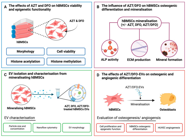
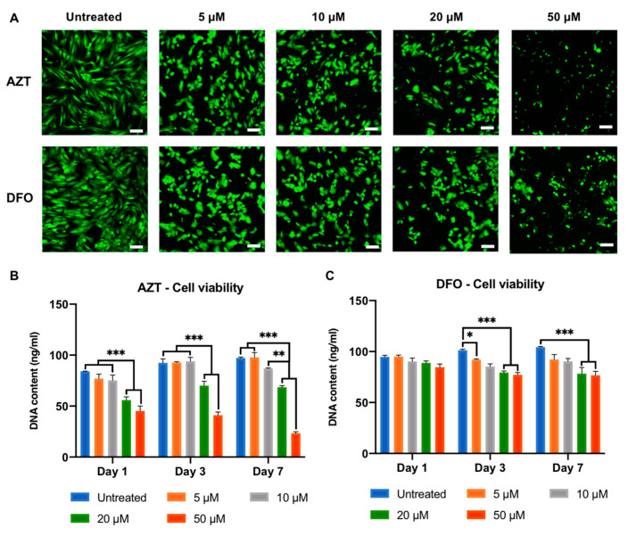
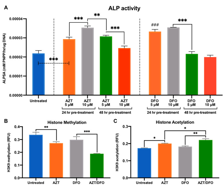
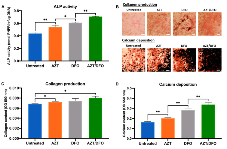
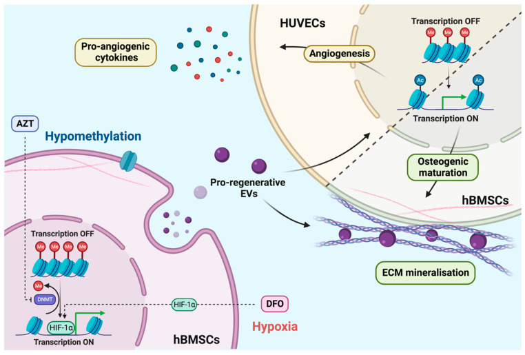
Mesenchymal stem cells (MSCs) are a promising cell population for regenerative medicine applications, where paracrine signalling through the extracellular vesicles (EVs) regulates bone tissue homeostasis and development. MSCs are known to reside in low oxygen tension, which promotes osteogenic differentiation via hypoxia-inducible factor-1α activation. Epigenetic reprogramming has emerged as a promising bioengineering strategy to enhance MSC differentiation. Particularly, the process of hypomethylation may enhance osteogenesis through gene activation. Therefore, this study aimed to investigate the synergistic effects of inducing hypomethylation and hypoxia on improving the therapeutic efficacy of EVs derived from human bone marrow MSCs (hBMSCs). The effects of the hypoxia mimetic agent deferoxamine (DFO) and the DNA methyltransferase inhibitor 5-azacytidine (AZT) on hBMSC viability was assessed by quantifying the DNA content. The epigenetic functionality was evaluated by assessing histone acetylation and histone methylation. hBMSC mineralisation was determined by quantifying alkaline phosphate activity, collagen production and calcium deposition. EVs were procured from AZT, DFO or AZT/DFO-treated hBMSCs over a two-week period, with EV size and concentration defined using transmission electron microscopy, nanoflow cytometry and dynamic light scattering. The effects of AZT-EVs, DFO-EVs or AZT/DFO-EVs on the epigenetic functionality and mineralisation of hBMSCs were evaluated. Moreover, the effects of hBMSC-EVs on human umbilical cord vein endothelial cells (HUVECs) angiogenesis was assessed by quantifying pro-angiogenic cytokine release. DFO and AZT caused a time-dose dependent reduction in hBMSC viability. Pre-treatment with AZT, DFO or AZT/DFO augmented the epigenetic functionality of the MSCs through increases in histone acetylation and hypomethylation. AZT, DFO and AZT/DFO pre-treatment significantly enhanced extracellular matrix collagen production and mineralisation in hBMSCs. EVs derived from AZT/DFO-preconditioned hBMSCs (AZT/DFO-EVs) enhanced the hBMSC proliferation, histone acetylation and hypomethylation when compared to EVs derived from AZT-treated, DFO-treated and untreated hBMSCs. Importantly, AZT/DFO-EVs significantly increased osteogenic differentiation and mineralisation of a secondary hBMSC population. Furthermore, AZT/DFO-EVs enhanced the pro-angiogenic cytokine release of HUVECs. Taken together, our findings demonstrate the considerable utility of synergistically inducing hypomethylation and hypoxia to improve the therapeutic efficacy of the MSC-EVs as a cell-free approach for bone regeneration.
Overview
The claims and statistics of this paper haven’t been checked yet.
Run the check — free, no account needed. It reads the open-access text and checks the sources it cites.
Integrity signals
Registry
Cites
Statistics
Figures
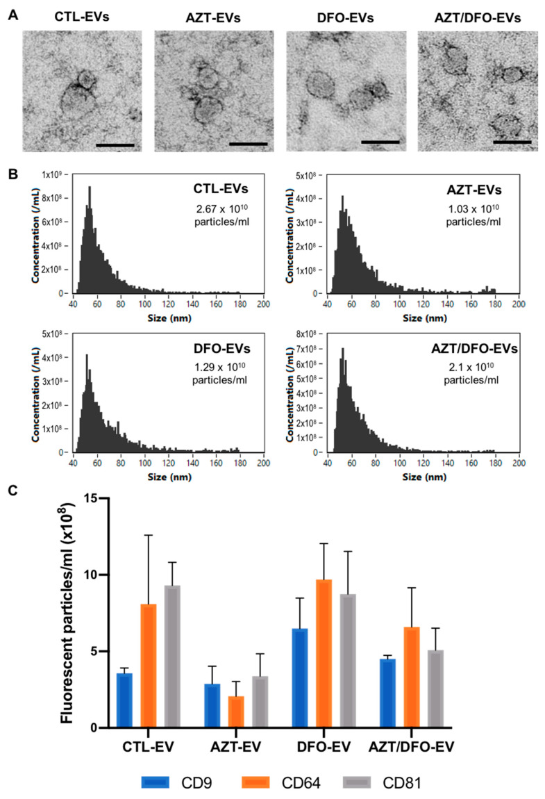
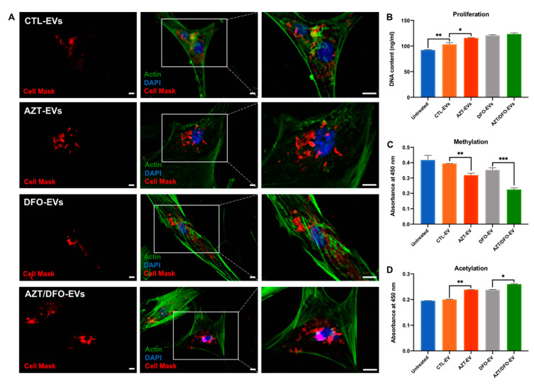
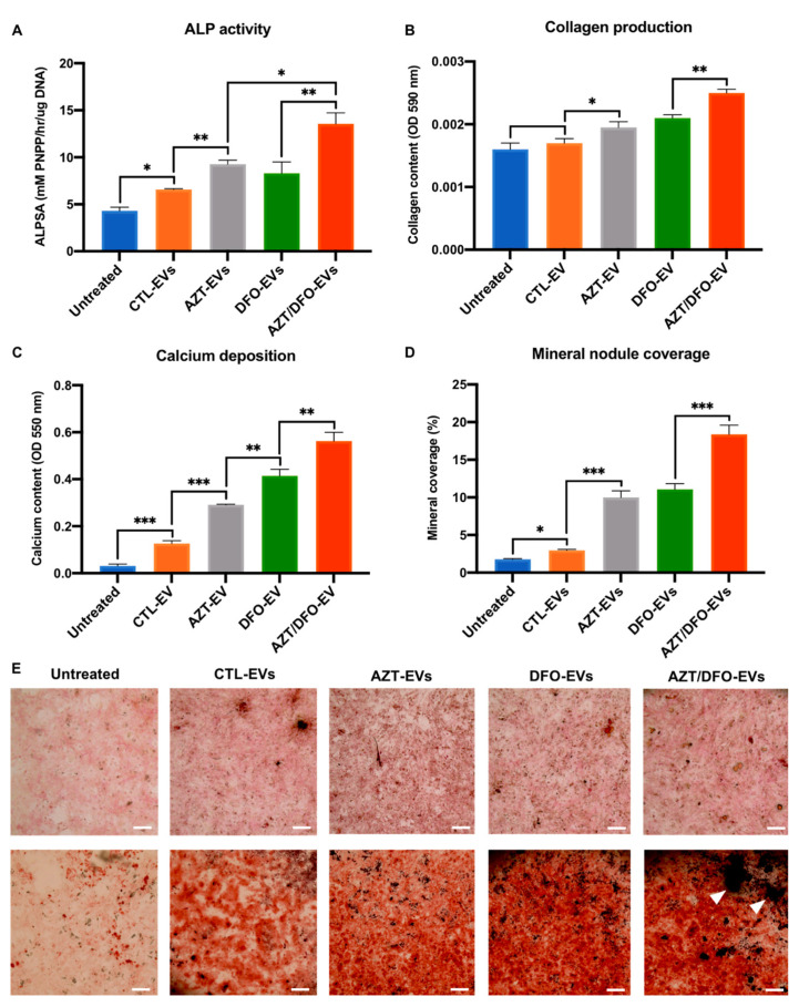
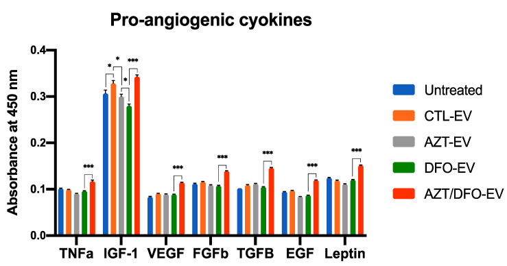
Verification
Claims the paper makes
The claims and statistics of this paper haven’t been checked yet.
Run the check — free, no account needed. It reads the open-access text and checks the sources it cites.
Claims other papers make about this one
Each is checked against this paper’s own text: does it say what the citing paper says it does?
The source backs part of the claim, not all of it — or backs it for a different population, condition or comparison.
In the citing paper
“the pre-treatment of BM-MSCs with a hypoxia inductor such as deferoxamine showed to enhance osteogenic differentiation of these cells and extracellular matrix mineralization through the decrease of histone methylation and increase of histone acetylation [131]”
From Mesenchymal stem cell secretome for regenerative medicine: Where do we stand? (2025) · cites this paper as [131]
Experimental — accuracy not yet measured
Self-check
Percentages, sums and reported differences re-computed from the paper’s own numbers.
The self-check needs the paper’s full text.
Citations
Cites
1 retracted
Jiang, Y., Zhang, J., Li, Z., & Jia, G. (2020) · Frontiers in Medicine · doi:10.3389/fmed.2020.577578 · Cited by 55
RetractedExtracellular vesicles: Exosomes, microvesicles, and friends
Raposo, G., & Stoorvogel, W. (2013) · Journal of Cell Biology · doi:10.1083/jcb.201211138 · Cited by 8,099
CpG island hypermethylation and tumor suppressor genes: a booming present, a brighter future
Esteller, M. (2002) · Oncogene · doi:10.1038/sj.onc.1205600 · Cited by 1,255
Stein, G. S., Lian, J. B., & Owen, T. A. (1990) · The FASEB Journal · doi:10.1096/fasebj.4.13.2210157 · Cited by 931
Wang, Y., Wan, C., Deng, L., Liu, X., Cao, X., Gilbert, S. R., Bouxsein, M., Monier‐Faugere, M., Guldberg, R. E., Gerstenfeld, L. C., et al. (2007) · Journal of Clinical Investigation · doi:10.1172/jci31581 · Cited by 748
Angiogenesis in bone regeneration
Hankenson, K. D., Dishowitz, M. I., Gray, C. F., & Schenker, M. L. (2011) · Injury · doi:10.1016/j.injury.2011.03.035 · Cited by 565
Exosomes and Cardiac Repair After Myocardial Infarction
Sahoo, S., & Losordo, D. W. (2014) · Circulation Research · doi:10.1161/circresaha.114.300639 · Cited by 532
Activation of the hypoxia-inducible factor-1α pathway accelerates bone regeneration
Wan, C., Gilbert, S. R., Wang, Y., Cao, X., Shen, X., Ramaswamy, G., Jacobsen, K. A., Al-Aql, Z. S., Eberhardt, A. W., Gerstenfeld, L. C., et al. (2008) · Proceedings of the National Academy of Sciences · doi:10.1073/pnas.0708474105 · Cited by 528
Liu, W., Li, L., Rong, Y., Qian, D., Chen, J., Zhou, Z. Y., Luo, Y., Jiang, D., Cheng, L., Zhao, S., et al. (2019) · Acta Biomaterialia · doi:10.1016/j.actbio.2019.12.020 · Cited by 485
Issa, J. J., & Kantarjian, H. M. (2009) · Clinical Cancer Research · doi:10.1158/1078-0432.ccr-08-2783 · Cited by 463
Cited by
Sorted by citations
Xu, G., Jin, J., Fu, Z., Wang, G., Lei, X., Xu, J., & Wang, J. (2025) · Signal Transduction and Targeted Therapy · doi:10.1038/s41392-025-02312-w · Cited by 320
Mesenchymal stem cell secretome for regenerative medicine: Where do we stand?
Trigo, C. M., Rodrigues, J. S., Camões, S. P., Solá, S., & Miranda, J. P. (2025) · Journal of Advanced Research · doi:10.1016/j.jare.2024.05.004 · Cited by 252
Mesenchymal Stem Cell-Derived Exosomal microRNAs in Cardiac Regeneration
Bhaskara, M. C., Anjorin, O., & Wang, M. (2023) · Cells · doi:10.3390/cells12242815 · Cited by 40
Chen, Y., Li, M., Yang, J., Chen, Y., & Wang, J. (2025) · Stem Cell Research & Therapy · doi:10.1186/s13287-025-04490-4 · Cited by 22
Extracellular vesicles as modifiers of epigenomic profiles
Zhou, H., Hu, S. C., & Yan, W. (2024) · Trends in Genetics · doi:10.1016/j.tig.2024.05.005 · Cited by 21
Huang, Y. L., & Xie, H. (2025) · International Journal of Nanomedicine · doi:10.2147/ijn.s522198 · Cited by 20
Yu, Q., Chen, B., Sun, E., Lu, X., Li, Z., Wang, R., & Fang, D. (2024) · Molecular Neurobiology · doi:10.1007/s12035-024-04118-3 · Cited by 17
Shi, H., Yang, Y., Xing, H., Jia, J., Xiong, W., Guo, S., & Yang, S. (2024) · Journal of Tissue Engineering · doi:10.1177/20417314241286606 · Cited by 15
Salama, A. N., Badr, E. A. E., Holah, N. S., Barbary, A. A. E., & Hessien, M. (2023) · Reproductive Sciences · doi:10.1007/s43032-023-01389-4 · Cited by 11
Advances in extracellular vesicle-based nanomedicine for regenerative orthopaedics
Izquierdo, S. G., Pilar, A. F., Rios, J. L., Lim, K. S., Toh, W. S., Liu, C., Gimona, M., Gawlitta, D., & Man, K. (2025) · Journal of Nanobiotechnology · doi:10.1186/s12951-025-03906-w · Cited by 13
Self-citationExtracellular vesicles in osteoimmunomodulation: Orchestrating immune-driven bone regeneration
Pol, F., Longoni, A., Levato, R., Gawlitta, D., & Man, K. (2025) · International Journal of Biological Macromolecules · doi:10.1016/j.ijbiomac.2025.149614 · Cited by 7
Self-citationShi, J., Liu, Z., Xie, J., Zhao, Z., Huang, X., & Cao, Z. (2025) · Acta Biomaterialia · doi:10.1016/j.actbio.2025.11.060 · Cited by 4
Song, W., Zhang, W., Hu, Y., Li, T., Lin, X., Liu, H., Odle, J., See, M. T., Wang, S., Cui, X., et al. (2026) · Poultry Science · doi:10.1016/j.psj.2026.106930 · Cited by 3
Filho, G. P. C., Tavares, P. D. A., de Jesus, A. F., Ciancaglini, P., Segundo, J. E. S., & Ramos, A. P. (2025) · Biophysical Reviews · doi:10.1007/s12551-025-01282-1 · Cited by 3
Recent advances of DNA methylation in osteonecrosis of the femoral head
Zhou, T., Liu, H., Li, H., Zhang, Y., & Li, X. (2025) · Frontiers in Genetics · doi:10.3389/fgene.2025.1526852 · Cited by 2
Zhu, H., Cheng, K., Tian, M., Peng, Y., Zhang, Y., Yan, H., Fan, S., Shang, B., Wu, J., Ding, H., et al. (2025) · iScience · doi:10.1016/j.isci.2025.113565 · Cited by 2
Stein, J. B., Arif, M., Cheung, H., & Lee, K. (2026) · Cell Biomaterials · doi:10.1016/j.celbio.2026.100495 · Cited by 2
Overview of biophysical approaches to study osteogenic proteins in lipidic interfaces
Hayann, L., de Melo, M. T., Cominal, J. G., Lima, I. S., Andrilli, L. H. D. S., Machado, J. P. M., de Sousa, J. D. R. S., Azevedo, P. A. B. S., Oliveira, M. F., Ramos, A. P., et al. (2026) · Biophysical Reviews · doi:10.1007/s12551-026-01455-6 · Cited by 1
TENG, H., Cho, M., MA, Z., Ji, X., Jin, S., ZHOU, Y., Xuan, M., Seong, H., LI, X., CHOI, S.-H., et al. (2024) · In Vivo · doi:10.21873/invivo.13825 · Cited by 1
Unnithan, A. R., Man, K., Kritika, Gethings, L. A., Hughes, C. J., Keenan, A., Heaney, L. M., Cox, S. C., Davies, O. G., & Haj, A. J. E. (2026) · Advanced Healthcare Materials · doi:10.1002/adhm.202504542 · Cited by 2
Self-citationDing, Y., Lin, F., & Liang, X. (2024) · World Journal of Stem Cells · doi:10.4252/wjsc.v16.i11.974
Salehy, S., Nokhbatolfoghahaei, H., & Khojasteh, A. (2025) · Biomaterials Advances · doi:10.1016/j.bioadv.2025.214661
Zhang, Y., Zhang, Y., Dong, Q., Zhang, Z., Ji, Z., Deng, Y., & Chen, Y. (2026) · BMC Oral Health · doi:10.1186/s12903-026-08189-7
Osteoimmunomodulatory Stem Cell Nanoghosts as a Novel Nanotherapeutic for Bone RegenerationPreprint
Micheli, G. A., Yang, T., Gawlitta, D., & Man, K. (2026) · bioRxiv (Cold Spring Harbor Laboratory) · doi:10.64898/2026.05.11.724218
Self-citationMechanosensing and mechanotransduction in bone cells
Kerdjoudj, H., & Schiavi, J. (2026) · Elsevier eBooks · doi:10.1016/b978-0-443-26739-0.00023-5
Metadata
- Authors
- Man, K., Brunet, M. Y., Lees, R., Peacock, B., & Cox, S. C.
- Affiliations
- University of Birmingham (GB)
- NanoFCM Co., Ltd., Nottingham NG90 6BH, UK
- Corresponding author
- Cox, Sophie C.
- Venue
- International Journal of Molecular Sciences
- Publisher
- MDPI AG
- Published
- 2023-04-20
- Volume
- 24
- Issue
- 8
- Pages
- 7564
- ISSN
- 1422-0067 (ISSN-L)
- PMC
- PMC10142722
- OpenAlex
- W4366590089
- Cited by
- 25
- References
- 75
- Citations by year
- 2026: 7 · 2025: 10 · 2024: 6 · 2023: 2
- FWCI
- 4.33
- Type
- journal article
- Language
- en
- Open access
- gold
- doi.org
- Licence
- Locations
- International Journal of Molecular Sciences (publishedVersion, cc-by)
- PubMed Central (publishedVersion, cc-by)
- PubMed (publishedVersion)
- University of Birmingham Research Portal (University of Birmingham) (publishedVersion, cc-by)
- Funders
- EPSRC (EP/S016589/1)
- Topics
- Life Sciences › Biochemistry, Genetics and Molecular Biology › Cell Biology › Mesenchymal stem cell research
- Life Sciences › Biochemistry, Genetics and Molecular Biology › Molecular Biology › Extracellular vesicles in disease
- Physical Sciences › Engineering › Biomedical Engineering › Bone Tissue Engineering Materials
- Keywords
- epigenetic reprogramming, DNA hypomethylation, hypoxia, bone marrow mesenchymal stem cells, azacitidine, deferoxamine, histone acetylation, histone methylation, osteogenic differentiation, bone regeneration, angiogenesis, paracrine signaling, DNA methyltransferases
- MeSH terms
- Extracellular Vesicles, Hypoxia, Cells, Cultured, DNA, Histones, Humans, Osteogenesis, Cytokines, Epigenesis, Genetic, Mesenchymal Stem Cells, Human Umbilical Vein Endothelial Cells
About these checks
Registry, retraction, Cites and statistics results are computed, not judged, with one exception: a printed reference that no index lists is read once by a model before it counts as a citation error. Claims are extracted by a model and kept only when their quote appears verbatim in the text; each cited source is then checked, with a model’s help, for whether it says what the claim relies on. Those verdicts can be wrong — read the claim’s quote and the cited source before relying on one.
Statistics and arithmetic are recomputed, not judged.
Confidence bands are shown only for fields calibrated on our annotated test set. None is calibrated yet, so no band is shown.
What counts as a claim ›How we measure confidence ›How we check this ›
Are you an author, or do you see a problem with this page? Request a review
