

The DOI matches its Crossref record; 24 of the 24 references it cites were found in a registry or an index.
Molecular Origins of the Mendelian Rare Diseases Reviewed by Orpha.net: A Structural Bioinformatics Investigation
Open accessPublished version of a preprint
Abstract
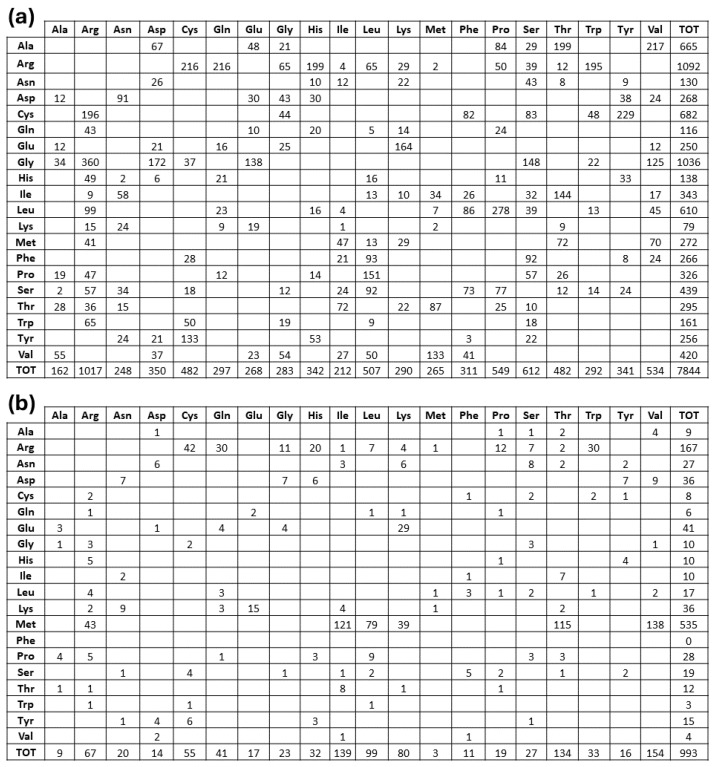
The study of rare diseases is important not only for the individuals affected but also for the advancement of medical knowledge and a deeper understanding of human biology and genetics. The wide repertoire of structural information now available from reliable and accurate prediction methods provides the opportunity to investigate the molecular origins of most of the rare diseases reviewed in the Orpha.net database. Thus, it has been possible to analyze the topology of the pathogenic missense variants found in the 2515 proteins involved in Mendelian rare diseases (MRDs), which form the database for our structural bioinformatics study. The amino acid substitutions responsible for MRDs showed different mutation site distributions at different three-dimensional protein depths. We then highlighted the depth-dependent effects of pathogenic variants for the 20,061 pathogenic variants that are present in our database. The results of this structural bioinformatics investigation are relevant, as they provide additional clues to mitigate the damage caused by MRD.
Overview
The claims and statistics of this paper haven’t been checked yet.
Run the check — free, no account needed. It reads the open-access text and checks the sources it cites.
Integrity signals
Registry
Cites
Statistics
Figures
Verification
Claims the paper makes
The claims and statistics of this paper haven’t been checked yet.
Run the check — free, no account needed. It reads the open-access text and checks the sources it cites.
Self-check
Percentages, sums and reported differences re-computed from the paper’s own numbers.
The self-check needs the paper’s full text.
Citations
Cites
Highly accurate protein structure prediction with AlphaFold
Jumper, J., Evans, R., Pritzel, A., Green, T., Figurnov, M., Ronneberger, O., Tunyasuvunakool, K., Bates, R., Žídek, A., Potapenko, A., et al. (2021) · Nature · doi:10.1038/s41586-021-03819-2 · Cited by 43,764
Berman, H. M. (2000) · Nucleic Acids Research · doi:10.1093/nar/28.1.235 · Cited by 40,196
Shrestha, S., & Offer, S. M. (2016) · Medical Epigenetics · doi:10.1159/000444713 · Cited by 8,914
UniProt: the Universal Protein Knowledgebase in 2023
The UniProt Consortium, Bateman, A., Martin, M.-J., Orchard, S., Magrane, M., Ahmad, S., Alpi, E., Bowler-Barnett, E. H., Britto, R., Bye-A-Jee, H., et al. (2023) · Nucleic Acids Research · doi:10.1093/nar/gkac1052 · Cited by 7,210
Protein complex prediction with AlphaFold-Multimer
Evans, R., O’Neill, M., Pritzel, A., Antropova, N., Senior, A., Green, T., Žídek, A., Bates, R., Blackwell, S., Yim, J., et al. (2021) · doi:10.1101/2021.10.04.463034 · Cited by 3,808
ClinVar: public archive of interpretations of clinically relevant variants
Landrum, M. J., Lee, J. M., Benson, M., Brown, G., Chao, C., Chitipiralla, S., Gu, B., Hart, J., Hoffman, D., Hoover, J., et al. (2016) · Nucleic Acids Research · doi:10.1093/nar/gkv1222 · Cited by 2,758
Amino Acid Difference Formula to Help Explain Protein Evolution
Grantham, R. (1974) · Science · doi:10.1126/science.185.4154.862 · Cited by 2,363
Accurate proteome-wide missense variant effect prediction with AlphaMissense
Cheng, J., Novati, G., Pan, J., Bycroft, C., Žemgulytė, A., Applebaum, T., Pritzel, A., Wong, L. H., Zielinski, M., Sargeant, T., et al. (2023) · Science · doi:10.1126/science.adg7492 · Cited by 2,163
Estimating cumulative point prevalence of rare diseases: analysis of the Orphanet database
Nguengang Wakap, S., Lambert, D. M., Olry, A., Rodwell, C., Gueydan, C., Lanneau, V., Murphy, D., Le Cam, Y., & Rath, A. (2020) · European Journal of Human Genetics · doi:10.1038/s41431-019-0508-0 · Cited by 1,752
Two types of amino acid substitutions in protein evolution
Miyata, T., Miyazawa, S., & Yasunaga, T. (1979) · Journal of Molecular Evolution · doi:10.1007/bf01732340 · Cited by 465
POPS: a fast algorithm for solvent accessible surface areas at atomic and residue level
Cavallo, L. (2003) · Nucleic Acids Research · doi:10.1093/nar/gkg601 · Cited by 222
The amino-acid mutational spectrum of human genetic disease
Vitkup, D., Sander, C., & Church, G. M. (2003) · Genome biology · doi:10.1186/gb-2003-4-11-r72 · Cited by 208
Clinical Practice Guidelines for Rare Diseases: The Orphanet Database
Pavan, S., Rommel, K., Marquina, M. E. M., Höhn, S., Serrière-Lanneau, V., & Rath, A. (2017) · PLoS ONE · doi:10.1371/journal.pone.0170365 · Cited by 202
Genetic medicines: treatment strategies for hereditary disorders
O’Connor, T. P., & Crystal, R. G. (2006) · Nature Reviews Genetics · doi:10.1038/nrg1829 · Cited by 189
Arginine residues at internal positions in a protein are always charged
Harms, M. J., Schlessman, J. L., Sue, G. R., & Bertrand García‐Moreno, E. (2011) · Proceedings of the National Academy of Sciences · doi:10.1073/pnas.1104808108 · Cited by 185
Non-randomness of Ammo-acid Changes in the Evolution of Homologous Proteins
Epstein, C. J. (1967) · Nature · doi:10.1038/215355a0 · Cited by 116
Häckel, M., Hinz, H., & Hedwig, G. R. (1999) · Biophysical Chemistry · doi:10.1016/s0301-4622(99)00104-0 · Cited by 107
Glycine Residues Appear to Be Evolutionarily Conserved for Their Ability to Inhibit Aggregation
Parrini, C., Taddei, N., Ramazzotti, M., Degl’Innocenti, D., Ramponi, G., Dobson, C. M., & Chiti, F. (2005) · Structure · doi:10.1016/j.str.2005.04.022 · Cited by 97
Teng, S., Srivastava, A. K., Schwartz, C. E., Alexov, E., & Wang, L. (2010) · International Journal of Computational Biology and Drug Design · doi:10.1504/ijcbdd.2010.038396 · Cited by 87
Disease-causing mutations in the human genome
Antonarakis, S. E., Krawczak, M., & Cooper, D. N. (2000) · European Journal of Pediatrics · doi:10.1007/pl00014395 · Cited by 80
Sharo, A. G., Zou, Y., Adhikari, A. N., & Brenner, S. E. (2023) · Genome Medicine · doi:10.1186/s13073-023-01199-y · Cited by 52
Pathogenic signal peptide variants in the human genome
Guarnizo, S. A. G., Kellogg, M. K., Miller, S. C., Tikhonova, E. B., Karamysheva, Z. N., & Karamyshev, A. L. (2023) · NAR Genomics and Bioinformatics · doi:10.1093/nargab/lqad093 · Cited by 34
Laddach, A., Ng, J. C.-F., & Fraternali, F. (2021) · PLoS Biology · doi:10.1371/journal.pbio.3001207 · Cited by 26
Bongini, P., Niccolai, N., Trezza, A., Mangiavacchi, G., Santucci, A., Spiga, O., Bianchini, M., & Gardini, S. (2020) · IEEE Transactions on Computational Biology and Bioinformatics · doi:10.1109/tcbb.2020.3033384 · Cited by 3
Self-citation
Metadata
- Authors
- Visibelli, A., Finetti, R., Niccolai, N., Spiga, O., & Santucci, A.
- Affiliations
- University of Siena (IT)
- Corresponding author
- Niccolai, Neri
- Venue
- International Journal of Molecular Sciences
- Publisher
- MDPI AG
- Published
- 2024-06-25
- Volume
- 25
- Issue
- 13
- Pages
- 6953
- ISSN
- 1422-0067 (ISSN-L)
- PMC
- PMC11241713
- OpenAlex
- W4400092293
- Cited by
- 1
- References
- 24
- Citations by year
- 2025: 1
- FWCI
- 0.27
- Type
- journal article
- Language
- en
- Open access
- gold
- doi.org
- Licence
- Locations
- International Journal of Molecular Sciences (publishedVersion, cc-by)
- Preprints.org (acceptedVersion, cc-by)
- PubMed Central (publishedVersion, cc-by)
- PubMed (publishedVersion)
- Use Siena air (University of Siena) (submittedVersion, cc-by)
- Topics
- Life Sciences › Biochemistry, Genetics and Molecular Biology › Genetics › Genomics and Rare Diseases
- Life Sciences › Biochemistry, Genetics and Molecular Biology › Molecular Biology › Protein Structure and Dynamics
- Life Sciences › Biochemistry, Genetics and Molecular Biology › Structural Biology › Enzyme Structure and Function
- Keywords
- protein structure, amino acid substitutions, structural bioinformatics
- MeSH terms
- Humans, Models, Molecular, Protein Conformation, Proteins, Computational Biology, Amino Acid Substitution, Mutation, Missense, Databases, Genetic, Rare Diseases
About these checks
Registry, retraction, Cites and statistics results are computed, not judged, with one exception: a printed reference that no index lists is read once by a model before it counts as a citation error. Claims are extracted by a model and kept only when their quote appears verbatim in the text; each cited source is then checked, with a model’s help, for whether it says what the claim relies on. Those verdicts can be wrong — read the claim’s quote and the cited source before relying on one.
Statistics and arithmetic are recomputed, not judged.
Confidence bands are shown only for fields calibrated on our annotated test set. None is calibrated yet, so no band is shown.
What counts as a claim ›How we measure confidence ›How we check this ›
Are you an author, or do you see a problem with this page? Request a review
Data sources
Bibliographic data: Semantic Scholar, OpenAlex, Crossref.
Contains information from the Semantic Scholar Academic Graph Datasets, made available under the ODC Attribution License (ODC-BY 1.0).
Semantic Scholar’s data is described in: Kinney, R., et al. (2023). The Semantic Scholar Open Data Platform. arXiv:2301.10140.
