

The DOI matches its Crossref record; 310 of the 312 references it cites were found in a registry or an index.
Bone Morphogenetic Protein-2 in Development and Bone Homeostasis
Open accessAbstract
Bone morphogenetic proteins (BMPs) are multi-functional growth factors belonging to the Transforming Growth Factor-Beta (TGF-β) superfamily. These proteins are essential to many developmental processes, including cardiogenesis, neurogenesis, and osteogenesis. Specifically, within the BMP family, Bone Morphogenetic Protein-2 (BMP-2) was the first BMP to be characterized and has been well-studied. BMP-2 has important roles during embryonic development, as well as bone remodeling and homeostasis in adulthood. Some of its specific functions include digit formation and activating osteogenic genes, such as Runt-Related Transcription Factor 2 (RUNX2). Because of its diverse functions and osteogenic potential, the Food and Drug Administration (FDA) approved usage of recombinant human BMP-2 (rhBMP-2) during spinal fusion surgery, tibial shaft repair, and maxillary sinus reconstructive surgery. However, shortly after initial injections of rhBMP-2, several adverse complications were reported, and alternative therapeutics have been developed to limit these side-effects. As the clinical application of BMP-2 is largely implicated in bone, we focus primarily on its role in bone. However, we also describe briefly the role of BMP-2 in development. We then focus on the structure of BMP-2, its activation and regulation signaling pathways, BMP-2 clinical applications, and limitations of using BMP-2 as a therapeutic. Further, this review explores other potential treatments that may be useful in treating bone disorders.
Overview
The claims and statistics of this paper haven’t been checked yet.
Run the check — free, no account needed. It reads the open-access text and checks the sources it cites.
Integrity signals
Registry
Cites
Statistics
Figures
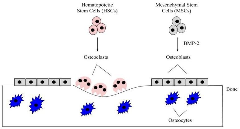
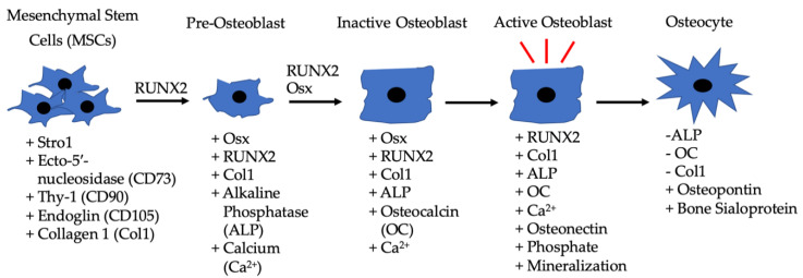
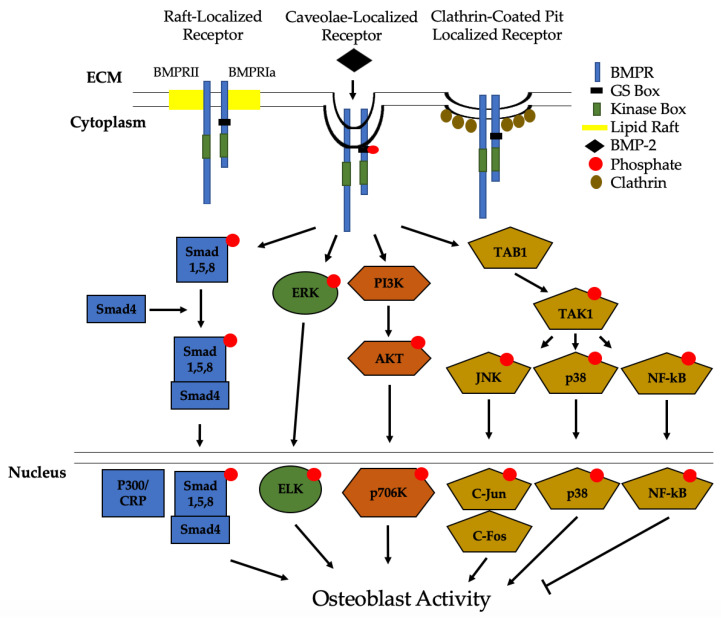
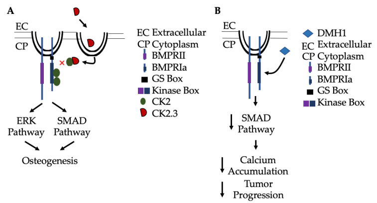
Verification
Claims the paper makes
The claims and statistics of this paper haven’t been checked yet.
Run the check — free, no account needed. It reads the open-access text and checks the sources it cites.
Claims other papers make about this one
Each is checked against this paper’s own text: does it say what the citing paper says it does?
The cited source says what the claim attributes to it.
In the citing paper
“BMP-2 has known roles in development [36], on tumorigenesis, [37] and in various body tissues [38–40].”
From Keratin Scaffold Formulation Impacts rhBMP-2 Biodistribution and Bone Regeneration in a Rat Femur Defect Model (2025) · cites this paper as [36]
Self-check
Percentages, sums and reported differences re-computed from the paper’s own numbers.
The self-check needs the paper’s full text.
Citations
Cites
1 retracted
Li, S., Peng, C., & Yang, Q. (2019) · Journal of bone oncology · doi:10.1016/j.jbo.2019.100217 · Cited by 14
RetractedBone: Formation by Autoinduction
Urist, M. R. (1965) · Science · doi:10.1126/science.150.3698.893 · Cited by 5,529
Neer, R. M., Arnaud, C. D., Zanchetta, J. R., Prince, R., Gaich, G. A., Reginster, J.-Y., Hodsman, A. B., Eriksen, E. F., Ish-Shalom, S., Genant, H. K., et al. (2001) · New England Journal of Medicine · doi:10.1056/nejm200105103441904 · Cited by 4,602
TGF-β signalling from cell membrane to nucleus through SMAD proteins
Heldin, C., Miyazono, K., & ten Dijke, P. (1997) · Nature · doi:10.1038/37284 · Cited by 3,697
Biology of Bone Tissue: Structure, Function, and Factors That Influence Bone Cells
Florencio-Silva, R., Sasso, G. R. D. S., Sasso-Cerri, E., Simões, M. J., & Cerri, P. S. (2015) · BioMed Research International · doi:10.1155/2015/421746 · Cited by 2,044
Chen, D., Zhao, M., & Mundy, G. R. (2004) · Growth Factors · doi:10.1080/08977190412331279890 · Cited by 1,921
Cummings, S. R., Eckert, S., Krueger, K. A., Grady, D., Powles, T. J., Cauley, J. A., Norton, L., Nickelsen, T., Bjarnason, N. H., Morrow, M., et al. (1999) · JAMA · doi:10.1001/jama.281.23.2189 · Cited by 1,872
TGF-β and BMP Signaling in Osteoblast Differentiation and Bone Formation
Chen, G., Deng, C., & Li, Y.-P. (2012) · International Journal of Biological Sciences · doi:10.7150/ijbs.2929 · Cited by 1,756
Khosla, S., Burr, D., Cauley, J., Dempster, D. W., Ebeling, P. R., Felsenberg, D., Gagel, R. F., Gilsanz, V., Guise, T., Koka, S., et al. (2007) · Journal of Bone and Mineral Research · doi:10.1359/jbmr.0707onj · Cited by 1,650
Research works printed but not linked: 2
- Steele, D.G., and Bramblett, C.A. (1988). The Anatomy and Biology of the Human Skeleton, Texas A&M University Press.
- (2020, April 23). The Basics of Bone in Health and Disease, Available online: https://www.ncbi.nlm.nih.gov/books/NBK45504/.
Cited by
Sorted by citations
Lu, P., Ruan, D., Huang, M., Tian, M., Zhu, K., Gan, Z., & Xiao, Z. (2024) · Signal Transduction and Targeted Therapy · doi:10.1038/s41392-024-01852-x · Cited by 762
Roles of mechanosensitive channel Piezo1/2 proteins in skeleton and other tissues
Qin, L., He, T., Chen, S., Yang, D., Yi, W., Cao, H., & Xiao, G. (2021) · Bone Research · doi:10.1038/s41413-021-00168-8 · Cited by 251
Progress of Wnt Signaling Pathway in Osteoporosis
Gao, Y., Chen, N., Fu, Z., & Zhang, Q. (2023) · Biomolecules · doi:10.3390/biom13030483 · Cited by 184
Li, Y., Zhu, J., Zhang, X., Li, Y., Zhang, S., Yang, L., Li, R., Wan, Q., Pei, X., Chen, J., et al. (2023) · ACS Applied Materials & Interfaces · doi:10.1021/acsami.2c23107 · Cited by 127
The Development of Molecular Biology of Osteoporosis
Gao, Y., Patil, S., & Jia, J. (2021) · International Journal of Molecular Sciences · doi:10.3390/ijms22158182 · Cited by 116
Metadata
- Authors
- Halloran, D., Durbano, H. W., & Nohe, A.
- Affiliations
- University of Delaware (US)
- Corresponding author
- Nohe, Anja
- Venue
- Journal of Developmental Biology
- Publisher
- MDPI AG
- Published
- 2020-09-13
- Volume
- 8
- Issue
- 3
- Pages
- 19
- ISSN
- 2221-3759 (ISSN-L)
- PMC
- PMC7557435
- OpenAlex
- W3085579791
- Cited by
- 348
- References
- 312
- Citations by year
- 2026: 57 · 2025: 86 · 2024: 79 · 2023: 42 · 2022: 55 · 2021: 27 · 2020: 1
- FWCI
- 2.99
- Type
- journal article
- Language
- en
- Open access
- gold
- doi.org
- Licence
- Locations
- Journal of Developmental Biology (publishedVersion, cc-by)
- PubMed Central (publishedVersion, cc-by)
- PubMed (publishedVersion)
- DOAJ (DOAJ: Directory of Open Access Journals) (submittedVersion, cc-by-sa)
- Topics
- Life Sciences › Biochemistry, Genetics and Molecular Biology › Molecular Biology › TGF-β signaling in diseases
- Life Sciences › Biochemistry, Genetics and Molecular Biology › Molecular Biology › Bone Metabolism and Diseases
- Life Sciences › Biochemistry, Genetics and Molecular Biology › Molecular Biology › Developmental Biology and Gene Regulation
- Keywords
- BMP-2, BMP signaling, TGF-beta superfamily, bone homeostasis, bone remodeling, bone formation, embryonic development, digit formation, Runx2, recombinant human BMP-2, bone graft substitutes, spinal fusion, maxillary sinus reconstruction, bone disease
About these checks
Registry, retraction, Cites and statistics results are computed, not judged, with one exception: a printed reference that no index lists is read once by a model before it counts as a citation error. Claims are extracted by a model and kept only when their quote appears verbatim in the text; each cited source is then checked, with a model’s help, for whether it says what the claim relies on. Those verdicts can be wrong — read the claim’s quote and the cited source before relying on one.
Statistics and arithmetic are recomputed, not judged.
Confidence bands are shown only for fields calibrated on our annotated test set. None is calibrated yet, so no band is shown.
What counts as a claim ›How we measure confidence ›How we check this ›
Are you an author, or do you see a problem with this page? Request a review
