

The DOI matches its Crossref record.
Antibiotic-Resistant Desulfovibrio Produces H2S from Supplements for Animal Farming
Open accessAbstract
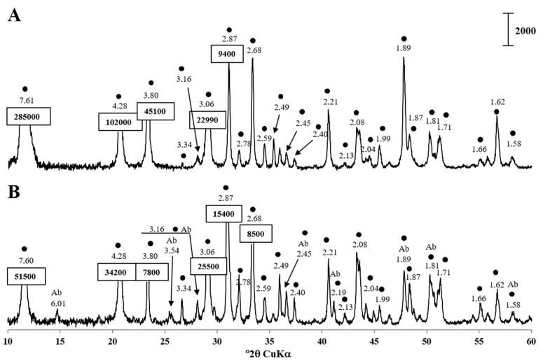
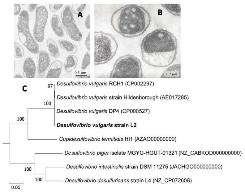
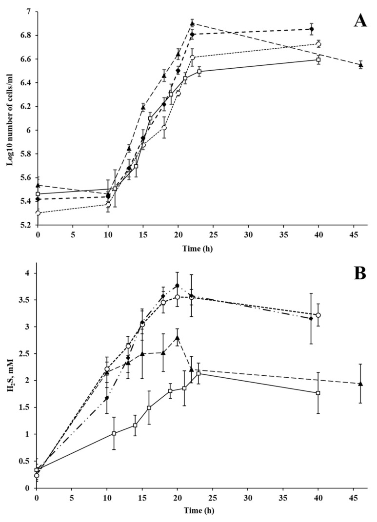
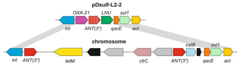
Sulphate-reducing bacteria, primarily Desulfovibrio, are responsible for the active generation of H2S in swine production waste. The model species for sulphate reduction studies, Desulfovibrio vulgaris strain L2, was previously isolated from swine manure characterized by high rates of dissimilatory sulphate reduction. The source of electron acceptors in low-sulphate swine waste for the high rate of H2S formation remains uncertain. Here, we demonstrate the ability of the L2 strain to use common animal farming supplements including L-lysine-sulphate, gypsum and gypsum plasterboards as electron acceptors for H2S production. Genome sequencing of strain L2 revealed the presence of two megaplasmids and predicted resistance to various antimicrobials and mercury, which was confirmed in physiological experiments. Most of antibiotic resistance genes (ARG) are carried by two class 1 integrons located on the chromosome and on the plasmid pDsulf-L2-2. These ARGs, predicted to confer resistance to beta-lactams, aminoglycosides, lincosamides, sulphonamides, chloramphenicol and tetracycline, were probably laterally acquired from various Gammaproteobacteria and Firmicutes. Resistance to mercury is likely enabled by two mer operons also located on the chromosome and on pDsulf-L2-2 and acquired via horizontal gene transfer. The second megaplasmid, pDsulf-L2-1, encoded nitrogenase, catalase and type III secretion system suggesting close contact of the strain with intestinal cells in the swine gut. The location of ARGs on mobile elements allows us to consider D. vulgaris strain L2 as a possible vector transferring antimicrobials resistance determinants between the gut microbiote and microbial communities in environmental biotopes.
Overview
The claims and statistics of this paper haven’t been checked yet.
Run the check — free, no account needed. It reads the open-access text and checks the sources it cites.
Integrity signals
Registry
Cites
Statistics
Figures
Verification
Claims the paper makes
The claims and statistics of this paper haven’t been checked yet.
Run the check — free, no account needed. It reads the open-access text and checks the sources it cites.
Claims other papers make about this one
Each is checked against this paper’s own text: does it say what the citing paper says it does?
The source backs part of the claim, not all of it — or backs it for a different population, condition or comparison.
In the citing paper
“The identification of mobile antimicrobial resistance genes and their potential impact on intestinal resistance also requires whole-genome sequencing and detailed characterization of antibiotic resistance genes [42,66,67,68].”
From Current Concepts in Probiotic Safety and Efficacy (2026) · cites this paper as [68]
Experimental — accuracy not yet measured
Self-check
Percentages, sums and reported differences re-computed from the paper’s own numbers.
The self-check needs the paper’s full text.
Citations
Cites
No references could be resolved yet.
Research works printed but not linked: 59
- Fox, 1994, Intracellular Campylobacter-like organism from ferrets and hamsters with proliferative bowel disease is a Desulfovibrio sp., J. Clin. Microbiol., 32, 1229, 10.1128/jcm.32.5.1229-1237.1994 · DOI
- Beerens, 1977, Sulfate-reducing anaerobic bacteria in human feces, Am. J. Clin. Nutr., 30, 1770, 10.1093/ajcn/30.11.1770 · DOI
- Gibson, 1988, Competition for hydrogen between sulphate-reducing bacteria and methanogenic bacteria from the human large intestine, J. Appl. Bacteriol., 65, 241, 10.1111/j.1365-2672.1988.tb01891.x · DOI
- Goldstein, 2003, Desulfovibrio desulfuricans bacteremia and review of human Desulfovibrio infections, J. Clin. Microbiol., 41, 2752, 10.1128/JCM.41.6.2752-2754.2003 · DOI
- Fite, 2004, Identification and quantitation of mucosal and faecal desulfovibrios using real time polymerase chain reaction, Gut, 53, 523, 10.1136/gut.2003.031245 · DOI
- Liu, 2013, Development of a group-specific PCR-based DGGE for analyzing sulfate-reducing bacteria in human gut, Chin. J. Microbiol., 25, 706
- Rey, 2013, Metabolic niche of a prominent sulfate-reducing human gut bacterium, Proc. Nat. Acad. Sci. USA, 110, 13582, 10.1073/pnas.1312524110 · DOI
- Barton, 2017, Sulfur Cycling and the Intestinal Microbiome, Dig. Dis. Sci., 62, 2241, 10.1007/s10620-017-4689-5 · DOI
- Pitcher, 2000, The contribution of sulphate reducing bacteria and 5-aminosalicylic acid to faecal sulphide in patients with ulcerative colitis, Gut, 46, 64, 10.1136/gut.46.1.64 · DOI
- Fiachra, 2010, Bacterial colonization of colonic crypt mucous gel and disease activity in ulcerative colitis, Ann. Surg., 252, 869, 10.1097/SLA.0b013e3181fdc54c · DOI
- Loubinoux, 2002, Sulfate-reducing bacteria in human feces and their association with inflammatory bowel diseases, FEMS Microbiol. Ecol., 40, 107, 10.1111/j.1574-6941.2002.tb00942.x · DOI
- Scanlan, 2009, Culture-independent analysis of desulfovibrios in the human distal colon of healthy, colorectal cancer and polypectomized individuals, FEMS Microbiol. Ecol., 69, 213, 10.1111/j.1574-6941.2009.00709.x · DOI
- Wolf, 2022, Diversity and distribution of sulfur metabolic genes in the human gut microbiome and their association with colorectal cancer, Microbiome, 10, 64, 10.1186/s40168-022-01242-x · DOI
- McDougall, 1997, Bacteremia caused by a recently described novel Desulfovibrio species, J. Clin. Microbiol., 35, 1805, 10.1128/jcm.35.7.1805-1808.1997 · DOI
- Loubinoux, 2000, Bacteremia caused by a strain of Desulfovibrio related to the provisionally named Desulfovibrio fairfieldensis, J. Clin. Microbiol., 38, 931, 10.1128/JCM.38.2.931-934.2000 · DOI
- Shukla, 2000, Desulfovibrio desulfuricans bacteremia in a dog, J. Clin. Microbiol., 38, 1701, 10.1128/JCM.38.4.1701-1702.2000 · DOI
- Hagiya, 2018, Desulfovibrio desulfuricans bacteremia: A case report and literature review, Anaerobe, 49, 112, 10.1016/j.anaerobe.2017.12.013 · DOI
- Lozniewski, 1999, First isolation of Desulfovibrio species as part of a polymicrobial infection from a brain abscess, Eur. J. Clin. Microbiol. Infect. Dis., 18, 602, 10.1007/s100960050357 · DOI
- Okamoto, Y., Miyabe, Y., Seki, M., Ushio, Y., Sato, K., Kasama, E., Akiyama, K., Karasawa, K., Uchida, K., and Kikuchi, K. (2022). First case of a renal cyst infection caused by Desulfovibrio: A case report and literature review. BMC Nephrol., 23. · DOI
- Lin, 2022, Pathogenic effects of Desulfovibrio in the gut on fatty liver in diet-induced obese mice and children with obesity, J. Gastroenterol., 57, 913, 10.1007/s00535-022-01909-0 · DOI
- Vasoo, 2014, Desulfovibrio legallii prosthetic shoulder joint infection and review of antimicrobial susceptibility and clinical characteristics of Desulfovibrio infections, J. Clin. Microbiol., 52, 3105, 10.1128/JCM.00083-14 · DOI
- Nakao, 2009, Susceptibilities of 23 Desulfovibrio isolates from humans. Antimicrob, Agents Chemother., 53, 5308, 10.1128/AAC.00630-09 · DOI
- Carattoli, 2013, Plasmids and the spread of resistance, Int. J. Med. Microbiol., 303, 298, 10.1016/j.ijmm.2013.02.001 · DOI
- Postgate, J.R. (1984). The Sulphate-Reducing Bacteria, Cambridge University Press. [2nd ed.].
- Heidelberg, 2004, The genome sequence of the anaerobic, sulfate-reducing bacterium Desulfovibrio vulgaris Hildenborough, Nat. Biotechnol., 22, 554, 10.1038/nbt959 · DOI
- Sheik, 2010, Desulfovibrio africanus subsp. uniflagellum subsp. nov., a sulfate-reducing bacterium from a uranium-contaminated subsurface aquifer, Int. J. Syst. Evol. Microbiol., 60, 880, 10.1099/ijs.0.006668-0 · DOI
- Whiteley, 2009, Characterization of pNC1, a small and mobilizable plasmid for use in genetic manipulation of Desulfovibrio africanus, J. Microbiol. Methods, 79, 23, 10.1016/j.mimet.2009.07.013 · DOI
- Rabus, 2015, A Post-Genomic View of the Ecophysiology, Catabolism and Biotechnological Relevance of Sulphate-Reducing Prokaryotes, Adv. Microb. Physiol., 66, 55, 10.1016/bs.ampbs.2015.05.002 · DOI
- Karnachuk, 2021, Microbial sulfate reduction by Desulfovibrio is an important source of hydrogen sulfide from a large swine finishing facility, Sci. Rep., 11, 10720, 10.1038/s41598-021-90256-w · DOI
- Karnachuk, 2002, Growth of sulfate-reducing bacteria with solid-phase electron acceptors, Appl. Microbiol. Biotechnol., 58, 482, 10.1007/s00253-001-0914-3 · DOI
- Li, 2019, Bioavailability of l-lysine sulfate relative to l-lysine HCl for growing-finishing pigs, Transl. Anim. Sci., 3, 1254, 10.1093/tas/txz094 · DOI
- Ran, 2019, Diversity and community pattern of sulfate-reducing bacteria in piglet gut, J. Anim. Sci. Biotechnol., 10, 40, 10.1186/s40104-019-0346-5 · DOI
- Holman, 2022, Novel Insights into the Pig Gut Microbiome Using Metagenome-Assembled Genomes, Microbiol. Spectr., 10, e0238022, 10.1128/spectrum.02380-22 · DOI
- Wylensek, 2020, A collection of bacterial isolates from the pig intestine reveals functional and taxonomic diversity, Nat. Commun., 11, 6389, 10.1038/s41467-020-19929-w · DOI
- Patel, 2020, Antibiotic Stewardship in Food-producing Animals: Challenges, Progress, and Opportunities, Clin. Ther., 42, 1649, 10.1016/j.clinthera.2020.07.004 · DOI
- Parajuli, P., Rajput, M.I., and Verma, N.K. (2019). Plasmids of Shigella flexneri serotype 1c strain Y394 provide advantages to bacteria in the host. BMC Microbiol., 19. · DOI
- Ikkert, 2013, Characterization of precipitates formed by H2S-producing, Cu-resistant Firmicute isolates of Tissierella from human gut and Desulfosporosinus from mine waste, Antonie Van Leeuwenhoek, 103, 1221, 10.1007/s10482-013-9900-x · DOI
- Balows, A., Trüper, H.G., Dworkin, M., Harder, W., and Schleifer, K.H. (1992). The Prokaryotes: A Handbook on the Biology of Bacteria: Ecophysiology, Isolation, Identification, Applications, Springer. · DOI
- Karnachuk, 2006, Distribution, diversity, and activity of sulfate-reducing bacteria in the water column in Gek-Gel Lake, Azerbaijan, Mikrobiologiia, 75, 101
- Karnachuk, 2019, Domestication of previously uncultivated ‘Candidatus Desulforudis audaxviator’ from a deep aquifer in Siberia sheds light on its physiology and evolution, ISME J., 13, 1947, 10.1038/s41396-019-0402-3 · DOI
- Salzberg, 2011, FLASH: Fast length adjustment of short reads to improve genome assemblies, Bioinformatics, 27, 2957, 10.1093/bioinformatics/btr507 · DOI · Paper page
- Kolmogorov, 2019, Assembly of long, error-prone reads using repeat graphs, Nat. Biotechnol., 37, 540, 10.1038/s41587-019-0072-8 · DOI · Paper page
- Walker, B.J., Abeel, T., Shea, T., Priest, M., Abouelliel, A., Sakthikumar, S., Cuomo, C.A., Zeng, Q., Wortman, J., and Young, S.K. (2014). Pilon: An integrated tool for comprehensive microbial variant detection and genome assembly improvement. PLoS ONE, 9. · DOI
- Brettin, 2015, RASTtk: A modular and extensible implementation of the RAST algorithm for building custom annotation pipelines and annotating batches of genomes, Sci. Rep., 5, 8365, 10.1038/srep08365 · DOI
- Waite, 2020, Proposal to reclassify the proteobacterial classes Deltaproteobacteria and Oligoflexia, and the phylum Thermodesulfobacteria into four phyla reflecting major functional capabilities, Int. J. Syst. Evol. Microbiol., 70, 5972, 10.1099/ijsem.0.004213 · DOI
- Walker, 2009, Contribution of mobile genetic elements to Desulfovibrio vulgaris genome plasticity, Environ. Microbiol., 11, 2244, 10.1111/j.1462-2920.2009.01946.x · DOI
- Konstantinidis, 2005, Genomic insights that advance the species definition for prokaryotes, Proc. Natl. Acad. Sci. USA, 102, 2567, 10.1073/pnas.0409727102 · DOI
- Jain, 2018, High throughput ANI analysis of 90K prokaryotic genomes reveals clear species boundaries, Nat. Commun., 9, 5114, 10.1038/s41467-018-07641-9 · DOI · Paper page
- Yoon, 2018, Pseudomonas syringae evades phagocytosis by animal cells via type III effector-mediated regulation of actin filament plasticity, Environ. Microbiol., 20, 3980, 10.1111/1462-2920.14426 · DOI
- Gillings, 2008, The evolution of class 1 integrons and the rise of antibiotic resistance, J. Bacteriol., 190, 5095, 10.1128/JB.00152-08 · DOI
- Nascimento, 2003, Operon mer: Bacterial resistance to mercury and potential for bioremediation of contaminated environments, Genet. Mol. Res., 2, 92
- Nie, Y., Xie, X.Q., Zhou, L., Guan, Q., Ren, Y., Mao, Y., Shi, J.S., Xu, Z.H., and Geng, Y. (2022). Desulfovibrio fairfieldensis-Derived Outer Membrane Vesicles Damage Epithelial Barrier and Induce Inflammation and Pyroptosis in Macrophages. Cells, 12. · DOI
- Wang, 2016, Escherichia coli Type III Secretion System 2 ATPase EivC Is Involved in the Motility and Virulence of Avian Pathogenic Escherichia coli, Front. Microbiol., 7, 1387, 10.3389/fmicb.2016.01387 · DOI
- Portaliou, 2016, Type III Secretion: Building and Operating a Remarkable Nanomachine, Trends Biochem. Sci., 41, 175, 10.1016/j.tibs.2015.09.005 · DOI
- Schneider, 2002, Genome wide identification of proteins secreted by the Hrp type III protein secretion system of Pseudomonas syringae pv. tomato DC3000, Proc. Natl. Acad. Sci. USA, 99, 7652, 10.1073/pnas.112183899 · DOI
- Osborn, 1997, Distribution, diversity and evolution of the bacterial mercury resistance (mer) operon, FEMS Microbiol. Rev., 19, 239, 10.1111/j.1574-6976.1997.tb00300.x · DOI
- Liu, 2011, Microfluidic fluorescence in situ hybridization and flow cytometry (μFlowFISH), Lab Chip, 11, 2673, 10.1039/c1lc20151d · DOI
- Feilberg, 2017, Contribution of livestock H2S to total sulfur emissions in a region with intensive animal production, Nat. Commun., 8, 1069, 10.1038/s41467-017-01016-2 · DOI
- Lei, H., Du, Q., Lu, N., Jiang, X., Li, M., Xia, D., and Long, K. (2023). Comparison of the microbiome-metabolome response to copper sulfate and copper glycinate in growing pigs. Animals, 13. · DOI
Cited by
Current Concepts in Probiotic Safety and Efficacy
Churin, A. A., Sokolyanskaya, L. O., Lukina, A. P., & Karnachuk, O. V. (2026) · Nutrients · doi:10.3390/nu18040696 · Cited by 31
“The identification of mobile antimicrobial resistance genes and their potential impact on intestinal resistance also requires whole-genome sequencing and detailed characterization of antibiotic resistance genes [42,66–68].”
Self-citation
Metadata
- Authors
- Karnachuk, O. V., Beletsky, A. V., Rakitin, A. L., Ikkert, O. P., Avakyan, M. R., Zyusman, V. S., Napilov, A., Mardanov, A. V., & Ravin, N. V.
- Affiliations
- National Research Tomsk State University (RU)
- Russian Academy of Sciences (RU)
- Corresponding author
- Karnachuk, Olga V.
- ORCID
- Venue
- Microorganisms
- Publisher
- MDPI AG
- Published
- 2023-03-25
- Volume
- 11
- Issue
- 4
- Pages
- 838
- ISSN
- 2076-2607 (ISSN-L)
- PMC
- PMC10146906
- OpenAlex
- W4361006555
- Cited by
- 13
- References
- 0
- Citations by year
- 2026: 4 · 2025: 4 · 2024: 3 · 2023: 2
- FWCI
- 1.11
- Type
- journal article
- Language
- en
- Open access
- gold
- doi.org
- Licence
- Locations
- Microorganisms (publishedVersion, cc-by)
- PubMed Central (publishedVersion, cc-by)
- PubMed (publishedVersion)
- DOAJ (DOAJ: Directory of Open Access Journals) (submittedVersion, cc-by-sa)
- Funders
- Ministry of Science (Agreement № 075-15-2021-1401)
- Topics
- Health Sciences › Medicine › Microbiology (medical) › Antibiotic Resistance in Bacteria
- Physical Sciences › Environmental Science › Pollution › Pharmaceutical and Antibiotic Environmental Impacts
- Physical Sciences › Energy › Renewable Energy, Sustainability and the Environment › Anaerobic Digestion and Biogas Production
- Keywords
- hydrogen sulfide production, sulfate-reducing bacteria, pig manure, antibiotic resistance genes, class 1 integron, megaplasmid, horizontal gene transfer, mercury resistance, mer operon, gypsum, beta-lactam resistance, aminoglycoside resistance, type III secretion system, nitrogenase, swine gut microbiota
About these checks
Registry, retraction, Cites and statistics results are computed, not judged, with one exception: a printed reference that no index lists is read once by a model before it counts as a citation error. Claims are extracted by a model and kept only when their quote appears verbatim in the text; each cited source is then checked, with a model’s help, for whether it says what the claim relies on. Those verdicts can be wrong — read the claim’s quote and the cited source before relying on one.
Statistics and arithmetic are recomputed, not judged.
Confidence bands are shown only for fields calibrated on our annotated test set. None is calibrated yet, so no band is shown.
What counts as a claim ›How we measure confidence ›How we check this ›
Are you an author, or do you see a problem with this page? Request a review
How to cite
Cite CoChat for the checks on this page, and the paper for its own findings.
- This page
- CoChat. (2026). Citation check: Antibiotic-Resistant Desulfovibrio Produces H2S from Supplements for Animal Farming. https://app.cochat.ai/papers/10.3390/microorganisms11040838
- The paper
- Karnachuk, O. V., Beletsky, A. V., Rakitin, A. L., Ikkert, O. P., Avakyan, M. R., Zyusman, V. S., Napilov, A., Mardanov, A. V., & Ravin, N. V. (2023). Antibiotic-Resistant Desulfovibrio Produces H2S from Supplements for Animal Farming. Microorganisms. https://doi.org/10.3390/microorganisms11040838
