

The DOI matches its Crossref record.
Characterization of Scardovia wiggsiae Biofilm by Original Scanning Electron Microscopy Protocol
Abstract
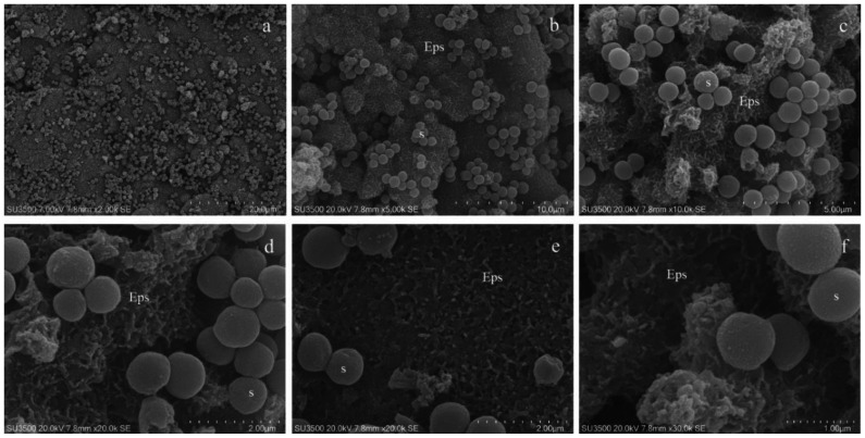
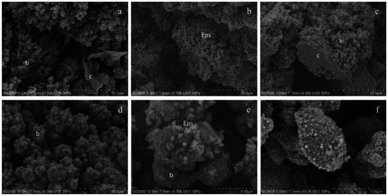
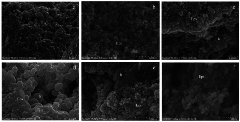
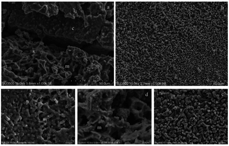
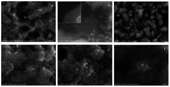
Early childhood caries (ECC) is a severe manifestation of carious pathology with rapid and disruptive progression. The ECC microbiota includes a wide variety of bacterial species, among which is an anaerobic newly named species, Scardovia wiggsiae, a previously unidentified Bifidobacterium. Our aim was to provide the first ultrastructural characterization of S. wiggsiae and its biofilm by scanning electron microscopy (SEM) using a protocol that faithfully preserved the biofilm architecture and allowed an investigation at very high magnifications (order of nanometers) and with the appropriate resolution. To accomplish this task, we analyzed Streptococcus mutans’ biofilm by conventional SEM and VP-SEM protocols, in addition, we developed an original procedure, named OsO4-RR-TA-IL, which avoids dehydration, drying and sputter coating. This innovative protocol allowed high-resolution and high-magnification imaging (from 10000× to 35000×) in high-vacuum and high-voltage conditions. After comparing three methods, we chose OsO4-RR-TA-IL to investigate S. wiggsiae. It appeared as a fusiform elongated bacterium, without surface specialization, arranged in clusters and submerged in a rich biofilm matrix, which showed a well-developed micro-canalicular system. Our results provide the basis for the development of innovative strategies to quantify the effects of different treatments, in order to establish the best option to counteract ECC in pediatric patients.
Overview
The claims and statistics of this paper haven’t been checked yet.
Run the check — free, no account needed. It reads the open-access text and checks the sources it cites.
Integrity signals
Registry
Cites
Statistics
Figures
Verification
Claims the paper makes
The claims and statistics of this paper haven’t been checked yet.
Run the check — free, no account needed. It reads the open-access text and checks the sources it cites.
Claims other papers make about this one
Each is checked against this paper’s own text: does it say what the citing paper says it does?
In the citing paper
“Of various methods available for qualitative structural BFI detection on abiotic or biotic surfaces, scanning electron microscopy (SEM)-based detection of BFI structure is considered the gold standard.20-22”
From SEMTWIST Quantification of Biofilm Infection in Human Chronic Wound Using Scanning Electron Microscopy and Machine Learning (2025) · cites this paper as [20]
Self-check
Percentages, sums and reported differences re-computed from the paper’s own numbers.
The self-check needs the paper’s full text.
Citations
Cites
No references could be resolved yet.
Research works printed but not linked: 70
- Berg, J.H., and Slayton, R.L. (2015). Early Childhood Oral Health, John Wiley & Sons, Inc.. [2nd ed.]. · DOI
- Douglass, 2004, A practical guide to infant oral health, Am. Fam. Physician, 70, 2113
- Marques, 2013, Impact of early childhood caries on the oral health-related quality of life of preschool children and their parents, Caries Res., 47, 211, 10.1159/000345534 · DOI
- Meyer, 2018, Early childhood caries: Epidemiology, aetiology, and prevention, Int. J. Dent., 2018, 1415873, 10.1155/2018/1415873 · DOI
- Gibbs, 1982, Oral flora of children with “nursing bottle caries”, J. Dent. Res., 61, 382, 10.1177/00220345820610020201 · DOI
- Berkowitz, 1984, Microbial characteristics of the human dental caries associated with prolonged bottle-feeding, Arch. Oral Biol., 29, 949, 10.1016/0003-9969(84)90097-9 · DOI
- Parisotto, 2010, Early childhood caries and mutans streptococci: A systematic review, Oral Health Prev. Dent., 8, 59
- Peterson, 2014, Functional expression of dental plaque microbiota, Front. Cell. Infect. Microbiol., 4, 108, 10.3389/fcimb.2014.00108 · DOI
- Li, 2015, Effect of antimicrobial interventions on the oral microbiota associated with early childhood caries, Pediatr. Dent., 37, 226
- Becker, 2002, Molecular analysis of bacterial species associated with childhood caries, J. Clin. Microbiol., 40, 1001, 10.1128/JCM.40.3.1001-1009.2002 · DOI · Paper page
- Lopman, 1996, The final pH of bacteria comprising the predominant flora on sound and carious human root and enamel surfaces, J. Dent. Res., 75, 1008, 10.1177/00220345960750040201 · DOI
- Downes, 2011, Scardovia wiggsiae sp. nov., isolated from the human oral cavity and clinical material, and emended descriptions of the genus Scardovia and Scardovia inopinata, Int. J. Syst. Evol. Microbiol., 61, 25, 10.1099/ijs.0.019752-0 · DOI
- Tanner, 2011, Cultivable anaerobic microbiota of severe early childhood caries, J. Clin. Microbiol., 49, 1464, 10.1128/JCM.02427-10 · DOI
- 2010, Identification of cultivable microrganisms from primary teeth with necrotic pulps, J. Clin. Pediatr. Dent., 34, 329, 10.17796/jcpd.34.4.20124lu111544377 · DOI
- Tanner, 2012, White-spot lesions and gingivitis microbiotas in orthodontic patients, J. Dent. Res., 91, 853, 10.1177/0022034512455031 · DOI
- Streiff, 2015, Screening and prevalence of the novel cariogenic pathogen scardovia wiggsiae among adult orthodontic and non-orthodontic patient saliva samples, Int. J. Dent. Oral Health, 1, 1
- Reyes, 2017, Prevalence of Scardovia wiggsiae among a pediatric Orthodontic patient population, EC Dent. Sci., 2, 203
- Kressirer, 2017, Scardovia wiggsiae and its potential role as a caries pathogen, J. Oral Biosci., 59, 135, 10.1016/j.job.2017.05.002 · DOI
- Larsen, 2017, Dental biofilm infections—An update, APMIS, 125, 376, 10.1111/apm.12688 · DOI
- Sutherland, 2001, Biofilm exopolysaccharides: A strong and sticky framework, Microbiology, 147, 3, 10.1099/00221287-147-1-3 · DOI
- Asahi, 2015, Simple observation of Streptococcus mutans biofilm by scanning electron microscopy using ionic liquids, AMB Express, 5, 6, 10.1186/s13568-015-0097-4 · DOI
- Weber, 2014, Comparison of SEM and VP-SEM imaging techniques with respect to Streptococcus mutans biofilm topography, FEMS Microbiol. Lett., 350, 175, 10.1111/1574-6968.12334 · DOI
- Priester, 2007, Enhanced visualization of microbial biofilms by staining and environmental scanning electron microscopy, J. Microbiol. Methods, 68, 577, 10.1016/j.mimet.2006.10.018 · DOI
- Donlan, 2002, Biofilms: Survival mechanisms of clinically relevant microorganisms, Clin. Microbiol. Rev., 15, 167, 10.1128/CMR.15.2.167-193.2002 · DOI · Paper page
- Kachlany, 2001, Structure and carbohydrate analysis of the exopolysaccharide capsule of Pseudomonas putida G7, Environ. Microbiol., 3, 774, 10.1046/j.1462-2920.2001.00248.x · DOI
- Fassel, 1999, Bacterial biofilms: Strategies for preparing glycocalyx for electron microscopy, Methods Enzym., 310, 194, 10.1016/S0076-6879(99)10017-X · DOI
- Little, 1991, Biofilms: An ESEM evaluation of artifacts introduced during SEM preparation, J. Ind. Microbiol., 8, 213, 10.1007/BF01576058 · DOI
- Reese, 2007, A novel TEM contrasting technique for extracellular polysaccharides in in vitro biofilms, Microsc. Res. Tech., 70, 816, 10.1002/jemt.20471 · DOI
- Casillo, 2017, Anti-biofilm activity of a long-chain fatty aldehyde from Antarctic Pseudoalteromonas haloplanktis TAC125 against Staphylococcus epidermidis biofilm, Front. Cell. Infect. Microbiol., 23, 46
- Chang, 1986, Biofilm loss during sample preparation for scanning electron microscopy, Water Res., 20, 1451, 10.1016/0043-1354(86)90145-4 · DOI
- Vitkov, 2005, Chlorhexidine-induced ultrastructural alterations in oral biofilm, Microsc. Res. Tech., 68, 85, 10.1002/jemt.20238 · DOI
- Vitkov, 2005, Bacterial internalization in periodontitis, Oral Microbiol. Immunol., 20, 317, 10.1111/j.1399-302X.2005.00233.x · DOI
- Vitkov, 2005, Ex vivo gingival-biofilm consortia, Lett. Appl. Microbiol., 41, 404, 10.1111/j.1472-765X.2005.01787.x · DOI
- Erlandsen, 2004, High-resolution visualization of the microbial glycocalyx with low-voltage scanning electron microscopy: Dependence on cationic dyes, J. Histochem. Cytochem., 52, 1427, 10.1369/jhc.4A6428.2004 · DOI
- Fulcher, 2001, Demonstration of biofilm in infectious crystalline keratopathy using ruthenium red and electron microscopy, Ophthalmology, 108, 1088, 10.1016/S0161-6420(01)00561-9 · DOI
- Luft, 1964, Electron microscopy of cell extraneous coats as revealed by ruthenium red staining, J. Cell Biol., 23, 54A
- Fassel, 1992, Comparison of alcian blue and ruthenium red effects on preservation of outer envelope ultrastructure in methanotrophic bacteria, Microsc. Res. Tech., 20, 87, 10.1002/jemt.1070200109 · DOI
- Fassel, 1999, Ruthenium red and the bacterial glycocalyx, Biotech. Histochem., 74, 194, 10.3109/10520299909047974 · DOI
- Carr, K.E., and Toner, P.G. (1982). Cell Structure: An Introduction to Biomedical Electron Microscopy, Churchill Livingstone. · DOI
- Murakami, 1983, Conductive staining of biological specimens for scanning electron microscopy with special reference to ligand-mediated osmium impregnation, Scan. Electron Microsc., 1, 235
- Murakami, 1974, A revised tannin-osmium method for non-coated SEM specimens, Arch. Histol. Jap., 36, 189, 10.1679/aohc1950.36.189 · DOI
- Murakami, 1973, A metal impregnation method of biological specimens for SEM, Arch. Histol. Jap., 35, 323, 10.1679/aohc1950.35.323 · DOI
- Murakami, 1976, Puncture perfusion of small tissue pieces for SEM, Arch. Histol. Jap., 39, 99, 10.1679/aohc1950.39.99 · DOI
- Murakami, 1977, Modified tannin osmium conductive staining method for non-coated SEM specimens. Its application to microdissection SEM of the spleen, Arch. Histol. Jap., 40, 35, 10.1679/aohc1950.40.35 · DOI
- Gunji, 1980, Conductive staining in SEM with especial reference to tissue transparency, Scanning, 3, 227, 10.1002/sca.4950030317 · DOI
- Arimoto, 2008, Development of new techniques for scanning electron microscope observation using ionic liquid, Electrochim. Acta, 53, 6228, 10.1016/j.electacta.2008.01.001 · DOI
- Tsuda, 2011, SEM observation of wet biological specimens pretreated with room-temperature ionic liquid, ChemBioChem, 12, 2547, 10.1002/cbic.201100476 · DOI
- Brodusch, 2014, Ionic liquid-based observation technique for nonconductive materials in the scanning electron microscope: Application to the characterization of a rare earth ore, Microsc. Res. Tech., 77, 225, 10.1002/jemt.22333 · DOI
- Imashuku, 2012, Possibility of scanning electron microscope observation and energy dispersive X-ray analysis in microscale region of insulating samples using diluted ionic liquid, Microsc. Microanal., 18, 365, 10.1017/S1431927611012669 · DOI
- Relucenti, 2018, The effect of postmastectomy radiation therapy on breast implants: material analysis on silicone and polyurethane prosthesis, Ann. Plast. Surg., 81, 228, 10.1097/SAP.0000000000001461 · DOI
- Relucenti, 2020, SEM BSE 3D image analysis of human incus bone affected by cholesteatoma ascribes to osteoclasts the bone erosion and VP-SEM dEDX analysis reveals new bone formation, Scanning, 2020, 9371516, 10.1155/2020/9371516 · DOI
- Relucenti, 2019, Ciliated cell observation by SEM on the surface of human incudo-malleolar-joint articular cartilage: Are they a new chondrocyte phenotype?, Acta Otolaryngol., 139, 439, 10.1080/00016489.2019.1575520 · DOI
- Timp, 2008, Electron microscopy of hydrated samples, Methods Cell Biol., 89, 391, 10.1016/S0091-679X(08)00614-6 · DOI
- Alhede, 2012, Combination of microscopic techniques reveals a comprehensive visual impression of biofilm structure and composition, FEMS Immunol. Med. Microbiol., 65, 335, 10.1111/j.1574-695X.2012.00956.x · DOI
- Araujo, 2003, Comparison of hexamethyldisilazane and critical point drying treatments for SEM analysis of anaerobic biofilms and granular sludge, J. Electron Microsc., 52, 429, 10.1093/jmicro/52.4.429 · DOI
- Piccolomini, 2008, Influence of temperature on biofilm formation by Listeria monocytogenes on various food-contact surfaces: Relationship with motility and cell surface hydrophobicity, J. Appl. Microbiol., 104, 1552, 10.1111/j.1365-2672.2007.03688.x · DOI
- Karcz, 2012, Application of lyophilization to prepare the nitrifying bacterial biofilm for imaging with scanning electron microscopy, Scanning, 34, 26, 10.1002/sca.20275 · DOI
- Bossù, M., Matassa, R., Relucenti, M., Iaculli, F., Salucci, A., Di Giorgio, G., Familiari, G., Polimeni, A., and Di Carlo, S. (2020). Morpho-Chemical Observations of Human Deciduous Teeth Enamel in Response to Biomimetic Toothpastes Treatment. Materials (Basel), 13. · DOI
- Saccucci, 2019, Enamel remineralization and repair results of Biomimetic Hydroxyapatite toothpaste on deciduous teeth: An effective option to fluoride toothpaste, J. Nanobiotechnol., 17, 17, 10.1186/s12951-019-0454-6 · DOI
- Redler, 2019, Ultrastructural Assessment of the Anterolateral Ligament, Orthop. J. Sports Med., 7, 2325967119887920, 10.1177/2325967119887920 · DOI
- Ishii, 2004, Two morphological types of cell appendages on a strongly adhesive bacterium, Acinetobacter sp. strain Tol 5, Appl. Environ. Microbiol., 70, 5026, 10.1128/AEM.70.8.5026-5029.2004 · DOI
- Weimer, 2006, Studies of the extracellular glycocalyx of the anaerobic cellulolytic bacterium Ruminococcus albus 7, Appl. Environ. Microbiol., 72, 7559, 10.1128/AEM.01632-06 · DOI
- Li, 2014, Insight into the roles of microbial extracellular polymer substances in metal biosorption, Bioresour. Technol., 160, 15, 10.1016/j.biortech.2013.11.074 · DOI
- Torimoto, 2010, New frontiers in materials science opened by ionic liquids, Adv. Mater., 22, 1196, 10.1002/adma.200902184 · DOI
- Kuwabata, 2006, Observation of ionic liquid by scanning electron microscope, Chem. Lett., 35, 600, 10.1246/cl.2006.600 · DOI
- Azeredo, 2017, Critical review on biofilm methods, Crit. Rev. Microbiol., 43, 313, 10.1080/1040841X.2016.1208146 · DOI
- Schatten, H. (2012). A method to visualize the micro architecture of glycoprotein matrices with scanning electron microscopy. Scanning Electron Microscopy for the Life Sciences, Cambridge University Press. · DOI
- Relucenti, 2005, Cumulus oophorus extracellular matrix in the human oocyte: A role for adhesive proteins, Ital. J. Anat. Embryol., 110, 219
- Familiari, 2001, The human zona pellucida and scanning electron microscopy. Reality or artifacts?, Ital. J. Anat. Embryol., 106, 33
- Familiari, 1992, A technique for exposure of the glycoproteic matrix (zona pellucida and mucus) for scanning electron microscopy, Microsc. Res. Tech., 23, 225, 10.1002/jemt.1070230305 · DOI
Cited by
Sorted by citations
Singh, S., Muniz De Oliveira, F., Wang, C., Kumar, M., Xuan, Y., DeMazumder, D., Sen, C. K., & Roy, S. (2025) · Advances in Wound Care · doi:10.1089/wound.2024.0291 · Cited by 6
Metadata
- Authors
- Bossù, M., Selan, L., Artini, M., Relucenti, M., Familiari, G., Papa, R., Vrenna, G., Spigaglia, P., Barbanti, F., Salucci, A., Di Giorgio, G., Rau, J. V., & Polimeni, A.
- Venue
- Microorganisms
- Publisher
- MDPI AG
- Published
- 2020-05-27
- Volume
- 8
- Issue
- 6
- Pages
- 807
- ISSN
- 2076-2607
- PMC
- PMC7355790
- Cited by
- 50
- References
- 0
- Language
- en
About these checks
Registry, retraction, Cites and statistics results are computed, not judged, with one exception: a printed reference that no index lists is read once by a model before it counts as a citation error. Claims are extracted by a model and kept only when their quote appears verbatim in the text; each cited source is then checked, with a model’s help, for whether it says what the claim relies on. Those verdicts can be wrong — read the claim’s quote and the cited source before relying on one.
Statistics and arithmetic are recomputed, not judged.
Confidence bands are shown only for fields calibrated on our annotated test set. None is calibrated yet, so no band is shown.
What counts as a claim ›How we measure confidence ›How we check this ›
Are you an author, or do you see a problem with this page? Request a review
