

The DOI matches its Crossref record; 42 of the 42 references it cites were found in a registry or an index.
Sevoflurane-Sulfobutylether-β-Cyclodextrin Complex: Preparation, Characterization, Cellular Toxicity, Molecular Modeling and Blood-Brain Barrier Transport Studies
Open accessAbstract
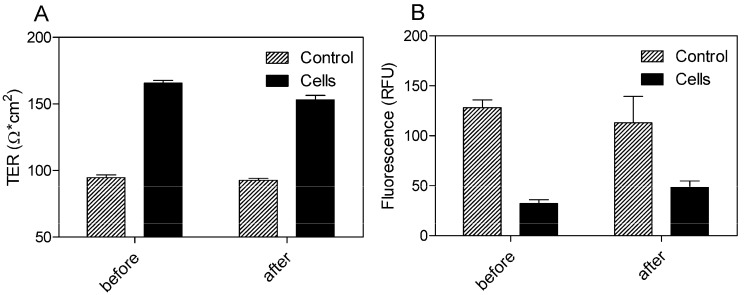
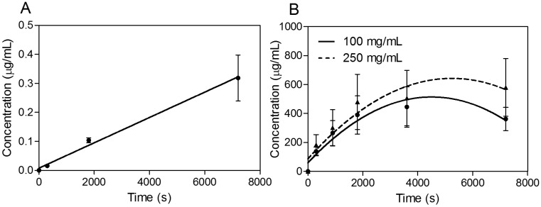
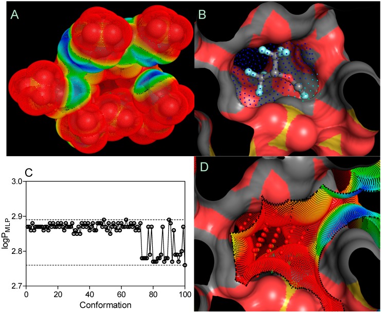
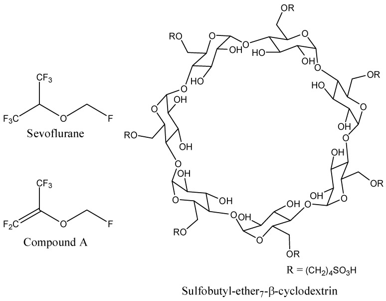
The objective of the present investigation was to study the ability of sulfobutylether-β-cyclodextrin (SBEβCD) to form an inclusion complex with sevoflurane (SEV), a volatile anesthetic with poor water solubility. The inclusion complex was prepared, characterized and its cellular toxicity and blood-brain barrier (BBB) permeation potential of the formulated SEV have also been examined for the purpose of controlled drug delivery. The SEV-SBEβCD complex was nontoxic to the primary brain microvascular endothelial (pEND) cells at a clinically relevant concentration of sevoflurane. The inclusion complex exhibited significantly higher BBB permeation profiles as compared with the reference substance (propranolol) concerning calculated apparent permeability values (Papp). In addition, SEV binding affinity to SBEβCD was confirmed by a minimal Gibbs free energy of binding (ΔGbind) value of -1.727 ± 0.042 kcal·mol-1 and an average binding constant (Kb) of 53.66 ± 9.24 mM indicating rapid drug liberation from the cyclodextrin amphiphilic cavity.
Overview
The claims and statistics of this paper haven’t been checked yet.
Run the check — free, no account needed. It reads the open-access text and checks the sources it cites.
Integrity signals
Registry
Cites
Statistics
Figures
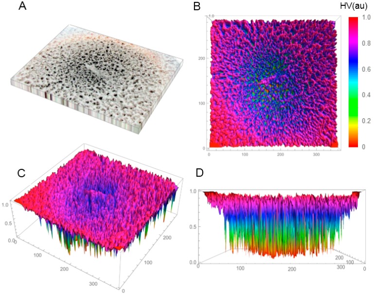
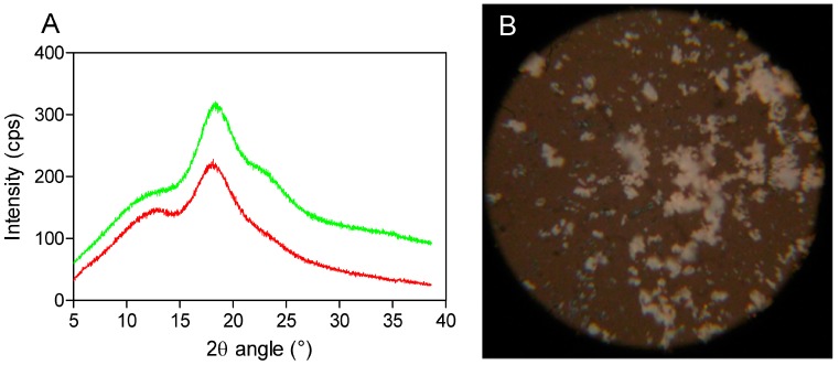
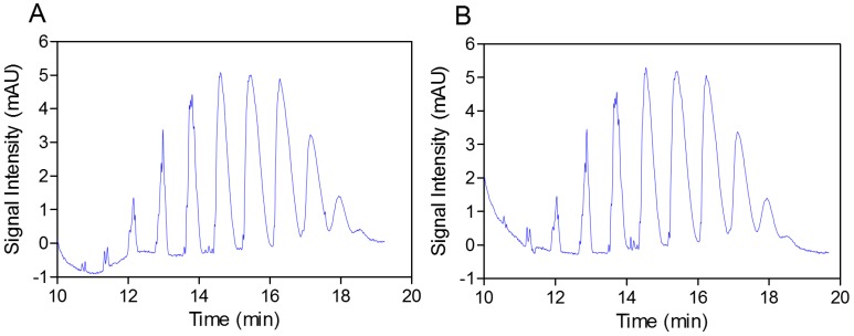
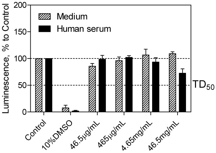
Verification
Claims the paper makes
The claims and statistics of this paper haven’t been checked yet.
Run the check — free, no account needed. It reads the open-access text and checks the sources it cites.
Claims other papers make about this one
Each is checked against this paper’s own text: does it say what the citing paper says it does?
The source backs part of the claim, not all of it — or backs it for a different population, condition or comparison.
In the citing paper
“For instance, it is known that cyclodextrins can be used for the preparation of formulations able to cross the BBB, administered through nasal or oral/parenteral routes [16,23,80,81].”
From Nanoemulsions for “Nose-to-Brain” Drug Delivery (2019) · cites this paper as [81]
Experimental — accuracy not yet measured
Self-check
Percentages, sums and reported differences re-computed from the paper’s own numbers.
The self-check needs the paper’s full text.
Citations
Cites
Iterative partial equalization of orbital electronegativity—a rapid access to atomic charges
Gasteiger, J., & Marsili, M. (1980) · Tetrahedron · doi:10.1016/0040-4020(80)80168-2 · Cited by 4,157
Ligand docking and binding site analysis with PyMOL and Autodock/Vina
Seeliger, D., & de Groot, B. L. (2010) · Journal of Computer-Aided Molecular Design · doi:10.1007/s10822-010-9352-6 · Cited by 2,648
Pharmaceutical Applications of Cyclodextrins. 1. Drug Solubilization and Stabilization
Loftsson, Þ., & Brewster, M. E. (1996) · Journal of Pharmaceutical Sciences · doi:10.1021/js950534b · Cited by 2,179
Pharmaceutical Applications of Cyclodextrins. III. Toxicological Issues and Safety Evaluation
Irie, T., & Uekama, K. (1997) · Journal of Pharmaceutical Sciences · doi:10.1021/js960213f · Cited by 925
Fischer, K. (1935) · Angewandte Chemie · doi:10.1002/ange.19350482605 · Cited by 627
Wallin, R., Regan, B. M., Napoli, M. D., et al. (1975) · Anesthesia & Analgesia · doi:10.1213/00000539-197511000-00021 · Cited by 332
Molecular Lipophilicity Potential, a tool in 3D QSAR: Method and applications
Gaillard, P., Carrupt, P., Testa, B., & Boudon, A. (1994) · Journal of Computer-Aided Molecular Design · doi:10.1007/bf00119860 · Cited by 281
Shityakov, S., & Foerster, C. Y. (2014) · Advances and Applications in Bioinformatics and Chemistry · doi:10.2147/aabc.s63749 · Cited by 227
Self-citationKiss, T., Fenyvesi, F., Bácskay, I., et al. (2010) · European Journal of Pharmaceutical Sciences · doi:10.1016/j.ejps.2010.04.014 · Cited by 203
Nalluri, B. N., Chowdary, K. P. R., Murthy, K. V. R., et al. (2003) · AAPS PharmSciTech · doi:10.1208/pt040102 · Cited by 154
Research works printed but not linked: 1
- Barash, P.G., Cullen, B.F., and Stoelting, R.K. (2001). Clinical Anesthesia, Lippincott Williams& Wilkins. [4th ed.].
Cited by
Sorted by citations
Nanoemulsions for “Nose-to-Brain” Drug Delivery
Bonferoni, M. C., Rossi, S., Sandri, G., Ferrari, F., Gavini, E., Rassu, G., & Giunchedi, P. (2019) · Pharmaceutics · doi:10.3390/pharmaceutics11020084 · Cited by 269
Metadata
- Authors
- Shityakov, S., Puskás, I., Pápai, K., Salvador, E., Roewer, N., Förster, C., & Broscheit, J.-A.
- Affiliations
- University of Würzburg (DE)
- CycloLab (Hungary) (HU)
- Sapiotec Ltd., 97078 Würzburg, Germany
- Corresponding author
- Shityakov, Sergey
- ORCID
- Venue
- Molecules
- Publisher
- MDPI AG
- Published
- 2015-06-03
- Volume
- 20
- Issue
- 6
- Pages
- 10264-10279
- ISSN
- 1420-3049 (ISSN-L)
- PMC
- PMC6272776
- OpenAlex
- W1584701817
- Cited by
- 26
- References
- 42
- Citations by year
- 2025: 1 · 2024: 1 · 2023: 2 · 2022: 4 · 2020: 1 · 2019: 4 · 2018: 4 · 2017: 4 · 2016: 3 · 2015: 2
- FWCI
- 2.40
- Type
- journal article
- Language
- en
- Open access
- gold
- dx.doi.org
- Licence
- Locations
- Molecules (publishedVersion, cc-by)
- Europe PMC (PubMed Central) (publishedVersion, other-oa)
- PubMed Central (publishedVersion, cc-by)
- PubMed (publishedVersion)
- Online Publication Service of Würzburg University (Würzburg University) (submittedVersion, cc-by)
- DOAJ (DOAJ: Directory of Open Access Journals) (submittedVersion, cc-by-sa)
- Funders
- Bundesministerium für Bildung und Forschung (EO1004, BMBF01)
- Julius-Maximilians-Universität Würzburg
- Topics
- Life Sciences › Pharmacology, Toxicology and Pharmaceutics › Pharmaceutical Science › Drug Solubulity and Delivery Systems
- Life Sciences › Neuroscience › Neurology › Barrier Structure and Function Studies
- Life Sciences › Pharmacology, Toxicology and Pharmaceutics › Pharmaceutical Science › Advanced Drug Delivery Systems
- Keywords
- sevoflurane, sulfobutylether-β-cyclodextrin, inclusion complexes, blood-brain barrier permeability, cytotoxicity, apparent permeability, binding affinity, drug solubility enhancement, volatile anesthetics, controlled drug delivery
- MeSH terms
- Sevoflurane, Animals, Blood-Brain Barrier, Brain, Capillary Permeability, Cell Survival, Delayed-Action Preparations, Drug Compounding, Methyl Ethers, Propranolol, Anesthetics, Inhalation, Endothelial Cells, beta-Cyclodextrins, Mice, Primary Cell Culture, Molecular Docking Simulation
About these checks
Registry, retraction, Cites and statistics results are computed, not judged, with one exception: a printed reference that no index lists is read once by a model before it counts as a citation error. Claims are extracted by a model and kept only when their quote appears verbatim in the text; each cited source is then checked, with a model’s help, for whether it says what the claim relies on. Those verdicts can be wrong — read the claim’s quote and the cited source before relying on one.
Statistics and arithmetic are recomputed, not judged.
Confidence bands are shown only for fields calibrated on our annotated test set. None is calibrated yet, so no band is shown.
What counts as a claim ›How we measure confidence ›How we check this ›
Are you an author, or do you see a problem with this page? Request a review
