

The DOI matches its Crossref record.
Small versus Large Iron Oxide Magnetic Nanoparticles: Hyperthermia and Cell Uptake Properties
Abstract
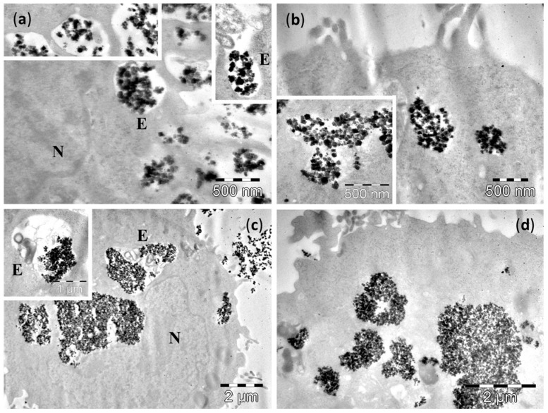
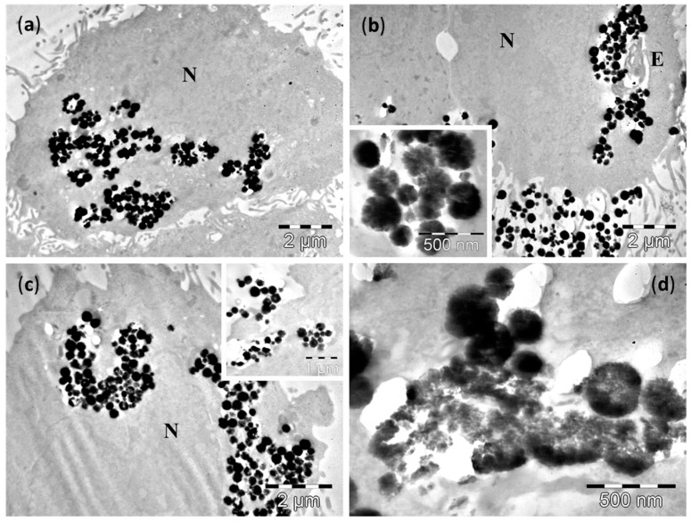
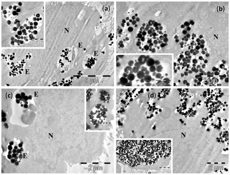
Efficient use of magnetic hyperthermia in clinical cancer treatment requires biocompatible magnetic nanoparticles (MNPs), with improved heating capabilities. Small (~34 nm) and large (~270 nm) Fe3O4-MNPs were synthesized by means of a polyol method in polyethylene-glycol (PEG) and ethylene-glycol (EG), respectively. They were systematically investigated by means of X-ray diffraction, transmission electron microscopy and vibration sample magnetometry. Hyperthermia measurements showed that Specific Absorption Rate (SAR) dependence on the external alternating magnetic field amplitude (up to 65 kA/m, 355 kHz) presented a sigmoidal shape, with remarkable SAR saturation values of ~1400 W/gMNP for the small monocrystalline MNPs and only 400 W/gMNP for the large polycrystalline MNPs, in water. SAR values were slightly reduced in cell culture media, but decreased one order of magnitude in highly viscous PEG1000. Toxicity assays performed on four cell lines revealed almost no toxicity for the small MNPs and a very small level of toxicity for the large MNPs, up to a concentration of 0.2 mg/mL. Cellular uptake experiments revealed that both MNPs penetrated the cells through endocytosis, in a time dependent manner and escaped the endosomes with a faster kinetics for large MNPs. Biodegradation of large MNPs inside cells involved an all-or-nothing mechanism.
Overview
The claims and statistics of this paper haven’t been checked yet.
Run the check — free, no account needed. It reads the open-access text and checks the sources it cites.
Integrity signals
Registry
Cites
Statistics
Figures
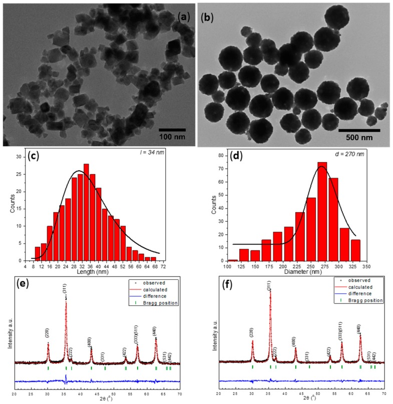
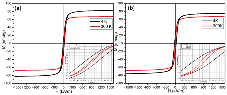
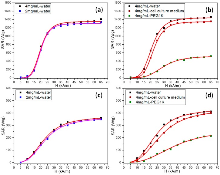
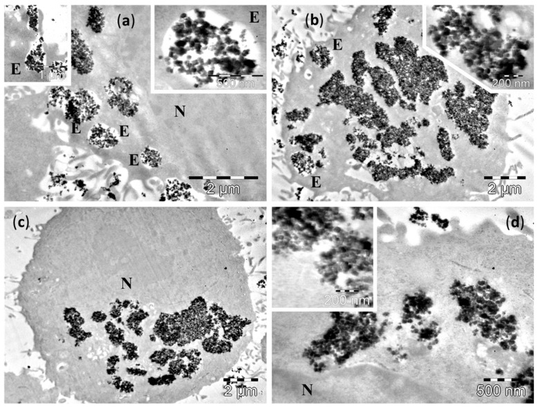
Verification
Claims the paper makes
The claims and statistics of this paper haven’t been checked yet.
Run the check — free, no account needed. It reads the open-access text and checks the sources it cites.
Claims other papers make about this one
Each is checked against this paper’s own text: does it say what the citing paper says it does?
In the citing paper
“it is well documented that SPIONs can accumulate in large amounts inside the cell, both in cancer cells and in normal cells [61,62].”
From Biomimetic Magnetoliposomes as Oxaliplatin Nanocarriers: In Vitro Study for Potential Application in Colon Cancer (2020) · cites this paper as [62]
Self-check
Percentages, sums and reported differences re-computed from the paper’s own numbers.
The self-check needs the paper’s full text.
Citations
Cites
No references could be resolved yet.
Research works printed but not linked: 46
- Farnaz, 2016, A biotechnological perspective on the application of iron oxide nanoparticles, Nano Res., 9, 2203, 10.1007/s12274-016-1131-9 · DOI
- Barakat, 2009, Magnetically modulated nanosystems: A unique drug-delivery platform, Nanomedicine, 4, 799, 10.2217/nnm.09.66 · DOI
- Riegler, 2010, Targeted magnetic delivery and tracking of cells using a magnetic resonance imaging system, Biomaterials, 31, 5366, 10.1016/j.biomaterials.2010.03.032 · DOI
- Gilchrist, 1957, Selective Inductive Heating of Lymph Nodes, J. Ann. Surg., 146, 596, 10.1097/00000658-195710000-00007 · DOI
- Goya, 2008, Magnetic nanoparticles for cancer therapy, Curr. Nanosci., 4, 1, 10.2174/157341308783591861 · DOI
- Perigo, E.A., Hemery, G., Sandre, O., Ortega, D., Garaio, E., Plazaola, F., and Teran, F.J. (2015). Fundamentals and advances in magnetic hyperthermia. Appl. Phys. Rev., 2. · DOI
- Walter, 2016, Design of iron oxide-based nanoparticles for MRI and magnetic hyperthermia, Nanomedicine, 11, 1889, 10.2217/nnm-2016-5001 · DOI · Paper page
- Rosensweig, 2002, Heating Magnetic Fluid with Alternating Magnetic Field, J. Magn. Magn. Mater., 252, 370, 10.1016/S0304-8853(02)00706-0 · DOI
- Hergt, 2006, The Effect of Field Parameters, Nanoparticle Properties and Immobilization on the Specific Heating Power in Magnetic Particle Hyperthermia, J. Phys. Condens. Matter, 18, S2935, 10.1088/0953-8984/18/38/S27 · DOI
- Hergt, 2007, Magnetic Particle Hyperthermia-Biophysical Limitations of a Visionary Tumour Therapy, J. Magn. Magn. Mater., 311, 187, 10.1016/j.jmmm.2006.10.1156 · DOI
- Wilczewska, 2012, Nanoparticles as drug delivery systems, Pharmacol. Rep., 64, 1020, 10.1016/S1734-1140(12)70901-5 · DOI · Paper page
- Ulrich, 2011, Efficacy and safety of intratumoral thermotherapy using magnetic iron oxide nanoparticles combined with external beam radiotherapy on patients with recurrent glioblastoma multiforme, J. Neuro-Oncol., 103, 317, 10.1007/s11060-010-0389-0 · DOI
- Hilger, 2005, Use of Magnetic Nanoparticle Heating in the Treatment of Breast Cancer, IEE Proc. Nanobiotechnol., 152, 33, 10.1049/ip-nbt:20055018 · DOI
- Stoner, 1948, A mechanism of magnetic hysteresis in heterogeneous alloys, Phil. Trans. R. Soc., A240, 599
- Carrey, 2011, Simple models for dynamic hysteresis loop calculations of magnetic single-domain nanoparticles: Application to magnetic hyperthermia optimization, J. Appl. Phys., 109, 083921, 10.1063/1.3551582 · DOI
- Song, 2004, Shape control and associated magnetic properties of spinel cobalt ferrite nanocrystals, J. Am. Chem. Soc., 126, 6164, 10.1021/ja049931r · DOI
- Das, 2016, Tunable high aspect ratio iron oxide nanorods for enhanced hyperthermia, J. Phys. Chem. C, 120, 10086, 10.1021/acs.jpcc.6b02006 · DOI
- Lv, 2015, Size dependent magnetic hyperthermia of octahedral Fe3O4 nanoparticles, RSC Adv., 5, 76764, 10.1039/C5RA12558H · DOI
- Chen, 2013, Maximizing hysteretic losses in magnetic ferrite nanoparticles via model-driven synthesis and materials optimization, ACS Nano, 7, 8990, 10.1021/nn4035266 · DOI
- Iacovita, 2015, Polyethylene glycol-mediated synthesis of cubic iron oxide nanoparticles with high heating power, Nanoscale Res. Lett., 10, 1, 10.1186/s11671-015-1091-0 · DOI
- Patil, 2015, In Vitro/in Vivo Toxicity Evaluation and Quantification of Iron Oxide Nanoparticles, Int. J. Mol. Sci., 16, 24417, 10.3390/ijms161024417 · DOI
- Markides, H., Rotherham, M., and El Haj, A.J. (2012). Biocompatibility and Toxicity of Magnetic Nanoparticles in Regenerative Medicine. J. Nanomater. · DOI
- Sahu, C.S., and Casciano, D.A. (2014). Handbook of Nanotoxicology, Nanomedicine and Stem Cell Use in Toxicology, John Wiley & Sons, Ltd. · DOI
- Arias, 2012, Magnetic Nanoparticles: Design and Characterization, Toxicity and Biocompatibility, Pharmaceutical and Biomedical Applications, Chem. Rev., 112, 5818, 10.1021/cr300068p · DOI · Paper page
- Deng, 2005, Monodisperse magnetic single-crystal ferrite microspheres, Angew. Chem. Int. Ed., 44, 2782, 10.1002/anie.200462551 · DOI
- Garaio, 2015, Specific absorption rate dependence on temperature in magnetic field hyperthermia measured by dynamic hysteresis losses (ac magnetometry), Nanotechnology, 26, 015704, 10.1088/0957-4484/26/1/015704 · DOI
- Sasaki, 1997, Radial Distribution of Electron Density in Magnetite, Fe3O4, Acta Crystallogr. Sect. B Struct. Sci., 53, 762, 10.1107/S0108768197007842 · DOI
- Noh, 2012, Nanoscale Magnetism Control via Surface and Exchange Anisotropy for Optimized Ferrimagnetic Hysteresis, Nano Lett., 12, 3716, 10.1021/nl301499u · DOI
- Daou, 2006, Hydrothermal Synthesis of Monodisperse Magnetite Nanoparticles, Chem. Mater., 18, 4399, 10.1021/cm060805r · DOI
- Shivashankar, 2014, Single crystalline magnetite, maghemite, and hematite nanoparticles with rich coercivity, RSC Adv., 4, 4105, 10.1039/C3RA45457F · DOI
- Andreu, 2015, Nano-objects for Addressing the Control of Nanoparticle Arrangement and Performance in Magnetic Hyperthermia, ACS Nano, 9, 1408, 10.1021/nn505781f · DOI
- Mehdaoui, 2011, Optimal Size of Nanoparticles for Magnetic Hyperthermia: A Combined Theoretical and Experimental Study, Adv. Funct. Mater., 21, 4573, 10.1002/adfm.201101243 · DOI
- Christiansen, M.G., Senko, A.W., Chen, R., Romero, G., and Anikeeva, P. (2014). Magnetically multiplexed heating of single domain nanoparticles. Appl. Phys. Lett., 104. · DOI
- Dennis, 2015, Internal Magnetic Structure of Nanoparticles Dominates Time-Dependent Relaxation Processes in a Magnetic Field, Adv. Funct. Mater., 25, 4300, 10.1002/adfm.201500405 · DOI
- Branquinho, 2014, Effect of magnetic dipolar interactions on nanoparticles heating efficiency: Implications for cancer hyperthermia, Sci. Rep., 3, 1
- Deatsch, 2014, Heating efficiency in magnetic hyperthermia, J. Mag. Mag. Mater., 354, 163, 10.1016/j.jmmm.2013.11.006 · DOI
- Ortega, 2015, High performance multi-core iron oxide nanoparticles for magnetic hyperthermia: Microwave assisted synthesis and the role of core-to-core interactions, Nanoscale, 7, 1768, 10.1039/C4NR06239F · DOI
- Riffle, 2009, Cell Uptake and in Vitro Toxicity of Magnetic Nanoparticles Suitable for Drug Delivery, Mol. Pharm., 6, 1417, 10.1021/mp900083m · DOI
- Mahmoudi, 2012, Assessing the in Vitro and in Vivo Toxicity of Superparamagnetic Iron Oxide Nanoparticles, Chem. Rev., 112, 2323, 10.1021/cr2002596 · DOI
- Chen, 2009, Effects of proteins from culture medium on surface property of silanes-functionalized magnetic nanoparticles, Nanoscale Res. Lett., 4, 204, 10.1007/s11671-008-9226-1 · DOI
- Ehrenberg, 2009, The influence of protein adsorption on nanoparticle association with cultured endothelial cells, Biomaterials, 30, 603, 10.1016/j.biomaterials.2008.09.050 · DOI · Paper page
- Rahman, 2013, Nanoparticle and protein corona, Protein-Nanoparticle Interactions: The Bio-Nano Interface, Volume 15, 21, 10.1007/978-3-642-37555-2_2 · DOI
- Mazuel, 2016, Massive Intracellular Biodegradation of Iron Oxide Nanoparticles Evidenced Magnetically at Single-Endosome and Tissue Levels, ACS Nano, 10, 7627, 10.1021/acsnano.6b02876 · DOI
- Vance, 2015, Nanotechnology in the real world: Redeveloping the nanomaterial consumer products inventory, Beilstein J. Nanotechnol., 6, 1769, 10.3762/bjnano.6.181 · DOI · Paper page
- Gautam, 2013, Synthesis of nanoparticles, their biocompatibility, and toxicity behavior for biomedical applications, J. Mater. Chem. B, 1, 5186, 10.1039/c3tb20738b · DOI
- Mou, 2015, Applications of Magnetic Nanoparticles in Targeted Drug Delivery Systems, J. Nanosci. Nanotechnol., 15, 54, 10.1166/jnn.2015.9585 · DOI
Cited by
Sorted by citations
Garcia-Pinel, B., Jabalera, Y., Ortiz, R., Cabeza, L., Jimenez-Lopez, C., Melguizo, C., & Prados, J. (2020) · Pharmaceutics · doi:10.3390/pharmaceutics12060589 · Cited by 40
Metadata
- Authors
- Iacovita, C., Florea, A., Dudric, R., Pall, E., Moldovan, A., Tetean, R., Stiufiuc, R., & Lucaciu, C.
- Venue
- Molecules
- Publisher
- MDPI AG
- Published
- 2016-10-13
- Volume
- 21
- Issue
- 10
- Pages
- 1357
- ISSN
- 1420-3049
- PMC
- PMC6274490
- Cited by
- 57
- References
- 0
- Language
- en
- Funders
- Romanian National Authority for Scientific Research, CNCSIS-UEFISCDI (PN-II-ID-PCE-2012-4-0531, PN-II-RU-TE-2014-4-1770)
About these checks
Registry, retraction, Cites and statistics results are computed, not judged, with one exception: a printed reference that no index lists is read once by a model before it counts as a citation error. Claims are extracted by a model and kept only when their quote appears verbatim in the text; each cited source is then checked, with a model’s help, for whether it says what the claim relies on. Those verdicts can be wrong — read the claim’s quote and the cited source before relying on one.
Statistics and arithmetic are recomputed, not judged.
Confidence bands are shown only for fields calibrated on our annotated test set. None is calibrated yet, so no band is shown.
What counts as a claim ›How we measure confidence ›How we check this ›
Are you an author, or do you see a problem with this page? Request a review
