

The DOI matches its Crossref record.
Biomimetic Magnetic Nanocarriers Drive Choline Kinase Alpha Inhibitor inside Cancer Cells for Combined Chemo-Hyperthermia Therapy
Abstract
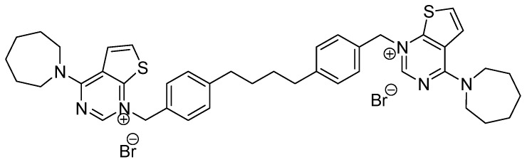
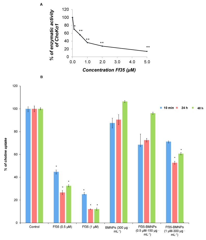
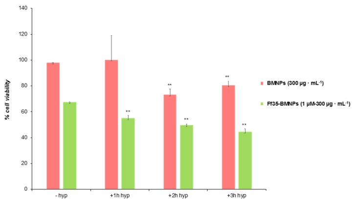
Choline kinase α1 (ChoKα1) has become an excellent antitumor target. Among all the inhibitors synthetized, the new compound Ff35 shows an excellent capacity to inhibit ChoKα1 activity. However, soluble Ff35 is also capable of inhibiting choline uptake, making the inhibitor not selective for ChoKα1. In this study, we designed a new protocol with the aim of disentangling whether the Ff35 biological action is due to the inhibition of the enzyme and/or to the choline uptake. Moreover, we offer an alternative to avoid the inhibition of choline uptake caused by Ff35, since the coupling of Ff35 to novel biomimetic magnetic nanoparticles (BMNPs) allows it to enter the cell through endocytosis without interacting with the choline transporter. This opens the possibility of a clinical use of Ff35. Our results indicate that Ff35-BMNPs nanoassemblies increase the selectivity of Ff35 and have an antiproliferative effect. Also, we demonstrate the effectiveness of the tandem Ff35-BMNPs and hyperthermia.
Overview
The claims and statistics of this paper haven’t been checked yet.
Run the check — free, no account needed. It reads the open-access text and checks the sources it cites.
Integrity signals
Registry
Cites
Statistics
Figures
Verification
Claims the paper makes
The claims and statistics of this paper haven’t been checked yet.
Run the check — free, no account needed. It reads the open-access text and checks the sources it cites.
Claims other papers make about this one
Each is checked against this paper’s own text: does it say what the citing paper says it does?
In the citing paper
“MamC-mediated biomimetic nanoparticles (here referred as BMNPs) have demonstrated their potential as promising drug nanocarriers [5,6,14,15]”
From Biomimetic Magnetoliposomes as Oxaliplatin Nanocarriers: In Vitro Study for Potential Application in Colon Cancer (2020) · cites this paper as [15]
In the citing paper
“Therefore, these MamC-mediated BMNPs have been proposed as efficient nanocarriers [5,6,14,15] of a number of molecules offering crucial advantages over the use of inorganically synthesized magnetic nanoparticles to this end.”
From Biomimetic Magnetoliposomes as Oxaliplatin Nanocarriers: In Vitro Study for Potential Application in Colon Cancer (2020) · cites this paper as [15]
Self-check
Percentages, sums and reported differences re-computed from the paper’s own numbers.
The self-check needs the paper’s full text.
Citations
Cites
No references could be resolved yet.
Research works printed but not linked: 28
- Siegel, 2018, Cancer statistics, 2018, CA Cancer J. Clin., 68, 7, 10.3322/caac.21442 · DOI
- Hanahan, 2000, The Hallmarks of Cancer, Cell, 100, 57, 10.1016/S0092-8674(00)81683-9 · DOI · Paper page
- Huang, 2015, Lipid metabolism, apoptosis and cancer therapy, Int. J. Mol. Sci., 16, 924, 10.3390/ijms16010924 · DOI
- Galbraith, 2018, Lipid pathway deregulation in advanced prostate cancer, Pharmacol. Res., 131, 177, 10.1016/j.phrs.2018.02.022 · DOI
- Glunde, 2011, Choline metabolism in malignant transformation, Nat. Rev. Cancer, 11, 835, 10.1038/nrc3162 · DOI
- Cao, 2016, Targeting choline phospholipid metabolism: GDPD5 and GDPD6 silencing decrease breast cancer cell proliferation, migration, and invasion, NMR Biomed., 29, 1098, 10.1002/nbm.3573 · DOI
- Iorio, 2016, Key Players in Choline Metabolic Reprograming in Triple-Negative Breast Cancer, Front. Oncol., 6, 205, 10.3389/fonc.2016.00205 · DOI
- Kennelly, 2017, The critical role of phosphatidylcholine and phosphatidylethanolamine metabolism in health and disease, Biochim. Biophys. Acta Biomembr., 1859, 1558, 10.1016/j.bbamem.2017.04.006 · DOI · Paper page
- Yalcin, 2010, Selective inhibition of choline kinase simultaneously attenuates MAPK and PI3K/AKT signaling, Oncogene, 29, 139, 10.1038/onc.2009.317 · DOI
- Mori, 2007, Choline Kinase Down-regulation Increases the Effect of 5-Fluorouracil in Breast Cancer Cells, Cancer Res., 67, 11284, 10.1158/0008-5472.CAN-07-2728 · DOI
- Chen, Z., Krishnamachary, B., and Bhujwalla, Z. (2016). Degradable Dextran Nanopolymer as a Carrier for Choline Kinase (ChoK) siRNA Cancer Therapy. Nanomaterials, 6. · DOI
- Baglioni, 2016, Design, synthesis, crystallization and biological evaluation of new symmetrical biscationic compounds as selective inhibitors of human Choline Kinase α1 (ChoKα1), Sci. Rep., 6, 23793, 10.1038/srep23793 · DOI
- Arlauckas, 2016, Choline kinase alpha—Putting the ChoK-hold on tumor metabolism, Prog. Lipid Res., 63, 28, 10.1016/j.plipres.2016.03.005 · DOI
- Lacal, 2015, Choline kinase as a precision medicine target for therapy in cancer, autoimmune diseases and malaria, Precis. Med., 2, 980
- Bioisoterism, 2017, Review-An Biological Modification, World J. Pharm. Pharm. Sci., 6, 1918
- Marco, 2019, Choline kinase inhibitors EB-3D and EB-3P interferes with lipid homeostasis in HepG2 cells, Sci. Rep., 9, 5109, 10.1038/s41598-019-40885-z · DOI
- Prozorov, 2013, Novel magnetic nanomaterials inspired by magnetotactic bacteria: Topical review, Mater. Sci. Eng. R Rep., 74, 133, 10.1016/j.mser.2013.04.002 · DOI · Paper page
- Peigneux, 2018, pH-Dependent Adsorption Release of Doxorubicin on MamC-Biomimetic Magnetite Nanoparticles, Langmuir, 34, 13713, 10.1021/acs.langmuir.8b03109 · DOI · Paper page
- Prozorov, 2015, Size control of In Vitro synthesized magnetite crystals by the MamC protein of Magnetococcus marinus strain MC-1, Appl. Microbiol. Biotechnol., 99, 5109, 10.1007/s00253-014-6326-y · DOI · Paper page
- 2010, Inorganic Magnetite Precipitation at 25 °C: A Low-Cost Inorganic Coprecipitation Method, J. Supercond. Nov. Magn., 24, 549
- Turiel, 2003, Assessment of the cross-reactivity and binding sites characterisation of a propazine-imprinted polymer using the Langmuir-Freundlich isotherm, Analyst, 128, 137, 10.1039/b210712k · DOI
- Carrasco, 2002, Hexadecylphosphocholine inhibits phosphatidylcholine biosynthesis and the proliferation of HepG2 cells, Eur. J. Biochem., 269, 4649, 10.1046/j.1432-1033.2002.03169.x · DOI
- Reynolds, 1963, The use of lead citrate at high pH as an electron-opaque stain in electron microscopy, J. Cell Biol., 17, 208, 10.1083/jcb.17.1.208 · DOI
- Iafisco, 2016, Superparamagnetic iron-doped nanocrystalline apatite as a delivery system for doxorubicin, J. Mater. Chem. B, 4, 57, 10.1039/C5TB01524C · DOI
- Geisow, 1984, pH in the endosome. Measurements during pinocytosis and receptor-mediated endocytosis, Exp. Cell Res., 150, 36, 10.1016/0014-4827(84)90699-2 · DOI
- Skowronek, 2007, Hyperthermia–description of a method and a review of clinical applications, Rep. Pract. Oncol. Radiother., 12, 267, 10.1016/S1507-1367(10)60065-X · DOI
- Lim, 2006, Cell cycle dependent apoptosis and cell cycle blocks induced by hyperthermia in HL-60 cells, Int. J. Hyperth., 22, 77, 10.1080/02656730500430538 · DOI
- Iglesias, G.R., Reyes-Ortega, F., Fernandez, B.L.C., and Delgado, Á.V. (2018). Hyperthermia-triggered gemcitabine release from polymer-coated magnetite nanoparticles. Polymers, 10. · DOI
Cited by
Sorted by citations
Garcia-Pinel, B., Jabalera, Y., Ortiz, R., Cabeza, L., Jimenez-Lopez, C., Melguizo, C., & Prados, J. (2020) · Pharmaceutics · doi:10.3390/pharmaceutics12060589 · Cited by 40
Self-citation
Metadata
- Authors
- Jabalera, Y., Sola-Leyva, A., Peigneux, A., Vurro, F., Iglesias, G. R., Vilchez-Garcia, J., Pérez-Prieto, I., Aguilar-Troyano, F. J., López-Cara, L. C., Carrasco-Jiménez, M. P., & Jimenez-Lopez, C.
- Venue
- Pharmaceutics
- Publisher
- MDPI AG
- Published
- 2019-08-12
- Volume
- 11
- Issue
- 8
- Pages
- 408
- ISSN
- 1999-4923
- PMC
- PMC6722936
- Cited by
- 27
- References
- 0
- Language
- en
- Funders
- Ministerio de Economía y Competitividad (CGL2013-46612, CGL2016-76723)
- Ramón y Cajal Programme (RYC-2014-16901)
- Andalusian regional government (CTS-236)
About these checks
Registry, retraction, Cites and statistics results are computed, not judged, with one exception: a printed reference that no index lists is read once by a model before it counts as a citation error. Claims are extracted by a model and kept only when their quote appears verbatim in the text; each cited source is then checked, with a model’s help, for whether it says what the claim relies on. Those verdicts can be wrong — read the claim’s quote and the cited source before relying on one.
Statistics and arithmetic are recomputed, not judged.
Confidence bands are shown only for fields calibrated on our annotated test set. None is calibrated yet, so no band is shown.
What counts as a claim ›How we measure confidence ›How we check this ›
Are you an author, or do you see a problem with this page? Request a review
How to cite
Cite CoChat for the checks on this page, and the paper for its own findings.
- This page
- CoChat. (2026). Citation check: Biomimetic Magnetic Nanocarriers Drive Choline Kinase Alpha Inhibitor inside Cancer Cells for Combined Chemo-Hyperthermia Therapy. https://app.cochat.ai/papers/10.3390/pharmaceutics11080408
- The paper
- Jabalera, Y., Sola-Leyva, A., Peigneux, A., Vurro, F., Iglesias, G. R., Vilchez-Garcia, J., Pérez-Prieto, I., Aguilar-Troyano, F. J., López-Cara, L. C., Carrasco-Jiménez, M. P., & Jimenez-Lopez, C. (2019). Biomimetic Magnetic Nanocarriers Drive Choline Kinase Alpha Inhibitor inside Cancer Cells for Combined Chemo-Hyperthermia Therapy. Pharmaceutics. https://doi.org/10.3390/pharmaceutics11080408
