

The DOI matches its Crossref record; 3 of the 3 references it cites were found in a registry or an index.
A Shortcut from Genome to Drug: The Employment of Bioinformatic Tools to Find New Targets for Gastric Cancer Treatment
Open accessPublished version of a preprint
Abstract
Gastric cancer (GC) is a highly heterogeneous, complex disease and the fifth most common cancer worldwide (about 1 million cases and 784,000 deaths worldwide in 2018). GC has a poor prognosis (the 5-year survival rate is less than 20%), but there is an effort to find genes highly expressed during tumor establishment and use the related proteins as targets to find new anticancer molecules. Data were collected from the Gene Expression Omnibus (GEO) bank to obtain three dataset matrices analyzing gastric tumor tissue versus normal gastric tissue and involving microarray analysis performed using the GPL570 platform and different sources. The data were analyzed using the GEPIA tool for differential expression and KMPlot for survival analysis. For more robustness, GC data from the TCGA database were used to corroborate the analysis of data from GEO. The genes found in in silico analysis in both GEO and TCGA were confirmed in several lines of GC cells by RT-qPCR. The AlphaFold Protein Structure Database was used to find the corresponding proteins. Then, a structure-based virtual screening was performed to find molecules, and docking analysis was performed using the DockThor server. Our in silico and RT-qPCR analysis results confirmed the high expression of the AJUBA, CD80 and NOLC1 genes in GC lines. Thus, the corresponding proteins were used in SBVS analysis. There were three molecules, one molecule for each target, MCULE-2386589557-0-6, MCULE-9178344200-0-1 and MCULE-5881513100-0-29. All molecules had favorable pharmacokinetic, pharmacodynamic and toxicological properties. Molecular docking analysis revealed that the molecules interact with proteins in critical sites for their activity. Using a virtual screening approach, a molecular docking study was performed for proteins encoded by genes that play important roles in cellular functions for carcinogenesis. Combining a systematic collection of public microarray data with a comparative meta-profiling, RT-qPCR, SBVS and molecular docking analysis provided a suitable approach for finding genes involved in GC and working with the corresponding proteins to search for new molecules with anticancer properties.
Overview
The claims and statistics of this paper haven’t been checked yet.
Run the check — free, no account needed. It reads the open-access text and checks the sources it cites.
Integrity signals
Registry
Cites
Statistics
Figures
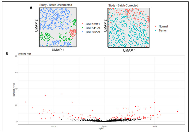
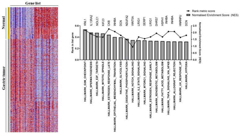
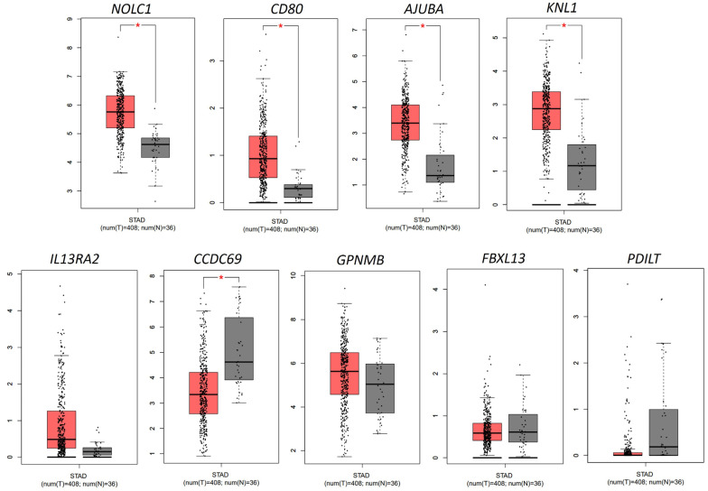
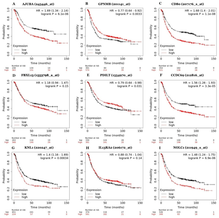
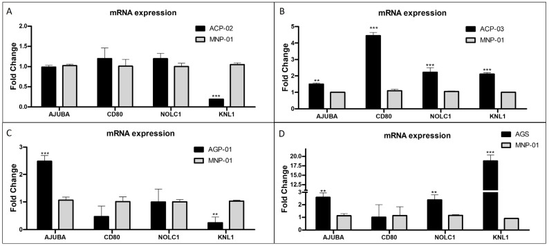
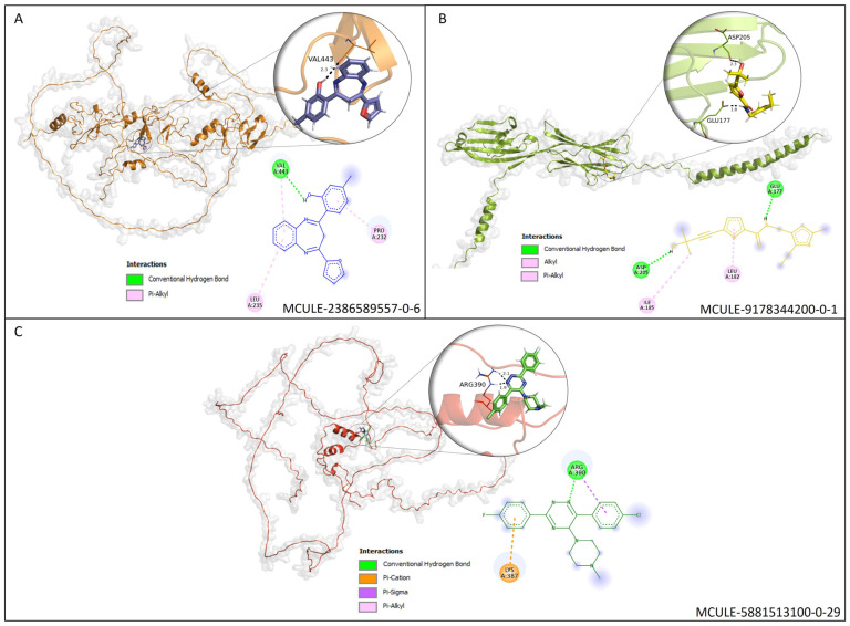
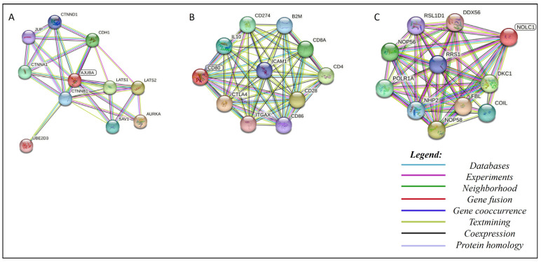
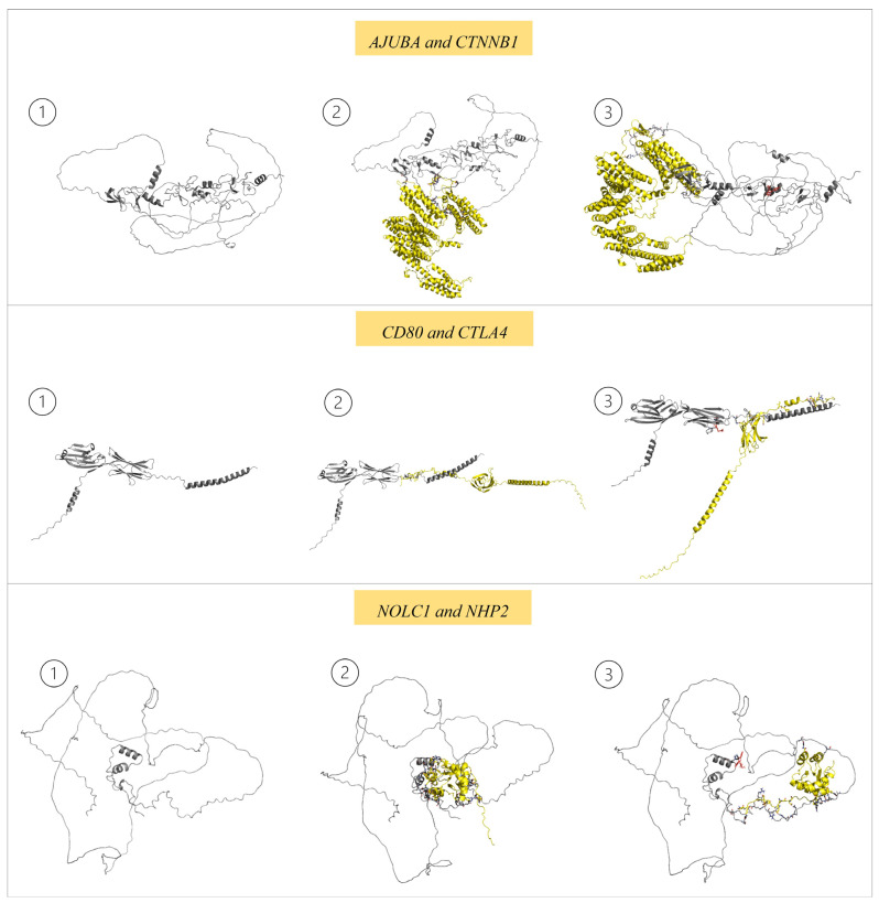
Verification
Claims the paper makes
The claims and statistics of this paper haven’t been checked yet.
Run the check — free, no account needed. It reads the open-access text and checks the sources it cites.
Self-check
Percentages, sums and reported differences re-computed from the paper’s own numbers.
The self-check needs the paper’s full text.
Citations
Cites
Subramanian, A., Tamayo, P., Mootha, V. K., Mukherjee, S., Ebert, B. L., Gillette, M. A., Paulovich, A., Pomeroy, S. L., Golub, T. R., Lander, E. S., et al. (2005) · Proceedings of the National Academy of Sciences · doi:10.1073/pnas.0506580102 · Cited by 53,625
Highly accurate protein structure prediction with AlphaFold
Jumper, J., Evans, R., Pritzel, A., Green, T., Figurnov, M., Ronneberger, O., Tunyasuvunakool, K., Bates, R., Žídek, A., Potapenko, A., et al. (2021) · Nature · doi:10.1038/s41586-021-03819-2 · Cited by 43,764
Lu, H., Zhou, Q., He, J., Jiang, Z., Peng, C., Tong, R., & Shi, J. (2020) · Signal Transduction and Targeted Therapy · doi:10.1038/s41392-020-00315-3 · Cited by 891
Research works printed but not linked: 45
- Smyth, 2020, Gastric Cancer, Lancet, 396, 635, 10.1016/S0140-6736(20)31288-5 · DOI
- Sung, 2021, Global Cancer Statistics 2020: GLOBOCAN Estimates of Incidence and Mortality Worldwide for 36 Cancers in 185 Countries, CA Cancer J. Clin., 71, 209, 10.3322/caac.21660 · DOI · Paper page
- Liu, 2022, Long Non-Coding RNA in Gastric Cancer: Mechanisms and Clinical Implications for Drug Resistance, Front. Oncol., 12, 841411, 10.3389/fonc.2022.841411 · DOI
- Lauren, 1965, The two histological main types of gastric carcinoma: Diffuse and so-called intestinal-type carcinoma. an attempt at a histo-clinical classification, Acta Pathol. Microbiol. Scand., 64, 31, 10.1111/apm.1965.64.1.31 · DOI
- Sasako, 2011, Five-Year Outcomes of a Randomized Phase III Trial Comparing Adjuvant Chemotherapy with S-1 versus Surgery Alone in Stage II or III Gastric Cancer, J. Clin. Oncol., 29, 4387, 10.1200/JCO.2011.36.5908 · DOI
- Leichman, 1992, Preoperative Systemic Chemotherapy Followed by Adjuvant Postoperative Intraperitoneal Therapy for Gastric Cancer: A University of Southern California Pilot Program, J. Clin. Oncol., 10, 1933, 10.1200/JCO.1992.10.12.1933 · DOI
- Ajani, 1999, Enhanced Staging and All Chemotherapy Preoperatively in Patients with Potentially Resectable Gastric Carcinoma, J. Clin. Oncol., 17, 2403, 10.1200/JCO.1999.17.8.2403 · DOI
- Guan, 2023, Gastric Cancer Treatment: Recent Progress and Future Perspectives, J. Hematol. Oncol., 16, 57, 10.1186/s13045-023-01451-3 · DOI
- Joshi, 2021, Current Treatment and Recent Progress in Gastric Cancer, CA Cancer J. Clin., 71, 264, 10.3322/caac.21657 · DOI
- Miller, 2015, Proteasome Inhibitors with Pyrazole Scaffolds from Structure-Based Virtual Screening, J. Med. Chem., 58, 2036, 10.1021/jm501344n · DOI
- Rohr, 2021, A Merged Microarray Meta-Dataset for Transcriptionally Profiling Colorectal Neoplasm Formation and Progression, Sci. Data, 8, 214, 10.1038/s41597-021-00998-5 · DOI
- Liberzon, 2011, Molecular Signatures Database (MSigDB) 3.0, Bioinformatics, 27, 1739, 10.1093/bioinformatics/btr260 · DOI · Paper page
- 2023, Discovery and Ranking of the Most Robust Prognostic Biomarkers in Serous Ovarian Cancer, GeroScience, 45, 1889, 10.1007/s11357-023-00742-4 · DOI
- Ribeiro, 2018, Gastric Cancer Cell Lines Have Different MYC-Regulated Expression Patterns but Share a Common Core of Altered Genes, Can. J. Gastroenterol. Hepatol., 2018, 5804376
- Riquelme, 2016, The Gene Expression Status of the PI3K/AKT/MTOR Pathway in Gastric Cancer Tissues and Cell Lines, Pathol. Oncol. Res., 22, 797, 10.1007/s12253-016-0066-5 · DOI
- Mesquita, 2021, Kinase Inhibitor Screening Reveals Aurora-a Kinase Is a Potential Therapeutic and Prognostic Biomarker of Gastric Cancer, J. Cell. Biochem., 122, 1376, 10.1002/jcb.30015 · DOI
- Bustin, S.A., Beaulieu, J.-F., Huggett, J., Jaggi, R., Kibenge, F.S.B., Olsvik, P.A., Penning, L.C., and Toegel, S. (2010). MIQE Precis: Practical Implementation of Minimum Standard Guidelines for Fluorescence-Based Quantitative Real-Time PCR Experiments. BMC Mol. Biol., 11. · DOI
- Varadi, 2022, AlphaFold Protein Structure Database: Massively Expanding the Structural Coverage of Protein-Sequence Space with High-Accuracy Models, Nucleic Acids Res., 50, D439, 10.1093/nar/gkab1061 · DOI · Paper page
- Kiss, 2012, http://Mcule.com: A Public Web Service for Drug Discovery, J. Cheminform., 4, 2012, 10.1186/1758-2946-4-S1-P17 · DOI
- Muteeb, G., Rehman, M.T., AlAjmi, M.F., Aatif, M., Farhan, M., and Shafi, S. (2022). Identification of a Potential Inhibitor (MCULE-8777613195-0-12) of New Delhi Metallo-β-Lactamase-1 (NDM-1) Using In Silico and In Vitro Approaches. Molecules, 27. · DOI
- Lipinski, 2012, Experimental and Computational Approaches to Estimate Solubility and Permeability in Drug Discovery and Development Settings, Adv. Drug Deliv. Rev., 64, 4, 10.1016/j.addr.2012.09.019 · DOI
- Lipinski, 2000, Drug-like Properties and the Causes of Poor Solubility and Poor Permeability, J. Pharmacol. Toxicol. Methods, 44, 235, 10.1016/S1056-8719(00)00107-6 · DOI
- Banerjee, 2018, ProTox-II: A Webserver for the Prediction of Toxicity of Chemicals, Nucleic Acids Res., 46, W257, 10.1093/nar/gky318 · DOI
- Daina, 2016, A BOILED-Egg To Predict Gastrointestinal Absorption and Brain Penetration of Small Molecules, ChemMedChem, 11, 1117, 10.1002/cmdc.201600182 · DOI
- Daina, 2017, SwissADME: A Free Web Tool to Evaluate Pharmacokinetics, Drug-Likeness and Medicinal Chemistry Friendliness of Small Molecules, Sci. Rep., 7, 42717, 10.1038/srep42717 · DOI · Paper page
- Almeida, 2014, A Dynamic Niching Genetic Algorithm Strategy for Docking Highly Flexible Ligands, Inf. Sci., 289, 206, 10.1016/j.ins.2014.08.002 · DOI
- Guedes, 2021, New Machine Learning and Physics-Based Scoring Functions for Drug Discovery, Sci. Rep., 11, 3198, 10.1038/s41598-021-82410-1 · DOI
- Inbar, 2005, Approaching the CAPRI Challenge with an Efficient Geometry-Based Docking, Proteins Struct. Funct. Bioinform., 60, 217, 10.1002/prot.20561 · DOI
- Housman, 2014, Drug Resistance in Cancer: An Overview, Cancers, 6, 1769, 10.3390/cancers6031769 · DOI · Paper page
- Borriello, A., Caldarelli, I., Basile, M.A., Bencivenga, D., Tramontano, A., Perrotta, S., della Ragione, F., and Oliva, A. (2011). The Tyrosine Kinase Inhibitor Dasatinib Induces a Marked Adipogenic Differentiation of Human Multipotent Mesenchymal Stromal Cells. PLoS ONE, 6. · DOI
- Yi, 2022, CCDC69 Is a Prognostic Marker of Breast Cancer and Correlates with Tumor Immune Cell Infiltration, Front. Surg., 9, 879921, 10.3389/fsurg.2022.879921 · DOI
- Prieto-Martínez, F.D., López-López, E., Eurídice Juárez-Mercado, K., and Medina-Franco, J.L. (2019). Silico Drug Design: Repurposing Techniques and Methodologies, Elsevier.
- Anthony, L.F.W., Kanding, B., and Selvan, R. (2020). Carbontracker: Tracking and Predicting the Carbon Footprint of Training Deep Learning Models. arXiv.
- Song, 2022, Therapeutic Exosomes Loaded with SERPINA5 Attenuated Endometrial Cancer Cell Migration via the Integrin Β1/FAK Signaling Pathway, Cell. Oncol., 45, 861, 10.1007/s13402-022-00687-4 · DOI
- Wu, 2021, Kaempferol Reverses Aerobic Glycolysis via MiR-339-5p-Mediated PKM Alternative Splicing in Colon Cancer Cells, J. Agric. Food Chem., 69, 3060, 10.1021/acs.jafc.0c07640 · DOI
- Jia, 2017, The LIM Protein AJUBA Promotes Colorectal Cancer Cell Survival through Suppression of JAK1/STAT1/IFIT2 Network, Oncogene, 36, 2655, 10.1038/onc.2016.418 · DOI
- Dommann, N., Sánchez-Taltavull, D., Eggs, L., Birrer, F., Brodie, T., Salm, L., Baier, F.A., Medova, M., Humbert, M., and Tschan, M.P. (2020). The LIM Protein AJUBA Augments Tumor Metastasis in Colon Cancer. Cancers, 12. · DOI
- Li, 1996, The Expression of Costimulatory Molecules CD80 and CD86 in Human Carcinoma Cell Lines: Its Regulation by Interferon Gamma and Interleukin-10, Cancer Immunol. Immunother., 43, 213, 10.1007/s002620050324 · DOI
- Horn, 2018, Soluble CD80 Protein Delays Tumor Growth and Promotes Tumor-Infiltrating Lymphocytes, Cancer Immunol. Res., 6, 59, 10.1158/2326-6066.CIR-17-0026 · DOI
- Feng, 2019, Low Expression of CD80 Predicts for Poor Prognosis in Patients with Gastric Adenocarcinoma, Future Oncol., 15, 473, 10.2217/fon-2018-0420 · DOI
- Peggs, 2010, Ipilimumab: Attenuation of an Inhibitory Immune Checkpoint Improves Survival in Metastatic Melanoma, Expert Rev. Anticancer Ther., 10, 1697, 10.1586/era.10.144 · DOI
- Zhai, 2023, Roles of NOLC1 in Cancers and Viral Infection, J. Cancer Res. Clin. Oncol., 149, 10593, 10.1007/s00432-023-04934-5 · DOI
- Kong, 2021, Knockdown of NOLC1 Inhibits PI3K-AKT Pathway to Improve the Poor Prognosis of Esophageal Carcinoma, J. Oncol., 2021, 9944132, 10.1155/2021/9944132 · DOI
- 2010, Protein-Protein Interactions and Cancer: Targeting the Central Dogma, Curr. Top. Med. Chem., 11, 258
- Bahmanyar, 2008, β-Catenin Is a Nek2 Substrate Involved in Centrosome Separation, Genes Dev., 22, 91, 10.1101/gad.1596308 · DOI
Metadata
- Authors
- Brito, D. M. S., Lima, O. G., Mesquita, F. P., da Silva, E. L., de Moraes, M. E. A., Burbano, R. M. R., Montenegro, R. C., & Souza, P. F. N.
- Affiliations
- Department of Biochemistry and Molecular Biology, Federal University of Ceará, Fortaleza 60020-181, Brazil
- Universidade Federal do Ceará (BR)
- Universidade Federal do Pará (BR)
- Corresponding author
- Souza, Pedro F. N.
- ORCID
- Venue
- Pharmaceutics
- Publisher
- MDPI AG
- Published
- 2023-09-12
- Volume
- 15
- Issue
- 9
- Pages
- 2303
- ISSN
- 1999-4923 (ISSN-L)
- PMC
- PMC10535099
- OpenAlex
- W4386638888
- Cited by
- 6
- References
- 3
- Citations by year
- 2026: 1 · 2025: 1 · 2024: 3 · 2023: 1
- FWCI
- 0.86
- Type
- journal article
- Language
- en
- Open access
- gold
- doi.org
- Licence
- Locations
- Pharmaceutics (publishedVersion, cc-by)
- Preprints.org (acceptedVersion, cc-by)
- PubMed Central (publishedVersion, cc-by)
- PubMed (publishedVersion)
- DOAJ (DOAJ: Directory of Open Access Journals) (submittedVersion, cc-by-sa)
- Funders
- National Council for Scientific and Technological Development (CNPq) (403493/2021-8, 305459/2019-8, 305003/2022-4, 151388/2022-9)
- CNPq (403493/2021-8, 305459/2019-8, 305003/2022-4, 151388/2022-9)
- Red Latinoamericana de Implementación y Validación de guias clínicas Farmacogenomicas (RELIVAF) (403493/2021-8, 305459/2019-8, 305003/2022-4, 151388/2022-9)
- Pró-Reitoria de Pesquisa e Pós-Graduação (PROPESP) from the Federal University of Pará (UFPA) (403493/2021-8, 305459/2019-8, 305003/2022-4, 151388/2022-9)
- Topics
- Life Sciences › Pharmacology, Toxicology and Pharmaceutics › Drug Discovery › Computational Drug Discovery Methods
- Health Sciences › Medicine › Oncology › Gastric Cancer Management and Outcomes
- Life Sciences › Biochemistry, Genetics and Molecular Biology › Molecular Biology › Gene expression and cancer classification
- Keywords
- gastric cancer, drug target identification, differentially expressed genes, Ajuba, CD80, NOLC1, structure-based virtual screening, molecular docking, anticancer drug discovery, gene expression profiling, prognostic factors
About these checks
Registry, retraction, Cites and statistics results are computed, not judged, with one exception: a printed reference that no index lists is read once by a model before it counts as a citation error. Claims are extracted by a model and kept only when their quote appears verbatim in the text; each cited source is then checked, with a model’s help, for whether it says what the claim relies on. Those verdicts can be wrong — read the claim’s quote and the cited source before relying on one.
Statistics and arithmetic are recomputed, not judged.
Confidence bands are shown only for fields calibrated on our annotated test set. None is calibrated yet, so no band is shown.
What counts as a claim ›How we measure confidence ›How we check this ›
Are you an author, or do you see a problem with this page? Request a review
