

The DOI matches its Crossref record.
Curcumin Disrupts a Positive Feedback Loop between ADMSCs and Cancer Cells in the Breast Tumor Microenvironment via the CXCL12/CXCR4 Axis
Open accessAbstract
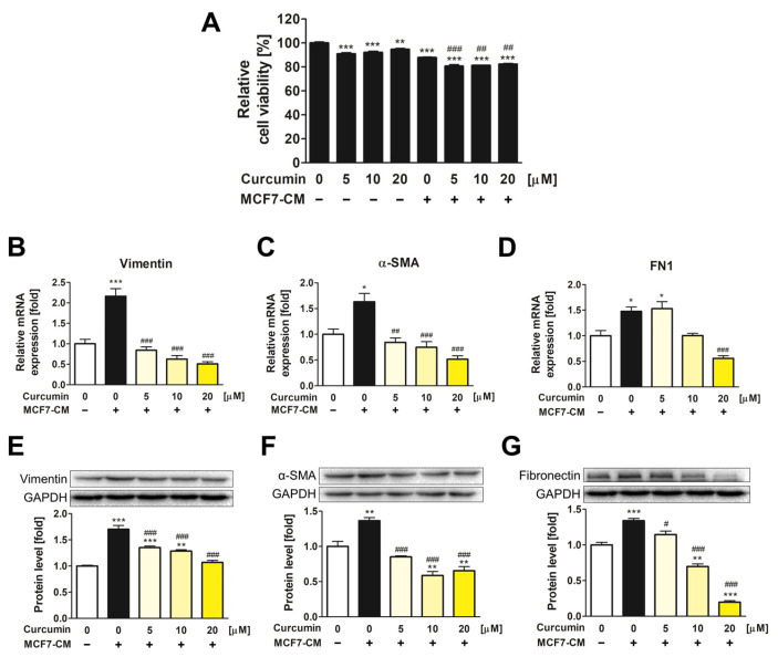
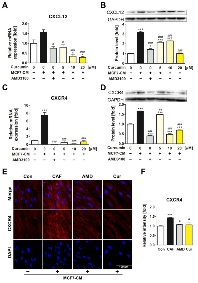
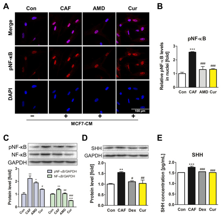
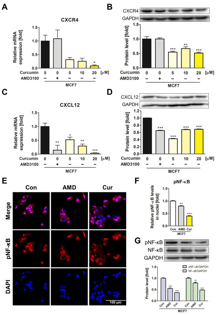
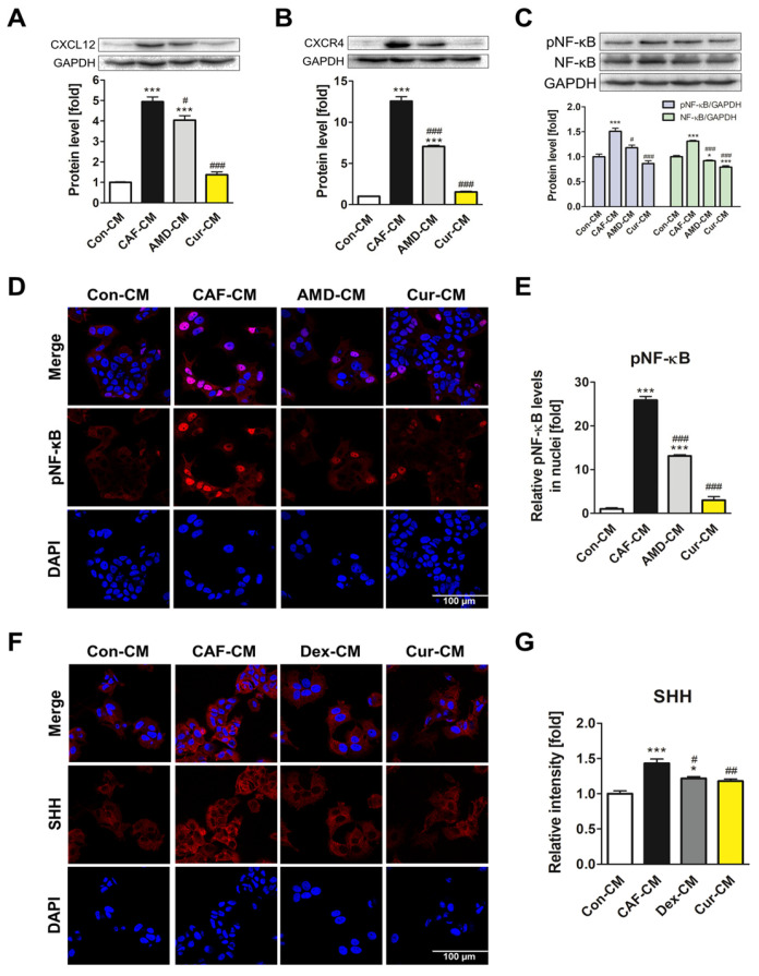
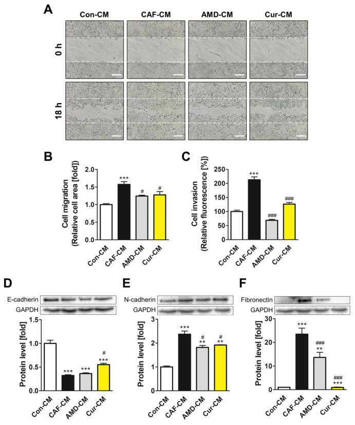
Adipose tissue has a significant impact on breast cancer initiation and progression owing to its substantial proportion in the breast. Adipose-derived mesenchymal stem cells (ADMSCs) are major players in the breast tumor microenvironment (TME) as they interact with cancer cells. The intricate interaction between ADMSCs and cancer cells not only drives the differentiation of ADMSCs into cancer-associated fibroblasts (CAFs) but also the metastasis of cancer cells, which is attributed to the CXCL12/CXCR4 axis. We investigated the effects of curcumin, a flavonoid known for CXCL12/CXCR4 axis inhibition, on breast TME by analyzing whether it can disrupt the ADMSC-cancer positive loop. Using MCF7 breast cancer cell-derived conditioned medium (MCF7-CM), we induced ADMSC transformation and verified that curcumin diminished the phenotypic change, inhibiting CAF marker expression. Additionally, curcumin suppressed the CXCL12/CXCR4 axis and its downstream signaling both in ADMSCs and MCF7 cells. The CM from ADMSCs, whose ADMSC-to-CAF transformation was repressed by the curcumin treatment, inhibited the positive feedback loop between ADMSCs and MCF7 as well as epithelial–mesenchymal transition in MCF7. Our study showed that curcumin is a potent anti-cancer agent that can remodel the breast TME, thereby restricting the ADMSC-cancer positive feedback loop associated with the CXCL12/CXCR4 axis.
Overview
The claims and statistics of this paper haven’t been checked yet.
Run the check — free, no account needed. It reads the open-access text and checks the sources it cites.
Integrity signals
Registry
Cites
Statistics
Figures
Verification
Claims the paper makes
The claims and statistics of this paper haven’t been checked yet.
Run the check — free, no account needed. It reads the open-access text and checks the sources it cites.
Claims other papers make about this one
Each is checked against this paper’s own text: does it say what the citing paper says it does?
The cited source says what the claim attributes to it.
In the citing paper
“The potential of curcumin to reduce metastasis of breast cancer is also evident in its inhibition of CXCL12/CXCR4 axis-mediated activation of the differentiation of adipose-derived MSCs (ADMSCs) into CAFs.292”
From Drug repurposing for cancer therapy (2024) · cites this paper as [292]
Self-check
Percentages, sums and reported differences re-computed from the paper’s own numbers.
The self-check needs the paper’s full text.
Citations
Cites
Still being checked.
Research works printed but not linked: 51
- Fletcher, 2017, Human breast adipose tissue: Characterization of factors that change during tumor progression in human breast cancer, J. Exp. Clin. Cancer Res., 36, 26, 10.1186/s13046-017-0494-4 · DOI
- Kothari, C., Diorio, C., and Durocher, F. (2020). The Importance of Breast Adipose Tissue in Breast Cancer. Int. J. Mol. Sci., 21. · DOI
- Miyazaki, 2021, Adipose-derived mesenchymal stem cells differentiate into heterogeneous cancer-associated fibroblasts in a stroma-rich xenograft model, Sci. Rep., 11, 4690, 10.1038/s41598-021-84058-3 · DOI
- Miyazaki, 2020, Adipose-derived mesenchymal stem cells differentiate into pancreatic cancer-associated fibroblasts in vitro, FEBS Open Bio, 10, 2268, 10.1002/2211-5463.12976 · DOI
- Strong, 2017, Obesity Enhances the Conversion of Adipose-Derived Stromal/Stem Cells into Carcinoma-Associated Fibroblast Leading to Cancer Cell Proliferation and Progression to an Invasive Phenotype, Stem Cells Int., 2017, 9216502, 10.1155/2017/9216502 · DOI
- Xue, 2022, Mesenchymal stem cells derived from adipose accelerate the progression of colon cancer by inducing a MT-CAFs phenotype via TRPC3/NF-KB axis, Stem Cell Res. Ther., 13, 335, 10.1186/s13287-022-03017-5 · DOI
- Lee, Y.T., Tan, Y.J., Falasca, M., and Oon, C.E. (2020). Cancer-Associated Fibroblasts: Epigenetic Regulation and Therapeutic Intervention in Breast Cancer. Cancers, 12. · DOI
- Ping, 2021, Cancer-associated fibroblasts: Overview, progress, challenges, and directions, Cancer Gene Ther., 28, 984, 10.1038/s41417-021-00318-4 · DOI
- Louault, K., Li, R.-R., and Declerck, Y.A. (2020). Cancer-Associated Fibroblasts: Understanding Their Heterogeneity. Cancers, 12. · DOI
- Wani, 2014, C-X-C motif chemokine 12/C-X-C chemokine receptor type 7 signaling regulates breast cancer growth and metastasis by modulating the tumor microenvironment, Breast Cancer Res., 16, R54, 10.1186/bcr3665 · DOI
- Zhou, 2019, Targeting CXCL12/CXCR4 Axis in Tumor Immunotherapy, Curr. Med. Chem., 26, 3026, 10.2174/0929867324666170830111531 · DOI
- Guo, 2016, CXCL12/CXCR4: A symbiotic bridge linking cancer cells and their stromal neighbors in oncogenic communication networks, Oncogene, 35, 816, 10.1038/onc.2015.139 · DOI
- Boimel, 2012, Contribution of CXCL12 secretion to invasion of breast cancer cells, Breast Cancer Res., 14, R23, 10.1186/bcr3108 · DOI
- Sun, 2010, CXCL12/CXCR4/CXCR7 chemokine axis and cancer progression, Cancer Metastasis Rev., 29, 709, 10.1007/s10555-010-9256-x · DOI
- Santagata, 2021, CXCR4 and CXCR7 Signaling Pathways: A Focus on the Cross-Talk Between Cancer Cells and Tumor Microenvironment, Front. Oncol., 11, 591386, 10.3389/fonc.2021.591386 · DOI
- Khare, T., Bissonnette, M., and Khare, S. (2021). CXCL12-CXCR4/CXCR7 Axis in Colorectal Cancer: Therapeutic Target in Preclinical and Clinical Studies. Int. J. Mol. Sci., 22. · DOI
- Lu, 2009, SDF-1alpha up-regulates interleukin-6 through CXCR4, PI3K/Akt, ERK, and NF-kappaB-dependent pathway in microglia, Eur. J. Pharmacol., 613, 146, 10.1016/j.ejphar.2009.03.001 · DOI
- Huang, 2009, Stromal cell-derived factor-1/CXCR4 enhanced motility of human osteosarcoma cells involves MEK1/2, ERK and NF-κB-dependent pathways, J. Cell. Physiol., 221, 204, 10.1002/jcp.21846 · DOI
- Xie, 2021, Emerging roles of the Hedgehog signaling pathway in inflammatory bowel disease, Cell Death Discov., 7, 314, 10.1038/s41420-021-00679-7 · DOI
- Douceau, S., Deutsch Guerrero, T., and Ferent, J. (2023). Establishing Hedgehog Gradients during Neural Development. Cells, 12. · DOI
- Patel, 2021, Role of aberrant Sonic hedgehog signaling pathway in cancers and developmental anomalies, J. Biomed. Res., 36, 1, 10.7555/JBR.35.20210139 · DOI
- Sleightholm, 2017, Emerging roles of the CXCL12/CXCR4 axis in pancreatic cancer progression and therapy, Pharmacol. Ther., 179, 158, 10.1016/j.pharmthera.2017.05.012 · DOI
- Kotha, R.R., and Luthria, D.L. (2019). Curcumin: Biological, Pharmaceutical, Nutraceutical, and Analytical Aspects. Molecules, 24. · DOI
- Li, 2017, Curcumin Attenuates Titanium Particle-Induced Inflammation by Regulating Macrophage Polarization In Vitro and In Vivo, Front. Immunol., 8, 55
- Ojo, 2022, Anticancer Properties of Curcumin Against Colorectal Cancer: A Review, Front. Oncol., 12, 881641, 10.3389/fonc.2022.881641 · DOI
- Gu, 2019, Curcumin inhibits liver metastasis of gastric cancer through reducing circulating tumor cells, Aging, 11, 1501, 10.18632/aging.101848 · DOI
- Zhao, R., Liu, J., Li, Z., Zhang, W., Wang, F., and Zhang, B. (2022). Recent Advances in CXCL12/CXCR4 Antagonists and Nano-Based Drug Delivery Systems for Cancer Therapy. Pharmaceutics, 14. · DOI
- Riggio, 2021, The lingering mysteries of metastatic recurrence in breast cancer, Br. J. Cancer, 124, 13, 10.1038/s41416-020-01161-4 · DOI
- Czerwiec, K., Zawrzykraj, M., Deptuła, M., Skoniecka, A., Tymińska, A., Zieliński, J., Kosiński, A., and Pikuła, M. (2023). Adipose-Derived Mesenchymal Stromal Cells in Basic Research and Clinical Applications. Int. J. Mol. Sci., 24. · DOI
- Frisbie, 2022, Carcinoma-Associated Mesenchymal Stem/Stromal Cells: Architects of the Pro-tumorigenic Tumor Microenvironment, Stem Cells, 40, 705, 10.1093/stmcls/sxac036 · DOI
- Zhang, 2023, CAFs orchestrates tumor immune microenvironment—A new target in cancer therapy?, Front. Pharmacol., 14, 1113378, 10.3389/fphar.2023.1113378 · DOI
- Geng, X., Chen, H., Zhao, L., Hu, J., Yang, W., Li, G., Cheng, C., Zhao, Z., Zhang, T., and Li, L. (2021). Cancer-Associated Fibroblast (CAF) Heterogeneity and Targeting Therapy of CAFs in Pancreatic Cancer. Front. Cell Dev. Biol., 9. · DOI
- Tan, 2020, CXCR4/TGF-β1 mediated self-differentiation of human mesenchymal stem cells to carcinoma-associated fibroblasts and promoted colorectal carcinoma development, Cancer Biol. Ther., 21, 248, 10.1080/15384047.2019.1685156 · DOI
- Hussain, 2020, The Roles of Stroma-Derived Chemokine in Different Stages of Cancer Metastases, Front. Immunol., 11, 598532, 10.3389/fimmu.2020.598532 · DOI
- Otsuka, 2011, CXCR4 Overexpression Is Associated with Poor Outcome in Females Diagnosed with Stage IV Non-small Cell Lung Cancer, J. Thorac. Oncol., 6, 1169, 10.1097/JTO.0b013e3182199a99 · DOI
- Batra, 2013, Anti-cancer potential of flavonoids: Recent trends and future perspectives, 3 Biotech, 3, 439, 10.1007/s13205-013-0117-5 · DOI
- Sharma, 2022, Advancements in clinical translation of flavonoid nanoparticles for cancer treatment, OpenNano, 8, 100074, 10.1016/j.onano.2022.100074 · DOI
- Zhang, 2016, Curcumin inhibits tumor epithelial-mesenchymal transition by downregulating the Wnt signaling pathway and upregulating NKD2 expression in colon cancer cells, Oncol. Rep., 35, 2615, 10.3892/or.2016.4669 · DOI
- Mellado, 2005, Activation of nuclear factor-κB in human prostate carcinogenesis and association to biochemical relapse, Br. J. Cancer, 93, 1285, 10.1038/sj.bjc.6602851 · DOI
- Li, 2018, NF-κB in pancreatic cancer: Its key role in chemoresistance, Cancer Lett., 421, 127, 10.1016/j.canlet.2018.02.011 · DOI
- Cai, 2011, NF-kappaB in Lung Tumorigenesis, Cancers, 3, 4258, 10.3390/cancers3044258 · DOI
- Smith, S.M., Lyu, Y.L., and Cai, L. (2014). NF-κB Affects Proliferation and Invasiveness of Breast Cancer Cells by Regulating CD44 Expression. PLoS ONE, 9. · DOI
- Olivera, 2012, Inhibition of the NF-κB signaling pathway by the curcumin analog, 3,5-bis(2-pyridinylmethylidene)-4-piperidone (EF31): Anti-inflammatory and anti-cancer properties, Int. Immunopharmacol., 12, 368, 10.1016/j.intimp.2011.12.009 · DOI
- Shi, 2020, The Role of the CXCL12/CXCR4/CXCR7 Chemokine Axis in Cancer, Front. Pharmacol., 11, 574667, 10.3389/fphar.2020.574667 · DOI
- Zielińska, K.A., and Katanaev, V.L. (2020). The Signaling Duo CXCL12 and CXCR4: Chemokine Fuel for Breast Cancer Tumorigenesis. Cancers, 12. · DOI
- Huber, 2004, NF-κB is essential for epithelial-mesenchymal transition and metastasis in a model of breast cancer progression, J. Clin. Investig., 114, 569, 10.1172/JCI200421358 · DOI
- Chartoumpekis, 2020, EMT Factors and Metabolic Pathways in Cancer, Front. Oncol., 10, 499, 10.3389/fonc.2020.00499 · DOI
- Tan, B.L., and Norhaizan, M.E. (2019). Curcumin Combination Chemotherapy: The Implication and Efficacy in Cancer. Molecules, 24. · DOI
- Xiao, 2015, Co-delivery of camptothecin and curcumin by cationic polymeric nanoparticles for synergistic colon cancer combination chemotherapy, J. Mater. Chem. B, 3, 7724, 10.1039/C5TB01245G · DOI
- Banerjee, 2017, Combinatorial effect of curcumin with docetaxel modulates apoptotic and cell survival molecules in prostate cancer, FBE, 9, 235
- Zhou, 2016, Curcumin Enhances the Effects of 5-Fluorouracil and Oxaliplatin in Inducing Gastric Cancer Cell Apoptosis Both In Vitro and In Vivo, Oncol. Res. Featur. Preclin. Clin. Cancer Ther., 23, 29
Cited by
Sorted by citations
Drug repurposing for cancer therapy
Xia, Y., Sun, M., Huang, H., & Jin, W.-L. (2024) · Signal Transduction and Targeted Therapy · doi:10.1038/s41392-024-01808-1 · Cited by 443
“292 In addition, Mao et al. demonstrated that curcumin suppressed proliferation of LGR5( + ) colorectal CSCs by not only promoting autophagy but also through transcriptional repression of the oncogenic transcription factor activating enhancer-binding protein 2A (TFAP2A)-regulated ECM pathway.”
Background
Metadata
- Authors
- Jang, B.-Y., Shin, M., Han, D.-H., & Sung, J.-S.
- Affiliations
- Dongguk University (KR)
- Corresponding author
- Sung, Jung-Suk
- Venue
- Pharmaceutics
- Publisher
- MDPI AG
- Published
- 2023-11-15
- Volume
- 15
- Issue
- 11
- Pages
- 2627
- ISSN
- 1999-4923 (ISSN-L)
- PMC
- PMC10675183
- OpenAlex
- W4388723961
- Cited by
- 19
- References
- 0
- Citations by year
- 2026: 5 · 2025: 6 · 2024: 7 · 2023: 1
- FWCI
- 1.60
- Type
- journal article
- Language
- en
- Open access
- gold
- doi.org
- Licence
- Locations
- Pharmaceutics (publishedVersion, cc-by)
- PubMed Central (publishedVersion, cc-by)
- PubMed (publishedVersion)
- DOAJ (DOAJ: Directory of Open Access Journals) (submittedVersion, cc-by-sa)
- Funders
- National Research Foundation of Korea (NRF) (2022R1A6A1A03053343)
- Ministry of Education (2022R1A6A1A03053343)
- Topics
- Life Sciences › Biochemistry, Genetics and Molecular Biology › Molecular Medicine › Curcumin's Biomedical Applications
- Health Sciences › Medicine › Oncology › Cancer Cells and Metastasis
- Life Sciences › Biochemistry, Genetics and Molecular Biology › Cell Biology › Mesenchymal stem cell research
- Keywords
- curcumin, adipose-derived stem cells, breast cancer, tumor microenvironment, CXCL12/CXCR4 axis, cancer-associated fibroblasts, epithelial-mesenchymal transition, MCF-7 cells, conditioned medium, metastasis, flavonoids
About these checks
Registry, retraction, Cites and statistics results are computed, not judged, with one exception: a printed reference that no index lists is read once by a model before it counts as a citation error. Claims are extracted by a model and kept only when their quote appears verbatim in the text; each cited source is then checked, with a model’s help, for whether it says what the claim relies on. Those verdicts can be wrong — read the claim’s quote and the cited source before relying on one.
Statistics and arithmetic are recomputed, not judged.
Confidence bands are shown only for fields calibrated on our annotated test set. None is calibrated yet, so no band is shown.
What counts as a claim ›How we measure confidence ›How we check this ›
Are you an author, or do you see a problem with this page? Request a review
