

The DOI matches its Crossref record.
Enhancement of tendon-bone healing following rotator cuff repair using hydroxyapatite with TGFβ1
Open accessAbstract
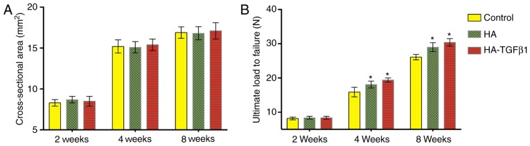
The formation of fibrocartilage at the healing site following a rotator cuff tear repair is a major problem in the field of tendon‑bone healing. The present study aimed to enhance the healing of the tendon‑bone interface following rotator‑cuff tear repair by the interposition of hydroxyapatite (HA) encapsulated with transforming growth factor β1 (TGFβ1). Using an acute rotator cuff repair model, rats were divided into three groups: i) Repair only (control); ii) HA group; and iii) HA‑TGFβ1 group. Animals were sacrificed at 2, 4 and 8 weeks following surgery. Micro‑computed tomography (CT), histomorphometric analyses and biomechanical tests were used to evaluate the supraspinatus tendon‑bone complex. The micro‑CT images revealed notable novel bone formation in the groups treated with HA‑TGFβ1. The histomorphometric analyses demonstrated improved fibrocartilage formation and collagen organization at the tendon‑bone interface. The HA‑TGFβ1 combination significantly improved the area of fibrocartilage, particularly at early time points (2 and 4 weeks). There was a significantly greater load‑to‑failure force achieved in the HA and HA‑TGFβ1 groups compared with the control group at 4 and 8 weeks. Augmentation of the ceramic powder with HA‑TGFβ1 at the tendon‑bone interface was demonstrated to strengthen the healing entheses, increase bone and fibrocartilage formation and improve collagen organization compared with surgical repair alone. Local application of HA‑TGFβ1 demonstrates potential in improving tendon‑bone healing following rotator cuff repair.
Overview
The claims and statistics of this paper haven’t been checked yet.
Run the check — free, no account needed. It reads the open-access text and checks the sources it cites.
Integrity signals
Registry
Cites
Statistics
Figures
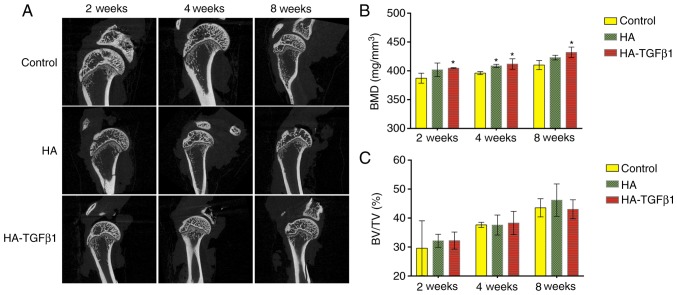
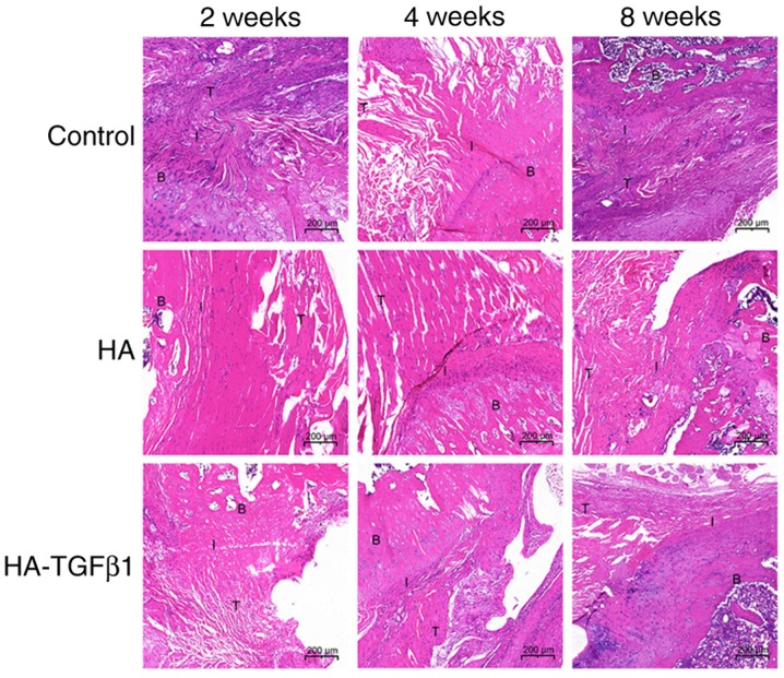
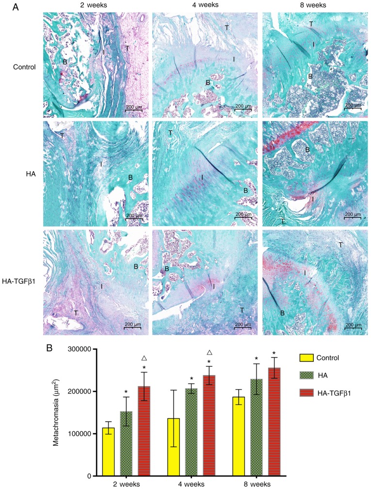
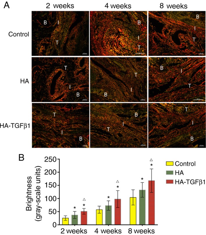
Verification
Claims the paper makes
The claims and statistics of this paper haven’t been checked yet.
Run the check — free, no account needed. It reads the open-access text and checks the sources it cites.
Claims other papers make about this one
Each is checked against this paper’s own text: does it say what the citing paper says it does?
The source backs part of the claim, not all of it — or backs it for a different population, condition or comparison.
In the citing paper
“Hydroxyapatite, a natural apatite mineral, has the capacity to interact with bone marrow mesenchymal stem cells (BMSCs) and enhance BMSC proliferation, differentiation, and mineralization through its action on various signaling pathways, including ERK/Sox9, BMP/Smad, Wnt/β-catenin, TGF-β, and MAPK [64,65,66,67,68].”
From Recent Advances in Silk Fibroin-Based Composites for Bone Repair Applications: A Review (2025) · cites this paper as [67]
Experimental — accuracy not yet measured
Self-check
Percentages, sums and reported differences re-computed from the paper’s own numbers.
The self-check needs the paper’s full text.
Citations
Cites
Still being checked.
Research works printed but not linked: 28
- Domb, 2008, High-tension double-row footprint repair compared with reduced-tension single-row repair for massive rotator cuff tears, J Bone Joint Surg Am, 90, S35, 10.2106/JBJS.H.00650 · DOI
- Nelson, 2008, Single-row modified mason-allen versus double-row arthroscopic rotator cuff repair: A biomechanical and surface area comparison, Arthroscopy, 24, 941, 10.1016/j.arthro.2008.03.011 · DOI
- Ozbaydar, 2008, A comparison of single-versus double-row suture anchor techniques in a simulated repair of the rotator cuff: An experimental study in rabbits, J Bone Joint Surg Br, 90, 1386, 10.1302/0301-620X.90B10.20862 · DOI
- Boileau, 2005, Arthroscopic repair of full-thickness tears of the supraspinatus: does the tendon really heal?, J Bone Joint Surg Am, 87, 1229, 10.2106/00004623-200506000-00007 · DOI
- Gerber, 2000, The results of repair of massive tears of the rotator cuff, J Bone Joint Surg Am, 82, 505, 10.2106/00004623-200004000-00006 · DOI
- Zumstein, 2008, The clinical and structural long-term results of open repair of massive tears of the rotator cuff, J Bone Joint Surg Am, 90, 2423, 10.2106/JBJS.G.00677 · DOI
- Li, 2011, Hydroxyapatite coating enhances polyethylene terephthalate artificial ligament graft osseointegration in the bone tunnel, Int Orthop, 35, 1561, 10.1007/s00264-010-1158-6 · DOI
- Huangfu, 2007, Tendon-bone healing enhancement using injectable tricalcium phosphate in a dog anterior cruciate ligament reconstruction model, Arthroscopy, 23, 455, 10.1016/j.arthro.2006.12.031 · DOI
- Zhao, 2014, Effect of the interposition of calcium phosphate materials on tendon-bone healing during repair of chronic rotator cuff tear, Am J Sports Med, 42, 1920, 10.1177/0363546514532781 · DOI
- Gulotta, 2009, Growth factors for rotator cuff repair, Clin Sports Med, 28, 13, 10.1016/j.csm.2008.09.002 · DOI
- Kim, 2011, The role of transforming growth factor beta isoforms in tendon-to-bone healing, Connect Tissue Res, 52, 87, 10.3109/03008207.2010.483026 · DOI
- Pryce, 2009, Recruitment and maintenance of tendon progenitors by TGFbeta signaling are essential for tendon formation, Development, 136, 1351, 10.1242/dev.027342 · DOI
- Manning, 2011, Sustained delivery of transforming growth factor beta three enhances tendon-to-bone healing in a rat model, J Orthop Res, 29, 1099, 10.1002/jor.21301 · DOI
- Arimura, 2017, TGF-β1 improves biomechanical strength by extracellular matrix accumulation without increasing the number of tenogenic lineage cells in a rat rotator cuff repair model, Am J Sports Med, 45, 2394, 10.1177/0363546517707940 · DOI
- Anaguchi, 2005, The effect of transforming growth factor-beta on mechanical properties of the fibrous tissue regenerated in the patellar tendon after resecting the central portion, Clin Biomech, 20, 959, 10.1016/j.clinbiomech.2005.05.012 · DOI
- Yamazaki, 2005, The effect of transforming growth factor-beta1 on intraosseous healing of flexor tendon autograft replacement of anterior cruciate ligament in dogs, Arthroscopy, 21, 1034, 10.1016/j.arthro.2005.05.011 · DOI
- Qi, 2013, Hydroxyapatite hierarchically nanostructured porous hollow microspheres: Rapid, sustainable microwave-hydrothermal synthesis by using creatine phosphate as an organic phosphorus source and application in drug delivery and protein adsorption, Chemistry, 19, 5332, 10.1002/chem.201203886 · DOI
- Galatz, 2006, Characteristics of the rat supraspinatus tendon during tendon-to-bone healing after acute injury, J Orthop Res, 24, 541, 10.1002/jor.20067 · DOI
- Canullo, 2013, Histological evaluation at different times after augmentation of extraction sites grafted with a magnesium-enriched hydroxyapatite: Double-blinded randomized controlled trial, Clin Oral Implants Res, 24, 398, 10.1111/clr.12035 · DOI
- Hamerschmidt, 2011, Hydroxyapatite granules used in the obliteration of mastoid cavities in rats, Braz J Otorhinolaryngol, 77, 315, 10.1590/S1808-86942011000300008 · DOI
- Gredes, 2015, Histological and molecular-biological analyses of poly(3-hydroxybutyrate) (PHB) patches for enhancement of bone regeneration, Ann Anat, 199, 36, 10.1016/j.aanat.2014.04.003 · DOI
- Kovacevic, 2011, Calcium-phosphate matrix with or without TGF-β3 improves tendon-bone healing after rotator cuff repair, Am J Sports Med, 39, 811, 10.1177/0363546511399378 · DOI
- Wang, 2017, Up-regulation of TGF-β promotes tendon-to-bone healing after anterior cruciate ligament reconstruction using bone marrow-derived mesenchymal stem cells through the TGF-β/MAPK signaling pathway in a new zealand white rabbit model, Cell Physiol Biochem, 41, 213, 10.1159/000456046 · DOI
- Li, 2015, Mesenchymal stem cells mitigate cirrhosis through BMP7, Cell Physiol Biochem, 35, 433, 10.1159/000369708 · DOI
- Tang, 2009, TGF-beta1-induced migration of bone mesenchymal stem cells couples bone resorption with formation, Nat Med, 15, 757, 10.1038/nm.1979 · DOI · Paper page
- Li, 2010, MEK/ERK and p38 MAPK regulate chondrogenesis of rat bone marrow mesenchymal stem cells through delicate interaction with TGF-beta1/Smads pathway, Cell Prolif, 43, 333, 10.1111/j.1365-2184.2010.00682.x · DOI
- Carpenter, 1998, Rotator cuff defect healing: A biomechanical and histologic analysis in an animal model, J Shoulder Elbow Surg, 7, 599, 10.1016/S1058-2746(98)90007-6 · DOI
- Soslowsky, 1995, Development and use of an animal model for investigations on rotator cuff disease, J Shoulder Elbow Surg, 5, 383, 10.1016/S1058-2746(96)80070-X · DOI
Cited by
Recent Advances in Silk Fibroin-Based Composites for Bone Repair Applications: A Review
Zhu, S., Zhang, Q., Xu, X., Liu, Z., Cheng, G., Long, D., Cheng, L., & Dai, F. (2025) · Polymers · doi:10.3390/polym17060772 · Cited by 36
Metadata
- Authors
- You, X., Shen, Y., Yu, W., & He, Y.
- Affiliations
- Department of Orthopedics, Shanghai Jiao Tong University Affiliated Sixth People's Hospital, Shanghai 200233, P.R. China
- State Key Laboratory of High Performance Ceramics and Superfine Microstructure, Shanghai Institute of Ceramics, Chinese Academy of Science, Shanghai 200050, P.R. China
- Shanghai Jiao Tong University (CN)
- Corresponding author
- He, Yaohua
- ORCID
- He, Yaohua
- Venue
- Molecular Medicine Reports
- Publisher
- Spandidos Publications
- Published
- 2018-01-26
- Volume
- 17
- Issue
- 4
- Pages
- 4981-4988
- ISSN
- 1791-2997 (ISSN-L)
- 1791-3004
- PMC
- PMC5865958
- OpenAlex
- W2787640574
- Cited by
- 15
- References
- 0
- Citations by year
- 2026: 2 · 2025: 4 · 2023: 2 · 2021: 4 · 2020: 1 · 2019: 1 · 2018: 1
- FWCI
- 1.82
- Type
- journal article
- Language
- en
- Open access
- hybrid
- doi.org
- Licence
- cc-by-nc-nd
- Locations
- Molecular Medicine Reports (publishedVersion, cc-by-nc-nd)
- PubMed Central (publishedVersion, cc-by-nc-nd)
- PubMed (publishedVersion)
- Funders
- National Natural Science Foundation of China (81271961)
- Topics
- Health Sciences › Medicine › Surgery › Shoulder Injury and Treatment
- Health Sciences › Medicine › Orthopedics and Sports Medicine › Tendon Structure and Treatment
- Physical Sciences › Engineering › Biomedical Engineering › Bone Tissue Engineering Materials
- Keywords
- rotator cuff repair, tendon-bone healing, hydroxyapatite, TGF-β1, fibrocartilage formation, supraspinatus tendon, enthesis, collagen organization, bone formation, rotator cuff tear
- MeSH terms
- Rotator Cuff Injuries, Animals, Biocompatible Materials, Collagen, Disease Models, Animal, Immunohistochemistry, Male, Time Factors, Wound Healing, Durapatite, Rats, Transforming Growth Factor beta1, X-Ray Microtomography
About these checks
Registry, retraction, Cites and statistics results are computed, not judged, with one exception: a printed reference that no index lists is read once by a model before it counts as a citation error. Claims are extracted by a model and kept only when their quote appears verbatim in the text; each cited source is then checked, with a model’s help, for whether it says what the claim relies on. Those verdicts can be wrong — read the claim’s quote and the cited source before relying on one.
Statistics and arithmetic are recomputed, not judged.
Confidence bands are shown only for fields calibrated on our annotated test set. None is calibrated yet, so no band is shown.
What counts as a claim ›How we measure confidence ›How we check this ›
Are you an author, or do you see a problem with this page? Request a review
Data sources
Bibliographic data: Semantic Scholar, OpenAlex, Crossref.
Contains information from the Semantic Scholar Academic Graph Datasets, made available under the ODC Attribution License (ODC-BY 1.0).
Semantic Scholar’s data is described in: Kinney, R., et al. (2023). The Semantic Scholar Open Data Platform. arXiv:2301.10140.
