

The DOI matches its Crossref record; 1 of the 1 references it cites were found in a registry or an index.
Pirfenidone suppresses TGF-β1-induced human intestinal fibroblasts activities by regulating proliferation and apoptosis via the inhibition of the Smad and PI3K/AKT signaling pathway
Open accessAbstract
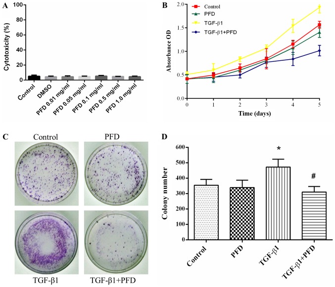
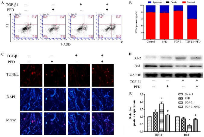
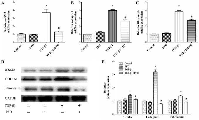
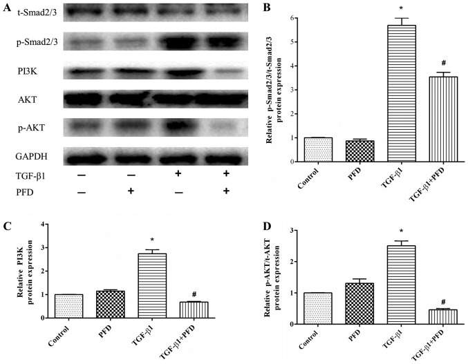
Intestinal fibroblasts, the main effector cells of intestinal fibrosis, are considered to be a good target for anti‑fibrotic therapy. The aim of the present study was to examine the effects of pirfenidone (PFD) on human intestinal fibroblasts (HIFs) stimulated by transforming growth factor (TGF)‑β1 and to explore the potential mechanism. Prior to stimulation with TGF‑β1 (10 ng/ml), HIFs were treated with or without PFD (1 mg/ml). Cell proliferation was determined by Cell Counting Kit (CCK)‑8 and colony formation assays, and cell apoptosis was assessed using flow cytometry and a TUNEL assay. Reverse transcription‑quantitative polymerase chain reaction and western blotting were performed to evaluate the mRNA and protein expressions of α‑smooth muscle actin (α‑SMA), collagen I and fibronectin. The protein expression of TGF‑β1/mothers against decapentaplegic homolog (Smad) and phosphoinositide 3‑kinase (PI3K)/protein kinase B (AKT) signaling pathways was evaluated by western blotting. CCK‑8 and colony formation assays demonstrated that PFD significantly inhibited cell proliferation in HIFs stimulated with TGF‑β1. Flow cytometry and TUNEL assays revealed that PFD treatment significantly enhanced apoptosis in TGF‑β1‑stimulated HIFs. In addition, PFD markedly reduced TGF‑β1‑induced HIF activities, such as myofibroblast differentiation (α‑SMA), and collagen production (collagen I and fibronectin). These effects of PFD were mediated by the inhibition of the TGF‑β1/Smad and PI3K/AKT signaling pathways. Therefore, the present study demonstrated that PFD reduced TGF‑β1‑induced fibrogenic activities of HIFs, suggesting that PFD may be a potential therapeutic agent for intestinal fibrosis.
Overview
The claims and statistics of this paper haven’t been checked yet.
Run the check — free, no account needed. It reads the open-access text and checks the sources it cites.
Integrity signals
Registry
Cites
Statistics
Figures
Verification
Claims the paper makes
The claims and statistics of this paper haven’t been checked yet.
Run the check — free, no account needed. It reads the open-access text and checks the sources it cites.
Claims other papers make about this one
Each is checked against this paper’s own text: does it say what the citing paper says it does?
The source backs part of the claim, not all of it — or backs it for a different population, condition or comparison.
In the citing paper
“Consistent with this, the small molecule pirfenidone (PFD), has been shown to reduce TGF-β signaling and inflammatory cytokine production in different murine experimental systems (135–137)”
From Targeting macrophages in liver fibrosis (2026) · cites this paper as [135]
Experimental — accuracy not yet measured
Self-check
Percentages, sums and reported differences re-computed from the paper’s own numbers.
The self-check needs the paper’s full text.
Citations
Cites
Conte, E., Gili, E., Fagone, E., Fruciano, M., Iemmolo, M., & Vancheri, C. (2014) · European Journal of Pharmaceutical Sciences · doi:10.1016/j.ejps.2014.02.014 · Cited by 368
Research works printed but not linked: 31
- Rieder, 2009, Intestinal fibrosis in IBD-a dynamic, multifactorial process, Nat Rev Gastroenterol Hepatol, 6, 228, 10.1038/nrgastro.2009.31 · DOI
- Stacey, 2014, Radiation-induced small bowel disease: Latest developments and clinical guidance, Ther Adv Chronic Dis, 5, 15, 10.1177/2040622313510730 · DOI
- Hamama, 2012, Therapeutic management of intestinal fibrosis induced by radiation therapy: From molecular profiling to new intervention strategies et vice et versa, Fibrogenesis Tissue Repair, 5, S13, 10.1186/1755-1536-5-S1-S13 · DOI
- Kendall, 2014, Fibroblasts in fibrosis: Novel roles and mediators, Front Pharmacol, 5, 123, 10.3389/fphar.2014.00123 · DOI · Paper page
- Zeisberg, 2013, Cellular mechanisms of tissue fibrosis. 1. Common and organ-specific mechanisms associated with tissue fibrosis, Am J Physiol Cell Physiol, 304, C216, 10.1152/ajpcell.00328.2012 · DOI
- Speca, 2012, Cellular and molecular mechanisms of intestinal fibrosis, World J Gastroenterol, 18, 3635, 10.3748/wjg.v18.i28.3635 · DOI
- Meng, 2016, TGF-β: The master regulator of fibrosis, Nat Rev Nephrol, 12, 325, 10.1038/nrneph.2016.48 · DOI · Paper page
- Li, 2017, DKK3 regulates cell proliferation, apoptosis and collagen synthesis in keloid fibroblasts via TGF-β1/Smad signaling pathway, Biomed Pharmacother, 91, 174, 10.1016/j.biopha.2017.03.044 · DOI
- Abe, 2012, Simvastatin attenuates intestinal fibrosis independent of the anti-inflammatory effect by promoting fibroblast/myofibroblast apoptosis in the regeneration/healing process from TNBS-induced colitis, Dig Dis Sci, 57, 335, 10.1007/s10620-011-1879-4 · DOI
- Azuma, 2005, Double-blind, placebo-controlled trial of pirfenidone in patients with idiopathic pulmonary fibrosis, Am J Respir Crit Care Med, 171, 1040, 10.1164/rccm.200404-571OC · DOI
- Di Sario, 2002, Effect of pirfenidone on rat hepatic stellate cell proliferation and collagen production, J Hepatol, 37, 584, 10.1016/S0168-8278(02)00245-3 · DOI · Paper page
- Miric, 2001, Reversal of cardiac and renal fibrosis by pirfenidone and spironolactone in streptozotocin-diabetic rat, Br J Pharmacol, 133, 687, 10.1038/sj.bjp.0704131 · DOI
- Shi, 2011, In vitro effects of pirfenidone on cardiac fibroblasts: Proliferation, myofibroblast differentiation, migration and cytokine secretion, PLoS One, 6, e28134, 10.1371/journal.pone.0028134 · DOI
- Duan, 2015, Pirfenidone attenuates bladder fibrosis and mitigates deterioration of bladder function in a rat model of partial bladder outlet obstruction, Mol Med Rep, 12, 3639, 10.3892/mmr.2015.3814 · DOI
- Lin, 2009, Effects of pirfenidone on proliferation, migration, and collagen contraction of human Tenon's fibroblasts in vitro, Invest Ophthalmol Vis Sci, 50, 3763, 10.1167/iovs.08-2815 · DOI
- Hewitson, 2001, Pirfenidone reduces in vitro rat renal fibroblast activation and mitogenesis, J Nephrol, 14, 453
- Chan, 2013, Therapeutic update in idiopathic pulmonary fibrosis, Clin Rev Allergy Immunol, 44, 65, 10.1007/s12016-010-8244-9 · DOI
- Cottin, 2013, The role of pirfenidone in the treatment of idiopathic pulmonary fibrosis, Respir Res, 14, S5, 10.1186/1465-9921-14-S1-S5 · DOI
- Takeda, 2014, Efficacy and safety of pirfenidone for idiopathic pulmonary fibrosis, Patient Prefer Adherence, 8, 361, 10.2147/PPA.S37233 · DOI
- Meier, 2016, Decreased fibrogenesis after treatment with pirfenidone in a newly developed mouse model of intestinal fibrosis, Inflamm Bowel Dis, 22, 569, 10.1097/MIB.0000000000000716 · DOI
- Li, 2016, Oral pirfenidone protects against fibrosis by inhibiting fibroblast proliferation and TGF-β signaling in a murine colitis model, Biochem Pharmacol, 117, 57, 10.1016/j.bcp.2016.08.002 · DOI
- Sun, 2018, Pirfenidone prevents radiation-induced intestinal fibrosis in rats by inhibiting fibroblast proliferation and differentiation and suppressing the TGF-β1/Smad/CTGF signaling pathway, Eur J Pharmacol, 822, 199, 10.1016/j.ejphar.2018.01.027 · DOI
- Livak, 2001, Analysis of relative gene expression data using real-time quantitative PCR and the 2(-Delta Delta C(T)) method, Methods, 25, 402, 10.1006/meth.2001.1262 · DOI · Paper page
- Ni, 2014, The effect of transforming growth factor β1 on the crosstalk between autophagy and apoptosis in the annulus fibrosus cells under serum deprivation, Cytokine, 70, 87, 10.1016/j.cyto.2014.07.249 · DOI
- Wu, 2017, Quercetin prevents hepatic fibrosis by inhibiting hepatic stellate cell activation and reducing autophagy via the TGF-β1/Smads and PI3K/Akt pathways, Sci Rep, 7, 9289, 10.1038/s41598-017-09673-5 · DOI
- Takano, 2015, Analysis of TGF-β1- and drug-induced epithelial-mesenchymal transition in cultured alveolar epithelial cell line RLE/Abca3, Drug Metab Pharmacokinet, 30, 111, 10.1016/j.dmpk.2014.10.007 · DOI
- Lee, 1998, Pirfenidone: A novel pharmacological agent that inhibits leiomyoma cell proliferation and collagen production, J Clin Endocrinol Metab, 83, 219, 10.1210/jcem.83.1.4503 · DOI
- Luna, 2011, Mesenchymal cell proliferation and programmed cell death: Key players in fibrogenesis and new targets for therapeutic intervention, Am J Physiol Gastrointest Liver Physiol, 300, G703, 10.1152/ajpgi.00504.2010 · DOI
- Derynck, 2003, Smad-dependent and Smad-independent pathways in TGF-beta family signalling, Nature, 425, 577, 10.1038/nature02006 · DOI
- Choi, 2012, Pirfenidone inhibits transforming growth factor-β1-induced fibrogenesis by blocking nuclear translocation of Smads in human retinal pigment epithelial cell line ARPE-19, Mol Vis, 18, 1010
- Downward, 2004, PI 3-kinase, Akt and cell survival, Semin Cell Dev Biol, 15, 177, 10.1016/j.semcdb.2004.01.002 · DOI
Cited by
Targeting macrophages in liver fibrosis
González del Barrio, L., Ipsen, D. H., Pfister, D. R., Hancz, D., & Scott, C. L. (2026) · Frontiers in Immunology · doi:10.3389/fimmu.2026.1835489 · Cited by 3
Metadata
- Authors
- Sun, Y., Zhang, Y., & Chi, P.
- Affiliations
- Union Hospital (CN)
- Corresponding author
- Chi, Pan
- Venue
- Molecular Medicine Reports
- Publisher
- Spandidos Publications
- Published
- 2018-08-22
- Volume
- 18
- Issue
- 4
- Pages
- 3907-3913
- ISSN
- 1791-2997 (ISSN-L)
- 1791-3004
- PMC
- PMC6131636
- OpenAlex
- W2888293125
- Cited by
- 50
- References
- 1
- Citations by year
- 2026: 3 · 2025: 7 · 2024: 3 · 2023: 7 · 2022: 5 · 2021: 10 · 2020: 10 · 2019: 5
- FWCI
- 2.22
- Type
- journal article
- Language
- en
- Open access
- hybrid
- doi.org
- Licence
- cc-by-nc-nd
- Locations
- Molecular Medicine Reports (publishedVersion, cc-by-nc-nd)
- Europe PMC (PubMed Central) (publishedVersion, other-oa)
- PubMed Central (publishedVersion, cc-by-nc-nd)
- PubMed (publishedVersion)
- Funders
- Fujian Provincial Department of Science and Technology (2015Y0058)
- Fujian Medical University
- Topics
- Life Sciences › Biochemistry, Genetics and Molecular Biology › Molecular Biology › TGF-β signaling in diseases
- Health Sciences › Medicine › Gastroenterology › Inflammatory Bowel Disease
- Health Sciences › Medicine › Pulmonary and Respiratory Medicine › Interstitial Lung Diseases and Idiopathic Pulmonary Fibrosis
- Keywords
- pirfenidone, intestinal fibrosis, intestinal fibroblasts, TGF-β1, Smad signaling, PI3K/Akt signaling, cell proliferation, apoptosis, myofibroblast differentiation, alpha-smooth muscle actin, type I collagen, fibronectin, antifibrotic therapy
- MeSH terms
- Cell Differentiation, Collagen, Fibroblasts, Humans, Intestines, Pyridones, Signal Transduction, Apoptosis, Phosphatidylinositol 3-Kinases, Cell Proliferation, Proto-Oncogene Proteins c-akt, Smad Proteins, Transforming Growth Factor beta1
About these checks
Registry, retraction, Cites and statistics results are computed, not judged, with one exception: a printed reference that no index lists is read once by a model before it counts as a citation error. Claims are extracted by a model and kept only when their quote appears verbatim in the text; each cited source is then checked, with a model’s help, for whether it says what the claim relies on. Those verdicts can be wrong — read the claim’s quote and the cited source before relying on one.
Statistics and arithmetic are recomputed, not judged.
Confidence bands are shown only for fields calibrated on our annotated test set. None is calibrated yet, so no band is shown.
What counts as a claim ›How we measure confidence ›How we check this ›
Are you an author, or do you see a problem with this page? Request a review
Data sources
Bibliographic data: Semantic Scholar, OpenAlex, Crossref.
Contains information from the Semantic Scholar Academic Graph Datasets, made available under the ODC Attribution License (ODC-BY 1.0).
Semantic Scholar’s data is described in: Kinney, R., et al. (2023). The Semantic Scholar Open Data Platform. arXiv:2301.10140.
