

The DOI matches its Crossref record; 11 of the 11 references it cites were found in a registry or an index.
Laryngeal Tuberculosis Mimicking Laryngeal Carcinoma on 18F-FDG PET/CT Imaging
Open accessAbstract
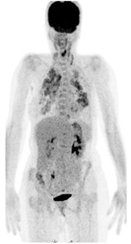
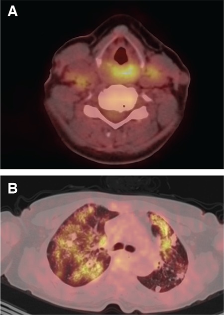
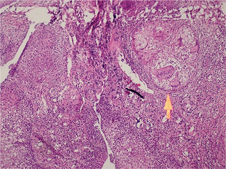
Laryngeal tuberculosis is a rare presentation of tuberculosis.It can mimic laryngeal carcinoma with its clinical and imaging findings.A 51-year old woman underwent 18 F-fluorodeoxyglucose positron emission tomography/computed tomography (PET/CT) imaging for clinically suspected carcinoma of the larynx.PET/CT revealed lung lesions consistent with tuberculosis in additional to hypermetabolic focus on larynx.The patient was histopathologically diagnosed with lung and laryngeal tuberculosis.
Overview
The claims and statistics of this paper haven’t been checked yet.
Run the check — free, no account needed. It reads the open-access text and checks the sources it cites.
Integrity signals
Registry
Cites
Statistics
Figures
Verification
Claims the paper makes
The claims and statistics of this paper haven’t been checked yet.
Run the check — free, no account needed. It reads the open-access text and checks the sources it cites.
Claims other papers make about this one
Each is checked against this paper’s own text: does it say what the citing paper says it does?
The source backs part of the claim, not all of it — or backs it for a different population, condition or comparison.
In the citing paper
“due to mostly absent pulmonary involvement and high clinical resemblance to neoplastic or chronic inflammatory conditions”
From Head and Neck Tuberculosis: A Rare Diagnosis and the Role of Surgical Biopsy and Histopathological Evaluation in Extrapulmonary Disease (2025) · cites this paper as [18]
Experimental — accuracy not yet measured
Self-check
Percentages, sums and reported differences re-computed from the paper’s own numbers.
The self-check needs the paper’s full text.
Citations
Cites
Pulmonary Tuberculoma Evaluated by Means of FDG PET: Findings in 10 Cases
Goo, J. M., Im, J.-G., Do, K., Yeo, J. S., Seo, J. B., Kim, H. Y., & Chung, J.-K. (2000) · Radiology · doi:10.1148/radiology.216.1.r00jl19117 · Cited by 353
Current clinical propensity of laryngeal tuberculosis: review of 60 cases
Lim, J., Kim, K.-M., Choi, E. C., Kim, Y.-H., Kim, H. S., & Choi, H. (2006) · European Archives of Oto-Rhino-Laryngology · doi:10.1007/s00405-006-0063-5 · Cited by 112
Laryngeal tuberculosis: an often forgotten diagnosis
Boscolo‐Rizzo, P., Mosto, M. C. D., Clari, M., Scotton, P. G., Vaglia, A., & Marchiori, C. (2003) · International Journal of Infectious Diseases · doi:10.1016/s1201-9712(03)90008-7 · Cited by 63
Yencha, M. W., Linfesty, R. L., & Blackmon, A. (2000) · American Journal of Otolaryngology · doi:10.1016/s0196-0709(00)85010-3 · Cited by 51
Smulders, Y. E., de Bondt, B., Lacko, M., et al. (2009) · Journal of Medical Case Reports · doi:10.1186/1752-1947-3-9288 · Cited by 46
Laryngeal tuberculosis: CT findings.
Moon, W. K., Han, M. H., Chang, K. H., et al. (1996) · American Journal of Roentgenology · doi:10.2214/ajr.166.2.8553964 · Cited by 43
Laryngeal involvement in patients with active pulmonary tuberculosis
Topak, M., Oysu, Ç., Yelken, K., Sahin‐Yilmaz, A., & Külekçi, M. (2007) · European Archives of Oto-Rhino-Laryngology · doi:10.1007/s00405-007-0459-x · Cited by 34
Epiglottic Tuberculosis in a Patient Treated with Steroids for Addison's Disease
Egeli, E., Oğhan, F., Alper, M., et al. (2003) · The Tohoku Journal of Experimental Medicine · doi:10.1620/tjem.201.119 · Cited by 29
CT Findings of Laryngeal Tuberculosis: Comparison to Laryngeal Carcinoma
Kim, M. D., Kim, D. I., Yune, H. Y., et al. (1997) · Journal of Computer Assisted Tomography · doi:10.1097/00004728-199701000-00006 · Cited by 29
Systemic Tuberculosis Presenting as an Epiglottic Mass Detected on F-18 FDG PET/CT
Fernandez, P., Guyot, M., Lazaro, E., et al. (2007) · Clinical Nuclear Medicine · doi:10.1097/rlu.0b013e318123f7fb · Cited by 13
Cited by
Mogoantă, C. A., Osman, A., Georgescu, A.-M., Mitroi, A. M., Busuioc, C. I., Tănase, I., Cioboată, R., Liliac, I. M., Cimpeanu, O. L., & Ciolofan, M. S. (2025) · Pathogens · doi:10.3390/pathogens14050479 · Cited by 2
“These cases may mimic squamous cell carcinoma [16,17] or granulomatous diseases such as sarcoidosis or Wegener’s granulomatosis [18].”
Metadata
- Authors
- Cengiz, A., Göksel, S., Başal, Y., Gülen, Ş. T., Döğer, F., & Yürekli, Y.
- Affiliations
- Adnan Menderes University (TR)
- Corresponding author
- Cengiz, Arzu
- Venue
- Malecular Imaging and Radionuclide Therapy
- Publisher
- Galenos Yayinevi
- Published
- 2018-06-08
- Volume
- 27
- Issue
- 2
- Pages
- 81-83
- ISSN
- 2146-1414 (ISSN-L)
- PMC
- PMC5996607
- OpenAlex
- W2807127984
- Cited by
- 18
- References
- 11
- Citations by year
- 2026: 3 · 2025: 1 · 2024: 2 · 2023: 4 · 2022: 2 · 2021: 3 · 2020: 2 · 2019: 1
- FWCI
- 1.56
- Type
- journal article
- Language
- en
- Open access
- diamond
- doi.org
- Locations
- Molecular Imaging and Radionuclide Therapy (publishedVersion)
- Europe PMC (PubMed Central) (publishedVersion, cc-by)
- PubMed Central (publishedVersion, cc-by)
- PubMed (publishedVersion)
- Topics
- Health Sciences › Medicine › Infectious Diseases › Diagnosis and treatment of tuberculosis
- Health Sciences › Medicine › Infectious Diseases › Tuberculosis Research and Epidemiology
- Health Sciences › Medicine › Pulmonary and Respiratory Medicine › Medical Imaging and Pathology Studies
- Keywords
- laryngeal tuberculosis, 18F-FDG PET/CT, pulmonary tuberculosis, differential diagnosis, laryngeal lesions
About these checks
Registry, retraction, Cites and statistics results are computed, not judged, with one exception: a printed reference that no index lists is read once by a model before it counts as a citation error. Claims are extracted by a model and kept only when their quote appears verbatim in the text; each cited source is then checked, with a model’s help, for whether it says what the claim relies on. Those verdicts can be wrong — read the claim’s quote and the cited source before relying on one.
Statistics and arithmetic are recomputed, not judged.
Confidence bands are shown only for fields calibrated on our annotated test set. None is calibrated yet, so no band is shown.
What counts as a claim ›How we measure confidence ›How we check this ›
Are you an author, or do you see a problem with this page? Request a review
Data sources
Bibliographic data: Semantic Scholar, OpenAlex, Crossref.
Contains information from the Semantic Scholar Academic Graph Datasets, made available under the ODC Attribution License (ODC-BY 1.0).
Semantic Scholar’s data is described in: Kinney, R., et al. (2023). The Semantic Scholar Open Data Platform. arXiv:2301.10140.
