Cited by 2

The DOI matches its Crossref record; 2 of the 2 references it cites were found in a registry or an index.

Serine 389 phosphorylation of 3-phosphoinositide-dependent kinase 1 by UNC-51-like kinase 1 affects its ability to regulate Akt and p70 S6kinase

Kim, Kidae, Park, Sung Goo, Park, Byoung Chul, Kim, Jeong-Hoon, Kim, Sunhong·2020·BMB Reports

Overview

The claims and statistics of this paper haven’t been checked yet.

Run the check — free, no account needed. It reads the open-access text and checks the sources it cites.

A few free checks a day · papers already verified open instantly · results are public

Integrity signals

Registry

Crossref · 10.5483/bmbrep.2020.53.7.299

Cites

2 of 2 references verified0 citation errors · 0 not checkable

Statistics

Figures

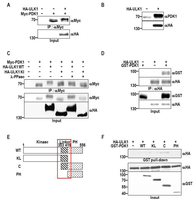
Fig. 1 ULK1 interacts with PDK1 and induces phosphorylation. (A) HEK293T cells were transfected with HA-ULK1 and Myc-PDK1 constructs. After 48 h of transfection, the cells were lysed, and the lysates were subjected to immunoprecipitation using anti-Myc antibody. The immune complexes were washed and the immunoblots for PDK1 were performed as described in Materials and Methods (upper panel). The same cell lysates were used for anti-HA immunoblot (lower panel). (B) HA-ULK1 was expressed in HEK293T cells. Whole cell lysate was analyzed by immunoblotting for the indicated proteins. (C) HEK293T cells transfected with Myc-PDK1 and HA-ULK1 wild-type (WT) or kinase-inactive (KI) mutant were lysed and subject to immunoprecipitation with anti-Myc antibody. The immune complexes were incubated with λ phosphatase, washed, and subjected to anti-Myc immunoblot as described in Materials and Methods (top panel). Anti-Myc and anti-HA immunoblots were conducted using the same cell lysates (middle and bottom panels, respectively). (D) HA-ULK1 and GST-PDK1 were expressed in the HEK293T cells. The cell lysates were subjected to immunoprecipitation using anti-HA antibody. Immunoblots were obtained with the indicated antibodies. (E) The scheme of PDK1 truncation mutants. (F) HEK293T cells transfected with HA-ULK1 and GST-PDK1 WT, kinase and linker (KL), C-terminal (C) or PH domain (PH) constructs were lysed, and the lysates were subjected to co-immunoprecipitation. The results shown are representative of three independent experiments.
Fig. 1 ULK1 interacts with PDK1 and induces phosphorylation. (A) HEK293T cells were transfected with HA-ULK1 and Myc-PDK1 constructs. After 48 h of transfection, the cells were lysed, and the lysates were subjected to immunoprecipitation using anti-Myc antibody. The immune complexes were washed and the immunoblots for PDK1 were performed as described in Materials and Methods (upper panel). The same cell lysates were used for anti-HA immunoblot (lower panel). (B) HA-ULK1 was expressed in HEK293T cells. Whole cell lysate was analyzed by immunoblotting for the indicated proteins. (C) HEK293T cells transfected with Myc-PDK1 and HA-ULK1 wild-type (WT) or kinase-inactive (KI) mutant were lysed and subject to immunoprecipitation with anti-Myc antibody. The immune complexes were incubated with λ phosphatase, washed, and subjected to anti-Myc immunoblot as described in Materials and Methods (top panel). Anti-Myc and anti-HA immunoblots were conducted using the same cell lysates (middle and bottom panels, respectively). (D) HA-ULK1 and GST-PDK1 were expressed in the HEK293T cells. The cell lysates were subjected to immunoprecipitation using anti-HA antibody. Immunoblots were obtained with the indicated antibodies. (E) The scheme of PDK1 truncation mutants. (F) HEK293T cells transfected with HA-ULK1 and GST-PDK1 WT, kinase and linker (KL), C-terminal (C) or PH domain (PH) constructs were lysed, and the lysates were subjected to co-immunoprecipitation. The results shown are representative of three independent experiments.CC BY-NC · PubMed Central
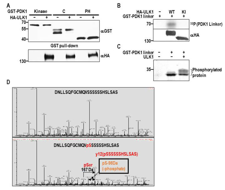
Fig. 2 ULK1 phosphorylates Ser389 of PDK1 linker region. (A) HA-ULK1 proteins were co-expressed with GST-PDK1 Kinase, C or PH proteins in HEK293T cells. The cell lysates were cleared and incubated with glutathione beads as described in Materials and Methods. Western blots for the precipitated proteins were obtained using anti-GST antibody (top panel). The anti-HA immunoblot (bottom panel) was also conducted using the same whole cell lysates. (B) HEK293T cells transfected with HA-ULK1 WT or KI were lysed and ULK1 assays were performed using GST-PDK1 linker protein as a substrate as described in Materials and Methods. The phosphorylated proteins were visualized by autoradiography (top panel). ULK1 expression was detected by the immunoblot obtained from the same whole cell lysates (bottom panel). The above results shown are representative of three independent experiments. (C) HEK293T cells were transfected with GST-PDK1 linker and HA-ULK1 WT. After 48 h of transfection, PDK1 linker proteins were pulled down and eluted as described in Materials and Methods. The fractions of eluted proteins were subjected to SDS-PAGE and the gel was stained by Coomassie R250 solution. (D) Phosphorylated proteins from (C) were analyzed by mass spectrometry as described in Materials and Methods.
Fig. 2 ULK1 phosphorylates Ser389 of PDK1 linker region. (A) HA-ULK1 proteins were co-expressed with GST-PDK1 Kinase, C or PH proteins in HEK293T cells. The cell lysates were cleared and incubated with glutathione beads as described in Materials and Methods. Western blots for the precipitated proteins were obtained using anti-GST antibody (top panel). The anti-HA immunoblot (bottom panel) was also conducted using the same whole cell lysates. (B) HEK293T cells transfected with HA-ULK1 WT or KI were lysed and ULK1 assays were performed using GST-PDK1 linker protein as a substrate as described in Materials and Methods. The phosphorylated proteins were visualized by autoradiography (top panel). ULK1 expression was detected by the immunoblot obtained from the same whole cell lysates (bottom panel). The above results shown are representative of three independent experiments. (C) HEK293T cells were transfected with GST-PDK1 linker and HA-ULK1 WT. After 48 h of transfection, PDK1 linker proteins were pulled down and eluted as described in Materials and Methods. The fractions of eluted proteins were subjected to SDS-PAGE and the gel was stained by Coomassie R250 solution. (D) Phosphorylated proteins from (C) were analyzed by mass spectrometry as described in Materials and Methods.CC BY-NC · PubMed Central
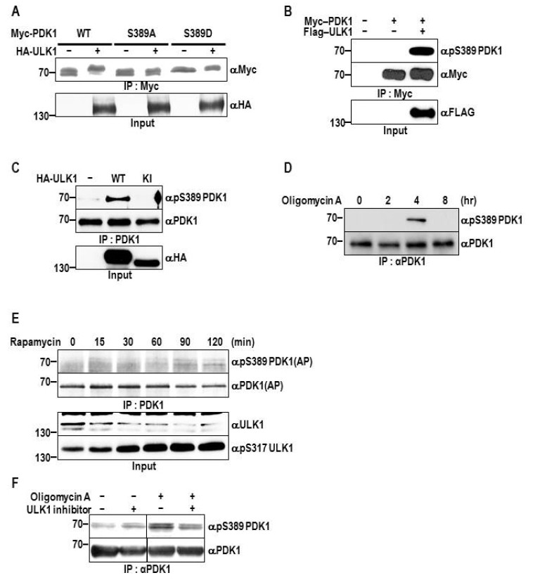
Fig. 3 ULK1 phosphorylates Ser389 of PDK1. (A) HEK293T cells were transfected with the indicated constructs. After 48 h of transfection, the cells were lysed and the lysates were subjected to immunoprecipitation using anti-Myc antibody (top panel). ULK1 immunoblot was performed using the same cell lysates (bottom panel). (B) Myc-PDK1 and Flag-ULK1 were overexpressed in the HEK293T cells. Immunoprecipitated Myc-PDK1 proteins were probed with an anti-pS389 PDK1 antibody (top panel). (C) HA-ULK1 WT and KI were overexpressed in the HEK293T cells and endogenous PDK1 proteins were immunoprecipitated using the anti-PDK1 antibody (top and middle panel). ULK1 expression was detected in the same cell lysates (bottom panel). (D) After the addition of 5 μM Oligomycin A to HEK293T cells at the indicated time points, the cells were lysed for immunoprecipitation of PDK1. The immune complexes were subjected to western blot using anti-pS389 and anti-PDK1 antibodies (top and bottom panel, respectively) (E) HEK293T cells were treated with 1 μM rapamycin at the indicated time points. Immunoprecipitated PDK1 proteins were probed with anti-pS389 and anti-PDK1 antibodies (top and upper middle panels, respectively). AP indicates alkaline phosphatase blotting. (F) HEK293T cells were treated with ULK1 inhibitor (1 μM, SBI-0206965) for 20 h, followed by addition of oligomycin A (5 μM) or DMSO for 4 h. Immunoprecipitated proteins using PDK1 antibody were analyzed via immunoblotting using the indicated antibodies. The above results shown are representative of three independent experiments.
Fig. 3 ULK1 phosphorylates Ser389 of PDK1. (A) HEK293T cells were transfected with the indicated constructs. After 48 h of transfection, the cells were lysed and the lysates were subjected to immunoprecipitation using anti-Myc antibody (top panel). ULK1 immunoblot was performed using the same cell lysates (bottom panel). (B) Myc-PDK1 and Flag-ULK1 were overexpressed in the HEK293T cells. Immunoprecipitated Myc-PDK1 proteins were probed with an anti-pS389 PDK1 antibody (top panel). (C) HA-ULK1 WT and KI were overexpressed in the HEK293T cells and endogenous PDK1 proteins were immunoprecipitated using the anti-PDK1 antibody (top and middle panel). ULK1 expression was detected in the same cell lysates (bottom panel). (D) After the addition of 5 μM Oligomycin A to HEK293T cells at the indicated time points, the cells were lysed for immunoprecipitation of PDK1. The immune complexes were subjected to western blot using anti-pS389 and anti-PDK1 antibodies (top and bottom panel, respectively) (E) HEK293T cells were treated with 1 μM rapamycin at the indicated time points. Immunoprecipitated PDK1 proteins were probed with anti-pS389 and anti-PDK1 antibodies (top and upper middle panels, respectively). AP indicates alkaline phosphatase blotting. (F) HEK293T cells were treated with ULK1 inhibitor (1 μM, SBI-0206965) for 20 h, followed by addition of oligomycin A (5 μM) or DMSO for 4 h. Immunoprecipitated proteins using PDK1 antibody were analyzed via immunoblotting using the indicated antibodies. The above results shown are representative of three independent experiments.CC BY-NC · PubMed Central
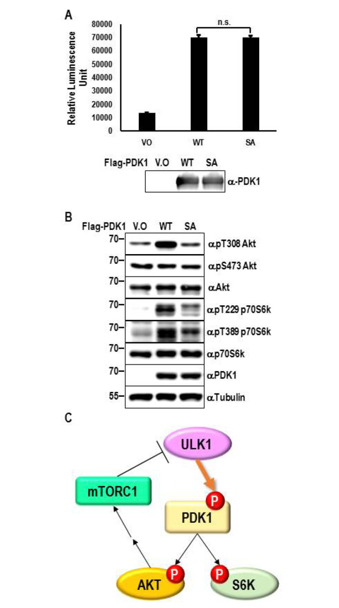
Fig. 4 Phosphorylation of PDK1 Ser389 regulates substrate phosphorylation. (A) Flag-PDK1 WT and S389A proteins were purified from HAP1 stable cells as described in Materials and Methods. PDK1 kinase activity was measured using ADP-GloTM PDK1 kinase assay kit according to the manufacturer’s instructions. The data represent means ± SD of three experiments. Statistical analysis was performed using Student’s t-test, and P-value < 0.05 was considered significant; however, the calculated P value was 0.69. n.s.: not significant (B) HAP1 PDK1 knockout cells stably expressing PDK1 WT and S389A mutant were lysed and the whole cell lysates were analyzed by immunoblotting using the indicated antibodies. (C) Negative feedback loop model between PDK1 and ULK1.
Fig. 4 Phosphorylation of PDK1 Ser389 regulates substrate phosphorylation. (A) Flag-PDK1 WT and S389A proteins were purified from HAP1 stable cells as described in Materials and Methods. PDK1 kinase activity was measured using ADP-GloTM PDK1 kinase assay kit according to the manufacturer’s instructions. The data represent means ± SD of three experiments. Statistical analysis was performed using Student’s t-test, and P-value < 0.05 was considered significant; however, the calculated P value was 0.69. n.s.: not significant (B) HAP1 PDK1 knockout cells stably expressing PDK1 WT and S389A mutant were lysed and the whole cell lysates were analyzed by immunoblotting using the indicated antibodies. (C) Negative feedback loop model between PDK1 and ULK1.CC BY-NC · PubMed Central

Verification

Claims the paper makes

The claims and statistics of this paper haven’t been checked yet.

Run the check — free, no account needed. It reads the open-access text and checks the sources it cites.

A few free checks a day · papers already verified open instantly · results are public

Claims other papers make about this one

Each is checked against this paper’s own text: does it say what the citing paper says it does?

Self-check

Percentages, sums and reported differences re-computed from the paper’s own numbers.

The self-check needs the paper’s full text.

Citations

Cites

Research works printed but not linked: 30

  1. 10.1074/jbc.274.13.8347 · DOI
  2. 10.1016/S0960-9822(98)70274-X · DOI
  3. 10.1016/S0960-9822(06)00122-9 · DOI
  4. 10.1126/science.279.5351.710 · DOI
  5. 10.1126/stke.2001.108.re17 · DOI
  6. 10.1074/jbc.274.38.27168 · DOI
  7. 10.1016/S0960-9822(98)00017-7 · DOI
  8. 10.1126/science.281.5385.2042 · DOI
  9. 10.1016/S0960-9822(06)00336-8 · DOI
  10. 10.1042/bj3420287 · DOI
  11. 10.1073/pnas.1019386108 · DOI
  12. 10.1073/pnas.2335486100 · DOI
  13. 10.1074/jbc.M001492200 · DOI
  14. 10.1126/scisignal.2000738 · DOI
  15. 10.1074/jbc.M304172200 · DOI
  16. 10.1083/jcb.150.6.1507 · DOI · Paper page
  17. 10.1006/geno.1998.5340 · DOI
  18. 10.1006/bbrc.1998.8546 · DOI
  19. 10.1016/S0378-1119(97)00084-X · DOI
  20. 10.1128/MCB.01344-09 · DOI
  21. 10.1038/sj.embor.7400917 · DOI
  22. 10.4161/auto.7.7.15491 · DOI · Paper page
  23. 10.4161/auto.7.10.16660 · DOI
  24. 10.1074/jbc.M602448200 · DOI
  25. 10.1093/emboj/cdg407 · DOI
  26. 10.1093/emboj/20.16.4380 · DOI · Paper page
  27. 10.1074/jbc.M909663199 · DOI
  28. 10.1074/jbc.M105916200 · DOI
  29. 10.1021/bi400488f · DOI
  30. 10.1016/j.cell.2017.02.004 · DOI · Paper page

Cited by

Sorted by citations

See all 2

Metadata

Authors
Kim, K., Park, S. G., Park, B. C., Kim, J.-H., & Kim, S.
Venue
BMB Reports
Published
2020-07-31
Volume
53
Issue
7
Pages
373-378
ISSN
1976-670X
Cited by
2
References
2
Language
en
Funders
Ministry of Science and ICT, South Korea (NRF-2019M3E5D4069882)

About these checks

Registry, retraction, Cites and statistics results are computed, not judged, with one exception: a printed reference that no index lists is read once by a model before it counts as a citation error. Claims are extracted by a model and kept only when their quote appears verbatim in the text; each cited source is then checked, with a model’s help, for whether it says what the claim relies on. Those verdicts can be wrong — read the claim’s quote and the cited source before relying on one.

Statistics and arithmetic are recomputed, not judged.

Confidence bands are shown only for fields calibrated on our annotated test set. None is calibrated yet, so no band is shown.

What counts as a claim ›How we measure confidence ›How we check this ›

Are you an author, or do you see a problem with this page? Request a review