

The DOI matches its Crossref record; 15 of the 15 references it cites were found in a registry or an index.
Predictive Value of Localized Stenosis of the Main Pancreatic Duct for Early Detection of Pancreatic Cancer
Open accessAbstract
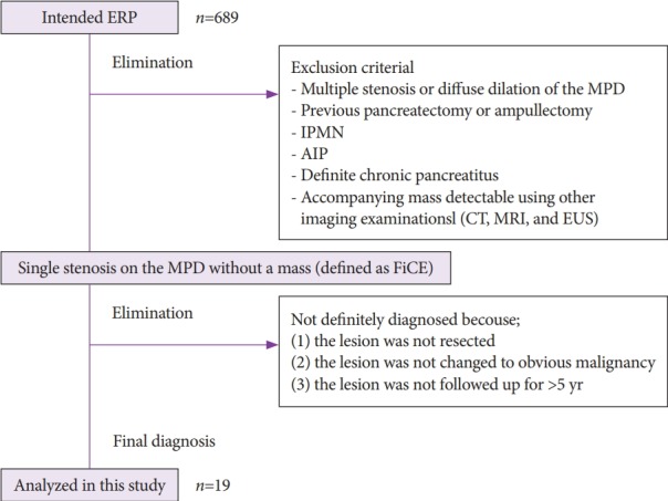
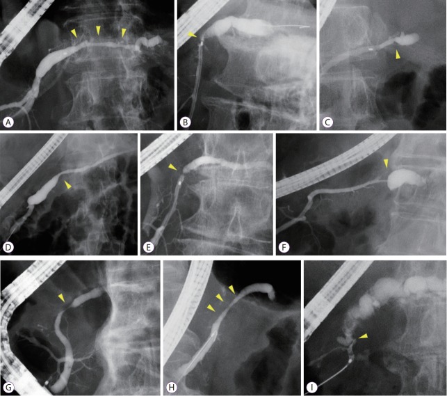
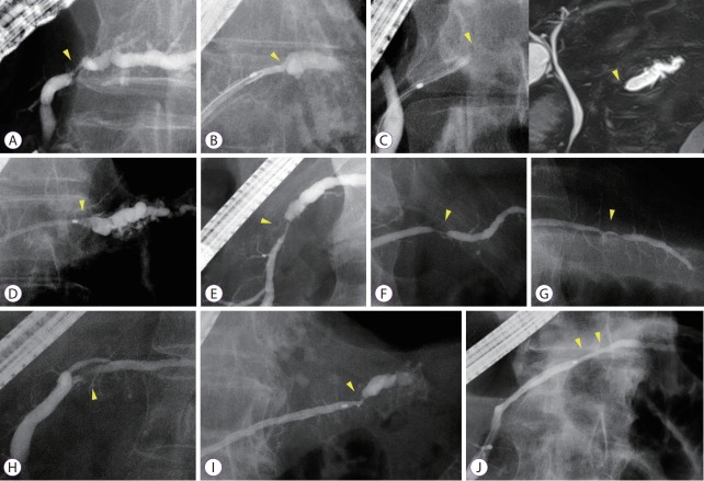
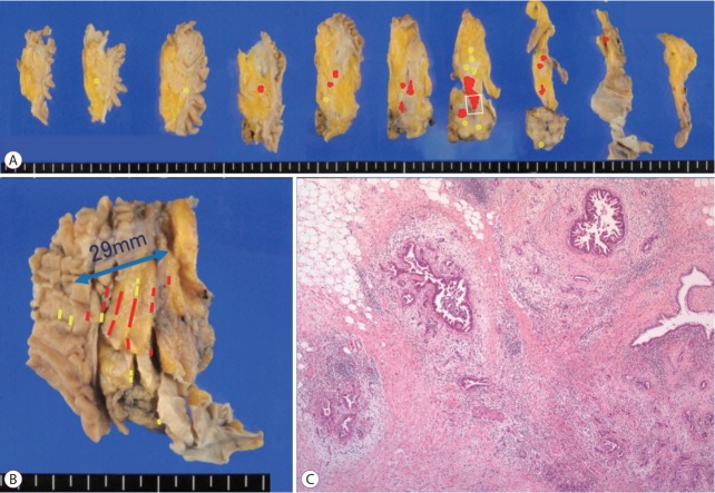
BACKGROUND/AIMS: In this study, we aimed to evaluate the predictive value of localized stenosis of the main pancreatic duct (MPD) for early detection of pancreatic cancer. METHODS: Among 689 patients who underwent endoscopic retrograde pancreatography from January 2008 to September 2018, 19 patients with MPD findings were enrolled. These patients showed findings for indicating suspicious pancreatic cancer at an early stage (FiCE); FiCE was defined as a single, localized stenosis in the MPD without a detectable mass (using any other imaging methods) and without other pancreatic diseases, such as definite chronic pancreatitis, intraductal papillary mucinous neoplasm, and autoimmune pancreatitis. Final diagnoses were established by examining resected specimens or through follow-up examinations after an interval of >5 years. RESULTS: Among 19 patients with FiCE, 11 underwent surgical resection and 8 were evaluated after a >5-year observation period. The final diagnosis of the MPD stenosis was judged to be pancreatic cancer in 9 patients (47%), including 3 with intraepithelial cancer, and to be a non-neoplastic change in 10. The sensitivity, specificity, and accuracy of preoperative pancreatic juice cytology were 75%, 100%, and 88%, respectively. CONCLUSION: The predictive value of FiCE for pancreatic cancer prevalence was 47%. Histological confirmation with pancreatic juice cytology is necessary before surgical resection.
Overview
The claims and statistics of this paper haven’t been checked yet.
Run the check — free, no account needed. It reads the open-access text and checks the sources it cites.
Integrity signals
Registry
Cites
Statistics
Figures
Verification
Claims the paper makes
The claims and statistics of this paper haven’t been checked yet.
Run the check — free, no account needed. It reads the open-access text and checks the sources it cites.
Claims other papers make about this one
Each is checked against this paper’s own text: does it say what the citing paper says it does?
The cited source says what the claim attributes to it.
In the citing paper
“In three case series studies of localized main pancreatic duct structure without a detectable mass, 47–54% of cases were actually diagnosed with pancreatic cancer (31-33).”
From Suspicious findings observed retrospectively on CT imaging performed before the diagnosis of pancreatic cancer (2023) · cites this paper as [31]
Self-check
Percentages, sums and reported differences re-computed from the paper’s own numbers.
The self-check needs the paper’s full text.
Citations
Cites
Siegel, R. L., Miller, K. D., & Jemal, A. (2018) · CA A Cancer Journal for Clinicians · doi:10.3322/caac.21442 · Cited by 16,632
Spencer, P. M., Cameron, J. L., & Lin, L. (2000) · Cited by 7,202
Endoscopic sphincterotomy complications and their management: an attempt at consensus
Cotton, P. B., Lehman, G. A., Vennes, J. A., Geenen, J. E., Russell, R. C. G., Meyers, W. C., Liguory, C., & Nickl, N. J. (1991) · Gastrointestinal Endoscopy · doi:10.1016/s0016-5107(91)70740-2 · Cited by 2,847
A lexicon for endoscopic adverse events: report of an ASGE workshop
Cotton, P. B., Eisen, G. M., Aabakken, L., Baron, T. H., Hutter, M. M., Jacobson, B. C., Mergener, K., Nemcek, A. A., Petersen, B. T., Petrini, J. L., et al. (2010) · Gastrointestinal Endoscopy · doi:10.1016/j.gie.2009.10.027 · Cited by 2,531
Kamisawa, T., Wood, L. D., Itoi, T., & Takaori, K. (2016) · The Lancet · doi:10.1016/s0140-6736(16)00141-0 · Cited by 2,212
Japan Pancreatic Cancer Registry; 30th Year Anniversary: Japan Pancreas Society
Egawa, S., Toma, H., Ohigashi, H., Okusaka, T., Nakao, A., Hatori, T., Maguchi, H., Yanagisawa, A., & Tanaka, M. (2012) · Pancreas · doi:10.1097/mpa.0b013e318258055c · Cited by 445
Multicenter study of early pancreatic cancer in Japan
Kanno, A., Masamune, A., Hanada, K., Maguchi, H., Shimizu, Y., Ueki, T., Hasebe, O., Ohtsuka, T., Nakamura, M., Takenaka, M., et al. (2018) · Pancreatology · doi:10.1016/j.pan.2017.11.007 · Cited by 291
Iiboshi, T., Hanada, K., Fukuda, T., Yonehara, S., Sasaki, T., & Chayama, K. (2012) · Pancreas · doi:10.1097/mpa.0b013e31823c0b05 · Cited by 145
Diagnostic strategies for early pancreatic cancer
Hanada, K., Okazaki, A., Hirano, N., Izumi, Y., Teraoka, Y., Ikemoto, J., Kanemitsu, K., Hino, F., Fukuda, T., & Yonehara, S. (2015) · Journal of Gastroenterology · doi:10.1007/s00535-014-1026-z · Cited by 121
Satoh, T., Kikuyama, M., Kawaguchi, S., Kanemoto, H., Muro, H., & Hanada, K. (2017) · Clinical Journal of Gastroenterology · doi:10.1007/s12328-017-0776-6 · Cited by 56
Research works printed but not linked: 1
- c2018, Cancer statistics in Japan; Table download [Internet]
Cited by
Endoscopic approach in the diagnosis of high‐grade pancreatic intraepithelial neoplasia
Hanada, K., Shimizu, A., Kurihara, K., Ikeda, M., Yamamoto, T., Okuda, Y., & Tazuma, S. (2022) · Digestive Endoscopy · doi:10.1111/den.14240 · Cited by 49
Sagami, R., Yamao, K., Nakahodo, J., Minami, R., Tsurusaki, M., Murakami, K., & Amano, Y. (2021) · Cancers · doi:10.3390/cancers13050945 · Cited by 45
Lee, H. S., Chae, W., Sung, M. J., Keum, J., Jo, J. H., Chung, M. J., Park, J. Y., Park, S. W., Song, S. Y., Park, E., et al. (2022) · The Journal of Clinical Endocrinology & Metabolism · doi:10.1210/clinem/dgac728 · Cited by 30
Miura, S., Kume, K., Kikuta, K., Hamada, S., Takikawa, T., Yoshida, N., Hongo, S., Tanaka, Y., Matsumoto, R., Sano, T., et al. (2020) · The Tohoku Journal of Experimental Medicine · doi:10.1620/tjem.252.63 · Cited by 22
Current Screening Strategies for Pancreatic Cancer
Vaněk, P., Urban, O., Zoundjiekpon, V., & Falt, P. (2022) · Biomedicines · doi:10.3390/biomedicines10092056 · Cited by 21
Metadata
- Authors
- Kanno, Y., Koshita, S., Ogawa, T., Kusunose, H., Masu, K., Sakai, T., Yonamine, K., Kawakami, Y., Fujii, Y., Miyamoto, K., Murabayashi, T., Kozakai, F., Horaguchi, J., Noda, Y., Oikawa, M., Okada, T., & Ito, K.
- Affiliations
- Sendai Medical Center (JP)
- Corresponding author
- Kanno, Yoshihide
- ORCID
- Venue
- Clinical Endoscopy
- Published
- 2019-11-30
- Volume
- 52
- Issue
- 6
- Pages
- 588-597
- ISSN
- 2234-2400 (ISSN-L)
- 2234-2443
- PMC
- PMC6900300
- OpenAlex
- W2991140622
- Cited by
- 23
- References
- 15
- Citations by year
- 2026: 4 · 2025: 3 · 2024: 3 · 2023: 3 · 2022: 4 · 2021: 3 · 2020: 2 · 2019: 1
- FWCI
- 1.04
- Type
- journal article
- Language
- en
- Open access
- gold
- doi.org
- Licence
- Locations
- Clinical Endoscopy (publishedVersion, cc-by-nc)
- PubMed Central (publishedVersion, cc-by-nc)
- PubMed (publishedVersion)
- DOAJ (DOAJ: Directory of Open Access Journals) (submittedVersion, cc-by-sa)
- Topics
- Health Sciences › Medicine › Oncology › Pancreatic and Hepatic Oncology Research
- Health Sciences › Medicine › Surgery › Pancreatitis Pathology and Treatment
- Health Sciences › Medicine › Gastroenterology › Gallbladder and Bile Duct Disorders
- Keywords
- pancreatic cancer, early detection, main pancreatic duct stenosis, endoscopic retrograde pancreatography, pancreatic juice cytology, diagnostic accuracy, surgical resection
About these checks
Registry, retraction, Cites and statistics results are computed, not judged, with one exception: a printed reference that no index lists is read once by a model before it counts as a citation error. Claims are extracted by a model and kept only when their quote appears verbatim in the text; each cited source is then checked, with a model’s help, for whether it says what the claim relies on. Those verdicts can be wrong — read the claim’s quote and the cited source before relying on one.
Statistics and arithmetic are recomputed, not judged.
Confidence bands are shown only for fields calibrated on our annotated test set. None is calibrated yet, so no band is shown.
What counts as a claim ›How we measure confidence ›How we check this ›
Are you an author, or do you see a problem with this page? Request a review
How to cite
Cite CoChat for the checks on this page, and the paper for its own findings.
- This page
- CoChat. (2026). Citation check: Predictive Value of Localized Stenosis of the Main Pancreatic Duct for Early Detection of Pancreatic Cancer. https://app.cochat.ai/papers/10.5946/ce.2019.018
- The paper
- Kanno, Y., Koshita, S., Ogawa, T., Kusunose, H., Masu, K., Sakai, T., Yonamine, K., Kawakami, Y., Fujii, Y., Miyamoto, K., Murabayashi, T., Kozakai, F., Horaguchi, J., Noda, Y., Oikawa, M., Okada, T., & Ito, K. (2019). Predictive Value of Localized Stenosis of the Main Pancreatic Duct for Early Detection of Pancreatic Cancer. Clinical Endoscopy. https://doi.org/10.5946/ce.2019.018
Data sources
Bibliographic data: Semantic Scholar, OpenAlex, Crossref.
Contains information from the Semantic Scholar Academic Graph Datasets, made available under the ODC Attribution License (ODC-BY 1.0).
Semantic Scholar’s data is described in: Kinney, R., et al. (2023). The Semantic Scholar Open Data Platform. arXiv:2301.10140.
