

The DOI matches its Crossref record; 83 of the 83 references it cites were found in a registry or an index.
Coral anthozoan-specific opsins employ a novel chloride counterion for spectral tuning
Open accessPublished version of a preprint
Abstract
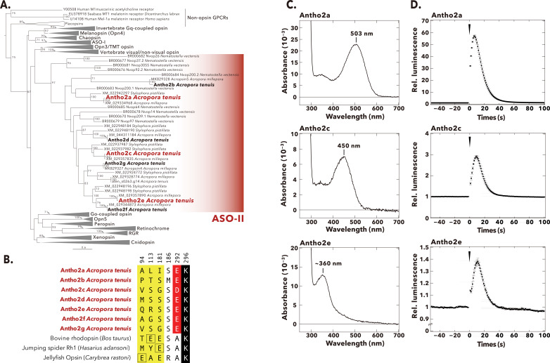
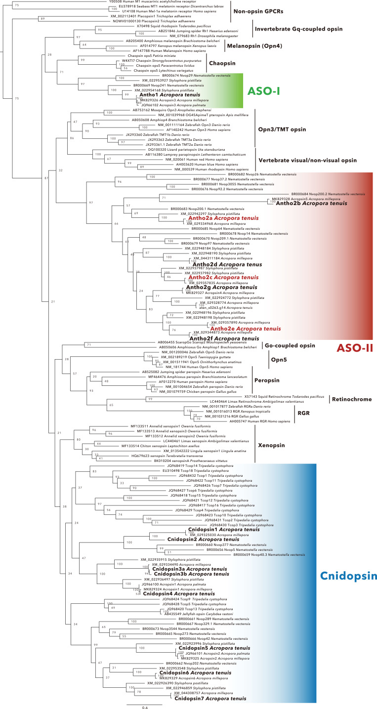
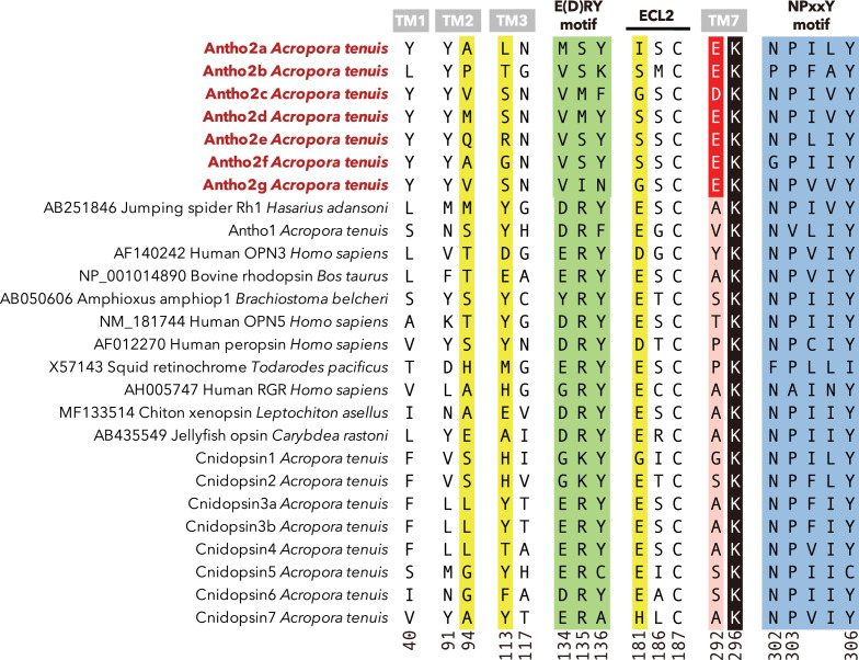
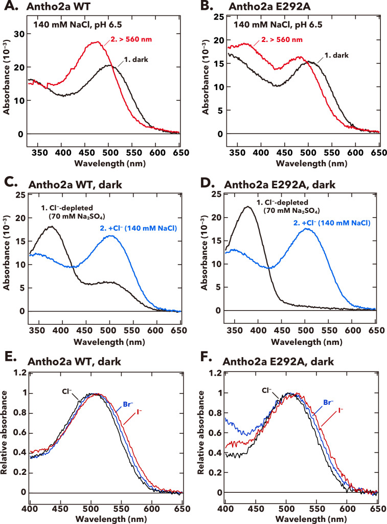
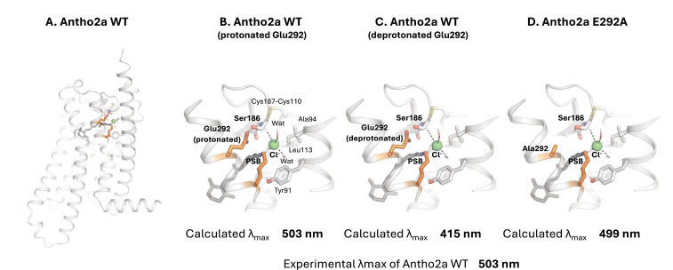
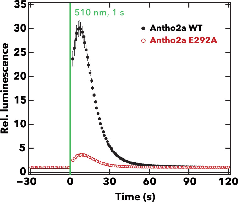
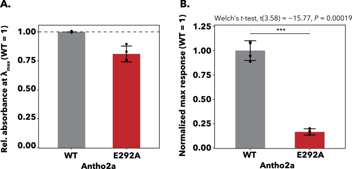
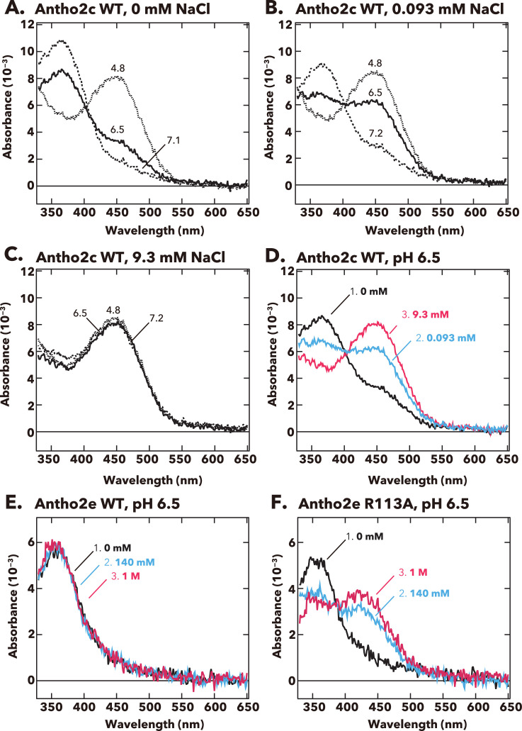
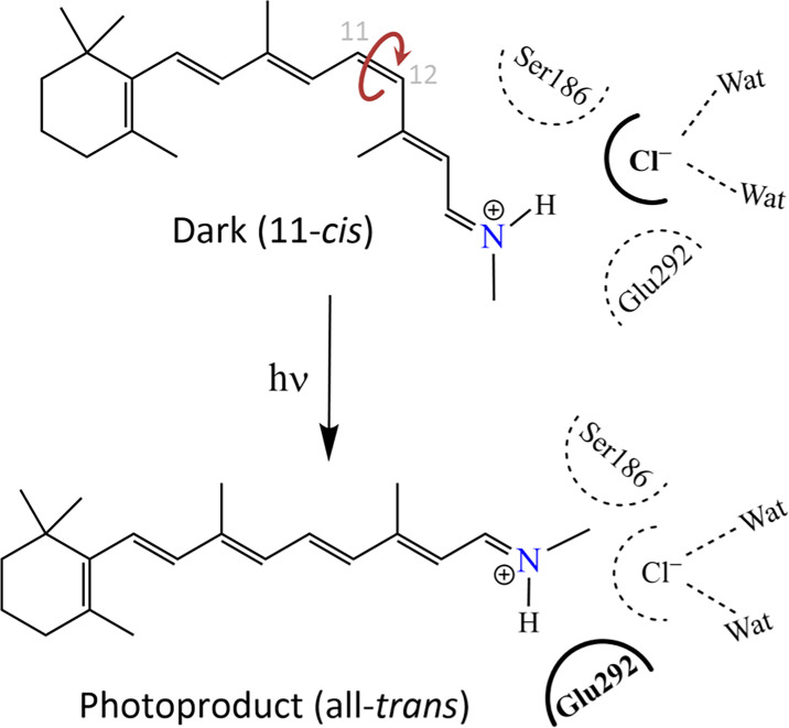
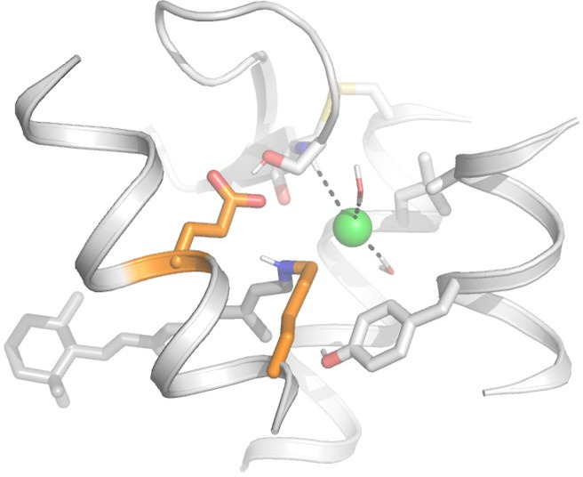
Animal opsins are G protein-coupled receptors that have evolved to sense light by covalently binding a retinal chromophore via a protonated (positively charged) Schiff base. A negatively charged amino acid in the opsin, acting as a counterion, stabilizes the proton on the Schiff base, which is essential for sensitivity to visible light. In this study, we investigate the spectroscopic properties of a unique class of opsins from a reef-building coral belonging to the anthozoan-specific opsin II group (ASO-II opsins), which intriguingly lack a counterion residue at any of established sites. Our findings reveal that, unlike other known animal opsins, the protonated state of the Schiff base in visible light-sensitive ASO-II opsins is highly dependent on exogenously supplied chloride ions (Cl − ). By using structural modeling and quantum mechanics/molecular mechanics (QM/MM) calculations to interpret spectroscopy data, we conclude that, in the dark state, ASO-II opsins employ environmental Cl − as their native counterion, while a nearby polar residue, Glu292 in its protonated neutral form, facilitates Cl − binding. In contrast, Glu292 plays a crucial role in maintaining the protonation state of the Schiff base in the light-activated protein, serving as the counterion in the photoproduct. Furthermore, Glu292 is involved in G protein activation of the ASO-II opsin, suggesting that this novel counterion system coordinates multiple functional properties.
Overview
The claims and statistics of this paper haven’t been checked yet.
Run the check — free, no account needed. It reads the open-access text and checks the sources it cites.
Integrity signals
Registry
Cites
Statistics
Figures
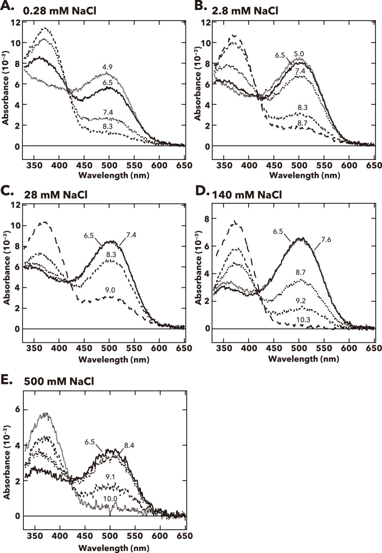
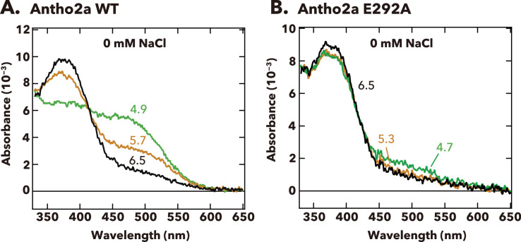
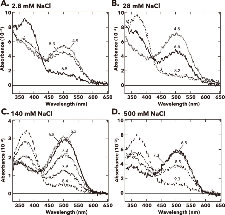
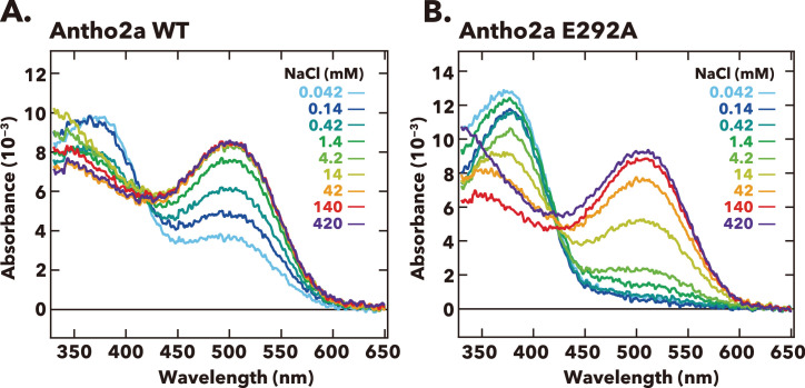
Verification
Claims the paper makes
The claims and statistics of this paper haven’t been checked yet.
Run the check — free, no account needed. It reads the open-access text and checks the sources it cites.
Self-check
Percentages, sums and reported differences re-computed from the paper’s own numbers.
The self-check needs the paper’s full text.
Citations
Cites
Density-functional exchange-energy approximation with correct asymptotic behavior
Becke, A. D. (1988) · Physical Review A · doi:10.1103/physreva.38.3098 · Cited by 53,711
MAFFT Multiple Sequence Alignment Software Version 7: Improvements in Performance and Usability
Katoh, K., & Standley, D. M. (2013) · Molecular Biology and Evolution · doi:10.1093/molbev/mst010 · Cited by 46,209
Highly accurate protein structure prediction with AlphaFold
Jumper, J., Evans, R., Pritzel, A., Green, T., Figurnov, M., Ronneberger, O., Tunyasuvunakool, K., Bates, R., Žídek, A., Potapenko, A., et al. (2021) · Nature · doi:10.1038/s41586-021-03819-2 · Cited by 43,764
Comparison of simple potential functions for simulating liquid water
Jorgensen, W. L., Chandrasekhar, J., Madura, J. D., Impey, R. W., & Klein, M. L. (1983) · The Journal of Chemical Physics · doi:10.1063/1.445869 · Cited by 41,298
Dunning, T. H. (1989) · The Journal of Chemical Physics · doi:10.1063/1.456153 · Cited by 31,648
Effect of the damping function in dispersion corrected density functional theory
Grimme, S., Ehrlich, S., & Goerigk, L. (2011) · Journal of Computational Chemistry · doi:10.1002/jcc.21759 · Cited by 28,221
Ryckaert, J.-P., Ciccotti, G., & Berendsen, H. J. C. (1977) · Journal of Computational Physics · doi:10.1016/0021-9991(77)90098-5 · Cited by 21,239
A new hybrid exchange–correlation functional using the Coulomb-attenuating method (CAM-B3LYP)
Yanai, T., Tew, D. P., & Handy, N. C. (2004) · Chemical Physics Letters · doi:10.1016/j.cplett.2004.06.011 · Cited by 14,976
trimAl: a tool for automated alignment trimming in large-scale phylogenetic analyses
Capella-Gutiérrez, S., Silla-Martínez, J. M., & Gabaldón, T. (2009) · Bioinformatics · doi:10.1093/bioinformatics/btp348 · Cited by 12,751
ff14SB: Improving the Accuracy of Protein Side Chain and Backbone Parameters from ff99SB
Maier, J. A., Martinez, C., Kasavajhala, K., Wickstrom, L., Hauser, K. E., & Simmerling, C. (2015) · Journal of Chemical Theory and Computation · doi:10.1021/acs.jctc.5b00255 · Cited by 10,830
Density-Functional Theory for Time-Dependent Systems
Runge, E., & Gross, E. K. U. (1984) · Physical Review Letters · doi:10.1103/physrevlett.52.997 · Cited by 8,914
Software update: The ORCA program system—Version 5.0
Neese, F. (2022) · WIREs Computational Molecular Science · doi:10.1002/wcms.1606 · Cited by 4,963
RAxML-NG: a fast, scalable and user-friendly tool for maximum likelihood phylogenetic inference
Kozlov, A. M., Darriba, D., Flouri, T., Morel, B., & Stamatakis, A. (2019) · Bioinformatics · doi:10.1093/bioinformatics/btz305 · Cited by 4,454
PROPKA3: Consistent Treatment of Internal and Surface Residues in Empirical p KaPredictions
Olsson, M. H. M., Søndergaard, C. R., Rostkowski, M., & Jensen, J. H. (2011) · Journal of Chemical Theory and Computation · doi:10.1021/ct100578z · Cited by 4,435
QM/MM Methods for Biomolecular Systems
Senn, H. M., & Thiel, W. (2009) · Angewandte Chemie International Edition · doi:10.1002/anie.200802019 · Cited by 2,684
ModelTest-NG: A New and Scalable Tool for the Selection of DNA and Protein Evolutionary Models
Darriba, D., Posada, D., Kozlov, A. M., Stamatakis, A., Morel, B., & Flouri, T. (2020) · Molecular Biology and Evolution · doi:10.1093/molbev/msz189 · Cited by 2,105
Søndergaard, C. R., Olsson, M. H. M., Rostkowski, M., & Jensen, J. H. (2011) · Journal of Chemical Theory and Computation · doi:10.1021/ct200133y · Cited by 1,966
Sherwood, P., de Vries, A. H., Guest, M. F., Schreckenbach, G., Catlow, C. R. A., French, S., Sokol, A. A., Bromley, S. T., Thiel, W., Turner, A. J., et al. (2003) · Journal of Molecular Structure THEOCHEM · doi:10.1016/s0166-1280(03)00285-9 · Cited by 1,058
Furche, F., Ahlrichs, R., Hättig, C., Klopper, W., Sierka, M., & Weigend, F. (2014) · WIREs Computational Molecular Science · doi:10.1002/wcms.1162 · Cited by 929
Hartree–Fock exchange fitting basis sets for H to Rn †
Weigend, F. (2007) · Journal of Computational Chemistry · doi:10.1002/jcc.20702 · Cited by 769
Glutamic acid-113 serves as the retinylidene Schiff base counterion in bovine rhodopsin.
Sakmar, T. P., Franke, R. R., & Khorana, H. G. (1989) · Proceedings of the National Academy of Sciences · doi:10.1073/pnas.86.21.8309 · Cited by 680
Terakita, A. (2005) · Genome biology · doi:10.1186/gb-2005-6-3-213 · Cited by 656
Self-citationStructure of the Light-Driven Chloride Pump Halorhodopsin at 1.8 Å Resolution
Kolbe, M., Besir, H., Essen, L., & Oesterhelt, D. (2000) · Science · doi:10.1126/science.288.5470.1390 · Cited by 543
Al‐Horani, F. A., Al-Moghrabi, S. M., & de Beer, D. (2002) · Marine Biology · doi:10.1007/s00227-002-0981-8 · Cited by 540
C hem S hell—a modular software package for QM / MM simulations
Metz, S., Kästner, J., Sokol, A. A., Keal, T. W., & Sherwood, P. (2013) · Wiley Interdisciplinary Reviews Computational Molecular Science · doi:10.1002/wcms.1163 · Cited by 531
Structure Optimizations for Excited States with Correlated Second-Order Methods: CC2 and ADC(2)
Hättig, C. (2005) · Advances in quantum chemistry · doi:10.1016/s0065-3276(05)50003-0 · Cited by 491
Effect of Carboxylic Acid Side Chains on the Absorption Maximum of Visual Pigments
Zhukovsky, E. A., & Oprian, D. D. (1989) · Science · doi:10.1126/science.2573154 · Cited by 477
A G protein-coupled receptor at work: the rhodopsin model
Hofmann, K. P., Scheerer, P., Hildebrand, P. W., Choe, H.-W., Park, J. H., Heck, M., & Ernst, O. P. (2009) · Trends in Biochemical Sciences · doi:10.1016/j.tibs.2009.07.005 · Cited by 376
Dryja, T. P., Berson, E. L., Rao, V. R., & Oprian, D. D. (1993) · Nature Genetics · doi:10.1038/ng0793-280 · Cited by 340
Bannwarth, C., & Grimme, S. (2014) · Computational and Theoretical Chemistry · doi:10.1016/j.comptc.2014.02.023 · Cited by 329
Nathans, J. (1990) · Biochemistry · doi:10.1021/bi00493a034 · Cited by 323
Levy, S., Elek, A., Grau‐Bové, X., Menéndez-Bravo, S., Iglesias, M., Tanay, A., Mass, T., & Sebé-Pedrós, A. (2021) · Cell · doi:10.1016/j.cell.2021.04.005 · Cited by 240
Metazoan opsin evolution reveals a simple route to animal vision
Feuda, R., Hamilton, S. C., McInerney, J. O., & Pisani, D. (2012) · Proceedings of the National Academy of Sciences · doi:10.1073/pnas.1204609109 · Cited by 193
Anion-induced wavelength regulation of absorption maxima of Schiff bases of retinal
Blatz, P. E., Mohler, J. H., & Navangul, H. V. (1972) · Biochemistry · doi:10.1021/bi00755a026 · Cited by 191
Jellyfish vision starts with cAMP signaling mediated by opsin-G s cascade
Koyanagi, M., Takano, K., Tsukamoto, H., Ohtsu, K., Tokunaga, F., & Terakita, A. (2008) · Proceedings of the National Academy of Sciences · doi:10.1073/pnas.0806215105 · Cited by 181
Self-citationImaging intracellular pH in a reef coral and symbiotic anemone
Venn, A. A., Tambutté, É., Lotto, S., Zoccola, D., Allemand, D., & Tambutté, S. (2009) · Proceedings of the National Academy of Sciences · doi:10.1073/pnas.0902894106 · Cited by 176
Diversity of animal opsin-based pigments and their optogenetic potential
Koyanagi, M., & Terakita, A. (2013) · Biochimica et Biophysica Acta (BBA) - Bioenergetics · doi:10.1016/j.bbabio.2013.09.003 · Cited by 170
Self-citationBiological and chemical characteristics of the coral gastric cavity
Agostini, S., Suzuki, Y., Higuchi, T., Casareto, B. E., Yoshinaga, K., Nakano, Y., & Fujimura, H. (2011) · Coral Reefs · doi:10.1007/s00338-011-0831-6 · Cited by 166
The last common ancestor of most bilaterian animals possessed at least 9 opsins
Ramirez, M. D., Pairett, A., Pankey, M. S., Serb, J., Speiser, D. I., Swafford, A., & Oakley, T. (2016) · Genome Biology and Evolution · doi:10.1093/gbe/evw248 · Cited by 164
Counterion displacement in the molecular evolution of the rhodopsin family
Terakita, A., Koyanagi, M., Tsukamoto, H., Yamashita, T., Miyata, T., & Shichida, Y. (2004) · Nature Structural & Molecular Biology · doi:10.1038/nsmb731 · Cited by 161
Self-citationEvolution and Functional Diversity of Jellyfish Opsins
Suga, H., Schmid, V. J., & Gehring, W. J. (2007) · Current Biology · doi:10.1016/j.cub.2007.11.059 · Cited by 160
Microelectrode characterization of coral daytime interior pH and carbonate chemistry
Cai, W., Ma, Y., Hopkinson, B. M., Grottoli, A. G., Warner, M. E., Ding, Q., Hu, X., Yuan, X., Schoepf, V., Xu, H., et al. (2016) · Nature Communications · doi:10.1038/ncomms11144 · Cited by 160
Crystal Structure of the Light-Driven Chloride Pump Halorhodopsin from Natronomonas pharaonis
Kouyama, T., Kanada, S., Takeguchi, Y., Narusawa, A., Murakami, M., & Ihara, K. (2009) · Journal of Molecular Biology · doi:10.1016/j.jmb.2009.11.061 · Cited by 151
Shinzato, C., Khalturin, K., Inoue, J. G., Zayasu, Y., Kanda, M., Kawamitsu, M., Yoshioka, Y., Yamashita, H., Suzuki, G., & Satoh, N. (2020) · Molecular Biology and Evolution · doi:10.1093/molbev/msaa216 · Cited by 150
Assembly of the cnidarian camera-type eye from vertebrate-like components
Kozmik, Z., Ruzickova, J., Jonasova, K., Matsumoto, Y., Vopalensky, P., Kozmikova, I., Strnad, H., Kawamura, S., Piatigorsky, J., Paces, V., et al. (2008) · Proceedings of the National Academy of Sciences · doi:10.1073/pnas.0800388105 · Cited by 147
Ultrafast structural changes direct the first molecular events of vision
Gruhl, T., Weinert, T., Rodrigues, M. J., Milne, C. J., Ortolani, G., Nass, K., Nango, E., Sen, S., Johnson, P. J. M., Cirelli, C., et al. (2023) · Nature · doi:10.1038/s41586-023-05863-6 · Cited by 145
Self-citationSakmar, T. P., Franke, R. R., & Khorana, H. G. (1991) · Proceedings of the National Academy of Sciences · doi:10.1073/pnas.88.8.3079 · Cited by 143
Terakita, A., Hara, R., & Hara, T. (1989) · Vision Research · doi:10.1016/0042-6989(89)90026-6 · Cited by 118
Self-citationTerakita, A., Yamashita, T., & Shichida, Y. (2000) · Proceedings of the National Academy of Sciences · doi:10.1073/pnas.260349597 · Cited by 115
Self-citationStudies on rhodopsin. 8. Retinylidenemethylamine, an indicator yellow analogue
Pitt, G. A. J., Collins, F. D., Morton, R. A., & Stok, P. (1955) · Biochemical Journal · doi:10.1042/bj0590122 · Cited by 112
Blue Halorhodopsin from Natronobacterium pharaonis: Wavelength Regulation by Anions
Scharf, B. E., & Engelhard, M. (1994) · Biochemistry · doi:10.1021/bi00187a002 · Cited by 103
Crystal structure of heliorhodopsin
Shihoya, W., Inoue, K., Singh, M. P., Konno, M., Hososhima, S., Yamashita, K., Ikeda, K., Higuchi, A., Izume, T., Okazaki, S., et al. (2019) · Nature · doi:10.1038/s41586-019-1604-6 · Cited by 102
Voolstra, C. R., Miller, D. J., Ragan, M. A., Hoffmann, A. A., Hoegh‐Guldberg, O., Bourne, D. G., Ball, E. E., Ying, H., Forêt, S., Takahashi, S., et al. (2015) · Frontiers in Marine Science · doi:10.3389/fmars.2015.00068 · Cited by 96
Dynamics and mechanism of a light-driven chloride pump
Mous, S. T., Gotthard, G., Ehrenberg, D., Sen, S., Weinert, T., Johnson, P. J. M., James, D., Nass, K., Furrer, A., Kekilli, D., et al. (2022) · Science · doi:10.1126/science.abj6663 · Cited by 95
Self-citationpKa of the protonated Schiff base of bovine rhodopsin. A study with artificial pigments
Steinberg, G., Ottolenghi, M., & Sheves, M. (1993) · Biophysical Journal · doi:10.1016/s0006-3495(93)81518-2 · Cited by 89
Prolific Origination of Eyes in Cnidaria with Co-option of Non-visual Opsins
Picciani, N., Kerlin, J. R., Sierra, N. C., Swafford, A. J. M., Ramirez, M. D., Roberts, N. G., Cannon, J. T., Daly, M., & Oakley, T. H. (2018) · Current Biology · doi:10.1016/j.cub.2018.05.055 · Cited by 81
Opsin activation as a cause of congenital night blindness
Jin, S., Cornwall, M. C., & Oprian, D. D. (2003) · Nature Neuroscience · doi:10.1038/nn1070 · Cited by 71
Hering, L., & Mayer, G. (2014) · Genome Biology and Evolution · doi:10.1093/gbe/evu193 · Cited by 69
Evolution and diversity of opsins
Terakita, A., Kawano‐Yamashita, E., & Koyanagi, M. (2011) · Wiley Interdisciplinary Reviews Membrane Transport and Signalling · doi:10.1002/wmts.6 · Cited by 66
Self-citationNagata, T., Koyanagi, M., Tsukamoto, H., Mutt, E., Schertler, G. F. X., Deupí, X., & Terakita, A. (2019) · Communications Biology · doi:10.1038/s42003-019-0409-3 · Cited by 63
Self-citationMitochondrial free [Ca2+] dynamics measured with a novel low-Ca2+ affinity aequorin probe
de la Fuente, S., Fonteríz, R. I., de la Cruz, P. J., Montero, M., & Álvarez, J. (2012) · Biochemical Journal · doi:10.1042/bj20120423 · Cited by 58
Cloning and expression of frog rhodopsin cDNA
Kayada, S., Hisatomi, O., & Tokunaga, F. (1995) · Comparative Biochemistry and Physiology Part B Biochemistry and Molecular Biology · doi:10.1016/0305-0491(94)00179-x · Cited by 52
Hosaka, T., Yoshizawa, S., Nakajima, Y., Ohsawa, N., Hato, M., DeLong, E. F., Kogure, K., Yokoyama, S., Kimura‐Someya, T., Iwasaki, W., et al. (2016) · Journal of Biological Chemistry · doi:10.1074/jbc.m116.728220 · Cited by 51
Frontiers in Multiscale Modeling of Photoreceptor Proteins
Mroginski, M. A., Adam, S., Amoyal, G. S., Barnoy, A., Bondar, A., Borin, V. A., Church, J. R., Domratcheva, T. M., Ensing, B., Fanelli, F., et al. (2020) · Photochemistry and Photobiology · doi:10.1111/php.13372 · Cited by 48
Self-citationShichida, Y., Kato, T., Sasayama, S., Fukada, Y., & Yoshizawa, T. (1990) · Biochemistry · doi:10.1021/bi00476a028 · Cited by 45
Photoreceptor Diversification Accompanies the Evolution of Anthozoa
Gornik, S. G., Bergheim, B. G., Morel, B., Stamatakis, A., Foulkes, N. S., & Guse, A. (2020) · Molecular Biology and Evolution · doi:10.1093/molbev/msaa304 · Cited by 45
Gerrard, E. J., Mutt, E., Nagata, T., Koyanagi, M., Flock, T., Lesca, E., Schertler, G. F. X., Terakita, A., Deupí, X., & Lucas, R. J. (2018) · Proceedings of the National Academy of Sciences · doi:10.1073/pnas.1721333115 · Cited by 42
Self-citationIdentification of a molecular pH sensor in coral
Barott, K. L., Barron, M. E., & Tresguerres, M. (2017) · Proceedings of the Royal Society B Biological Sciences · doi:10.1098/rspb.2017.1769 · Cited by 41
Koyanagi, M., Shen, B., Nagata, T., Sun, L., Wada, S., Kamimura, S., Kage‐Nakadai, E., & Terakita, A. (2022) · Proceedings of the National Academy of Sciences · doi:10.1073/pnas.2204341119 · Cited by 40
Self-citationHomolWat: a web server tool to incorporate ‘homologous’ water molecules into GPCR structures
Mayol, E., García‐Recio, A., Tiemann, J. K. S., Hildebrand, P. W., Guixà-González, R., Olivella, M., & Cordomí, A. (2020) · Nucleic Acids Research · doi:10.1093/nar/gkaa440 · Cited by 34
Besaw, J. E., Ou, W., Morizumi, T., Eger, B. T., Vásquez, J. D. S., Chu, J. H. Y., Harris, A., Brown, L. S., Miller, R. J. D., & Ernst, O. P. (2020) · Journal of Biological Chemistry · doi:10.1074/jbc.ra120.014118 · Cited by 34
Multiple opsins in a reef-building coral, Acropora millepora
Mason, B. M., Koyanagi, M., Sugihara, T., Iwasaki, M., Slepak, V. Z., Miller, D. J., Sakai, Y., & Terakita, A. (2023) · Scientific Reports · doi:10.1038/s41598-023-28476-5 · Cited by 29
Self-citationMizuno, M., Nakajima, A., Kandori, H., & Mizutani, Y. (2018) · The Journal of Physical Chemistry A · doi:10.1021/acs.jpca.7b12332 · Cited by 28
Photo-movement in the sea anemone Aiptasia influenced by light quality and symbiotic association
Foo, S. A., Liddell, L. C., Grossman, A. R., & Caldeira, K. (2019) · Coral Reefs · doi:10.1007/s00338-019-01866-w · Cited by 26
Sinha, A., Brunette, A. M. J., Fay, J. F., Schafer, C. T., & Farrens, D. (2014) · Biochemistry · doi:10.1021/bi401534y · Cited by 25
Convergent evolutionary counterion displacement of bilaterian opsins in ciliary cells
Sakai, K., Ikeuchi, H., Fujiyabu, C., Imamoto, Y., & Yamashita, T. (2022) · Cellular and Molecular Life Sciences · doi:10.1007/s00018-022-04525-6 · Cited by 22
Besaw, J. E., Reichenwallner, J., Guzman, P. D., et al. (2022) · Scientific Reports · doi:10.1038/s41598-022-17716-9 · Cited by 21
[4] Folding and assembly of rhodopsin from expressed fragments
Ridge, K. D., & Abdulaev, N. G. (2000) · Methods in enzymology on CD-ROM/Methods in enzymology · doi:10.1016/s0076-6879(00)15834-3 · Cited by 20
Church, J. R., Olsen, J. M. H., & Schapiro, I. (2021) · Molecules · doi:10.3390/molecules27010071 · Cited by 20
Kishimoto, M., Gornik, S. G., Foulkes, N. S., & Guse, A. (2023) · Scientific Reports · doi:10.1038/s41598-023-44583-9 · Cited by 11
Photosensitivities of Rhodopsin Mutants with a Displaced Counterion
Tsutsui, K., & Shichida, Y. (2010) · Biochemistry · doi:10.1021/bi101020p · Cited by 9
Correction for Goldberg, Complex nature of malaria parasite hemoglobin degredation
(2013) · Proceedings of the National Academy of Sciences · doi:10.1073/pnas.1305990110 · Cited by 8
Molecular basis underlying the specificity of an antagonist AA92593 for mammalian melanopsins
Obayashi, K., Zou, R., Kawaguchi, T., Mori, T., & Tsukamoto, H. (2025) · Journal of Biological Chemistry · doi:10.1016/j.jbc.2025.108461 · Cited by 6
Research works printed but not linked: 17
- Case, 2016, AMBER 2016
- Sakai Y, Sen S, Sugihara T, Kakeyama Y, Iwasaki M, Schertler GFX, Deupi X, Koyanagi M, Terakita A. 2025. Acropora tenuis Cnidopsin1 CDS. NCBI Nucleotide. https://www.ncbi.nlm.nih.gov/nuccore/?term=LC844924. LC844924.
- Sakai Y, Sen S, Sugihara T, Kakeyama Y, Iwasaki M, Schertler GFX, Deupi X, Koyanagi M, Terakita A. 2025. Acropora tenuis Cnidopsin2 CDS. NCBI Nucleotide. https://www.ncbi.nlm.nih.gov/nuccore/?term=LC844925. LC844925.
- Sakai Y, Sen S, Sugihara T, Kakeyama Y, Iwasaki M, Schertler GFX, Deupi X, Koyanagi M, Terakita A. 2025. Acropora tenuis Cnidopsin3a CDS. NCBI Nucleotide. https://www.ncbi.nlm.nih.gov/nuccore/?term=LC844926. LC844926.
- Sakai Y, Sen S, Sugihara T, Kakeyama Y, Iwasaki M, Schertler GFX, Deupi X, Koyanagi M, Terakita A. 2025. Acropora tenuis Cnidopsin3b CDS. NCBI Nucleotide. https://www.ncbi.nlm.nih.gov/nuccore/?term=LC844927. LC844927.
- Sakai Y, Sen S, Sugihara T, Kakeyama Y, Iwasaki M, Schertler GFX, Deupi X, Koyanagi M, Terakita A. 2025. Acropora tenuis Cnidopsin4 CDS. NCBI Nucleotide. https://www.ncbi.nlm.nih.gov/nuccore/?term=LC844928. LC844928.
- Sakai Y, Sen S, Sugihara T, Kakeyama Y, Iwasaki M, Schertler GFX, Deupi X, Koyanagi M, Terakita A. 2025. Acropora tenuis Cnidopsin5 CDS. NCBI Nucleotide. https://www.ncbi.nlm.nih.gov/nuccore/?term=LC844929. LC844929.
- Sakai Y, Sen S, Sugihara T, Kakeyama Y, Iwasaki M, Schertler GFX, Deupi X, Koyanagi M, Terakita A. 2025. Acropora tenuis Cnidopsin6 CDS. NCBI Nucleotide. https://www.ncbi.nlm.nih.gov/nuccore/?term=LC844930. LC844930.
- Sakai Y, Sen S, Sugihara T, Kakeyama Y, Iwasaki M, Schertler GFX, Deupi X, Koyanagi M, Terakita A. 2025. Acropora tenuis Cnidopsin7 CDS. NCBI Nucleotide. https://www.ncbi.nlm.nih.gov/nuccore/?term=LC844931. LC844931.
- Sakai Y, Sen S, Sugihara T, Kakeyama Y, Iwasaki M, Schertler GFX, Deupi X, Koyanagi M, Terakita A. 2025. Acropora tenuis Antho2a CDS. NCBI Nucleotide. https://www.ncbi.nlm.nih.gov/nuccore/?term=LC844932. LC844932.
- Sakai Y, Sen S, Sugihara T, Kakeyama Y, Iwasaki M, Schertler GFX, Deupi X, Koyanagi M, Terakita A. 2025. Acropora tenuis Antho2b CDS. NCBI Nucleotide. https://www.ncbi.nlm.nih.gov/nuccore/?term=LC844933. LC844933.
- Sakai Y, Sen S, Sugihara T, Kakeyama Y, Iwasaki M, Schertler GFX, Deupi X, Koyanagi M, Terakita A. 2025. Acropora tenuis Antho2c CDS. NCBI Nucleotide. https://www.ncbi.nlm.nih.gov/nuccore/?term=LC844934. LC844934.
- Sakai Y, Sen S, Sugihara T, Kakeyama Y, Iwasaki M, Schertler GFX, Deupi X, Koyanagi M, Terakita A. 2025. Acropora tenuis Antho2d CDS. NCBI Nucleotide. https://www.ncbi.nlm.nih.gov/nuccore/?term=LC844935. LC844935.
- Sakai Y, Sen S, Sugihara T, Kakeyama Y, Iwasaki M, Schertler GFX, Deupi X, Koyanagi M, Terakita A. 2025. Acropora tenuis Antho2e CDS. NCBI Nucleotide. https://www.ncbi.nlm.nih.gov/nuccore/?term=LC844936. LC844936.
- Sakai Y, Sen S, Sugihara T, Kakeyama Y, Iwasaki M, Schertler GFX, Deupi X, Koyanagi M, Terakita A. 2025. Acropora tenuis Antho2f CDS. NCBI Nucleotide. https://www.ncbi.nlm.nih.gov/nuccore/?term=LC844937. LC844937.
- Sakai Y, Sen S, Sugihara T, Kakeyama Y, Iwasaki M, Schertler GFX, Deupi X, Koyanagi M, Terakita A. 2025. Acropora tenuis Antho2g CDS. NCBI Nucleotide. https://www.ncbi.nlm.nih.gov/nuccore/?term=LC844938. LC844938.
- Sakai Y, Sen S, Sugihara T, Kakeyama Y, Iwasaki M, Schertler GFX, Deupi X, Koyanagi M, Terakita A. 2025. Acropora tenuis Antho1 CDS. NCBI Nucleotide. https://www.ncbi.nlm.nih.gov/nuccore/?term=LC844939. LC844939.
Metadata
- Authors
- Sakai, Y., Sen, S., Sugihara, T., Kakeyama, Y., Iwasaki, M., Schertler, G. F., Deupi, X., Koyanagi, M., & Terakita, A.
- Affiliations
- Osaka Metropolitan University (JP)
- SIB Swiss Institute of Bioinformatics (CH)
- Paul Scherrer Institute (CH)
- Swiss Institute of Bioinformatics (SIB)
- Corresponding author
- Koyanagi, Mitsumasa
- Terakita, Akihisa
- Venue
- eLife
- Publisher
- eLife Sciences Publications, Ltd
- Published
- 2025-09-01
- Volume
- 14
- ISSN
- 2050-084X (ISSN-L)
- PMC
- PMC12401545
- OpenAlex
- W4408360919
- Cited by
- 2
- References
- 83
- Citations by year
- 2025: 2
- Type
- posted content
- Language
- en
- Open access
- gold
- doi.org
- Licence
- cc-by
- creativecommons.org/licenses/by/4.0/ (vor)
- creativecommons.org/licenses/by/4.0/ (am)
- creativecommons.org/licenses/by/4.0/ (tdm)
- Locations
- eLife (publishedVersion, cc-by)
- Europe PMC (PubMed Central) (publishedVersion, cc-by)
- PubMed (publishedVersion)
- PubMed Central (publishedVersion, cc-by)
- DORA PSI (Paul Scherrer Institute) (submittedVersion, cc-by)
- DOAJ (DOAJ: Directory of Open Access Journals) (submittedVersion, cc-by-sa)
- Funders
- Ministry of Education, Culture, Sports, Science and Technology (Grants-in-Aid for Scientific Research 23H02516, Grants-in-Aid for Scientific Research 22H02663, Grants-in-Aid for Scientific Research JP20J01841)
- Japan Science and Technology Agency (Core Research for Evolutional Science and Technology (CREST) Grant JPMJCR1753)
- Swiss National Science Foundation (project grant 192780)
- Horizon 2020 Framework Programme (10.3030/951644, Marie Skłodowska-Curie grant agreement 884104 (PSI-FELLOW-III-3i))
- Topics
- Life Sciences › Neuroscience › Cellular and Molecular Neuroscience › Photoreceptor and optogenetics research
- Life Sciences › Biochemistry, Genetics and Molecular Biology › Molecular Biology › Receptor Mechanisms and Signaling
- Physical Sciences › Materials Science › Materials Chemistry › Photochromic and Fluorescence Chemistry
- Keywords
- chloride counterion, Schiff base protonation, retinal chromophore, spectral tuning, G protein activation, photoproducts, G protein-coupled receptors, reef-building corals, photosensitivity
- MeSH terms
- Animals, Chlorides, Light, Protons, Schiff Bases, Anthozoa, Opsins
About these checks
Registry, retraction, Cites and statistics results are computed, not judged, with one exception: a printed reference that no index lists is read once by a model before it counts as a citation error. Claims are extracted by a model and kept only when their quote appears verbatim in the text; each cited source is then checked, with a model’s help, for whether it says what the claim relies on. Those verdicts can be wrong — read the claim’s quote and the cited source before relying on one.
Statistics and arithmetic are recomputed, not judged.
Confidence bands are shown only for fields calibrated on our annotated test set. None is calibrated yet, so no band is shown.
What counts as a claim ›How we measure confidence ›How we check this ›
Are you an author, or do you see a problem with this page? Request a review
Data sources
Bibliographic data: Semantic Scholar, OpenAlex, Crossref.
Contains information from the Semantic Scholar Academic Graph Datasets, made available under the ODC Attribution License (ODC-BY 1.0).
Semantic Scholar’s data is described in: Kinney, R., et al. (2023). The Semantic Scholar Open Data Platform. arXiv:2301.10140.
