

The DOI matches its Crossref record; 1 of the 1 references it cites were found in a registry or an index.
Genome-wide identification and characterization of heat shock protein family 70 provides insight into its divergent functions on immune response and development of Paralichthys olivaceus
Abstract
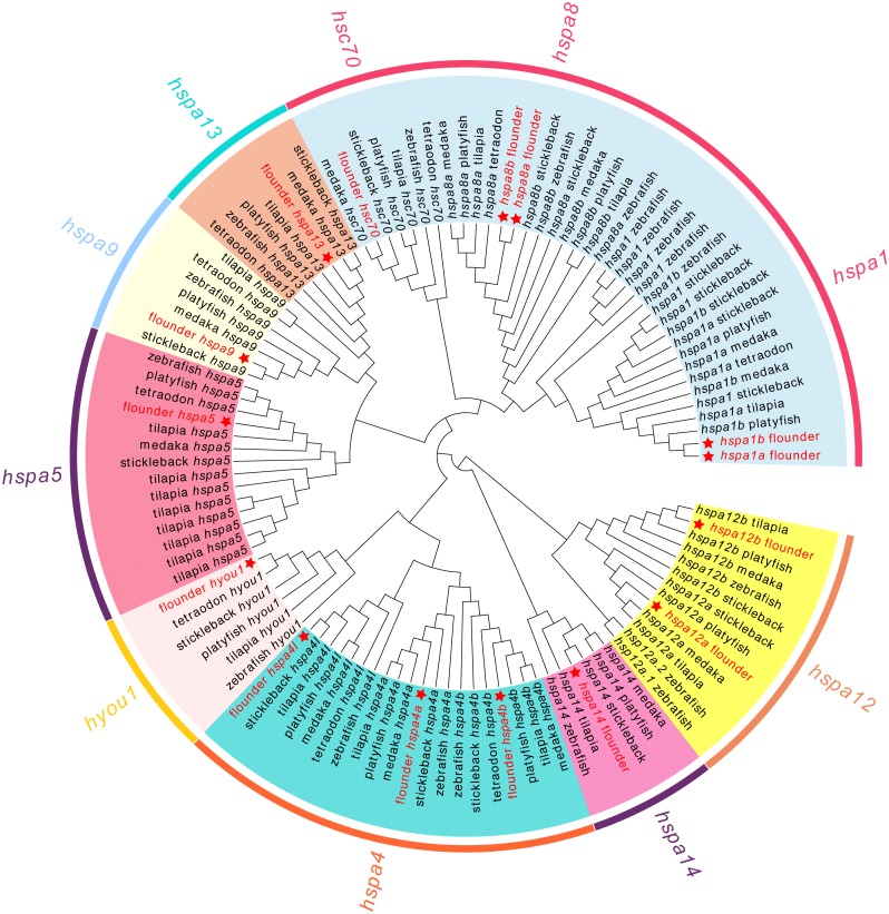
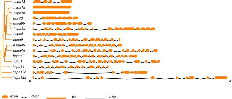
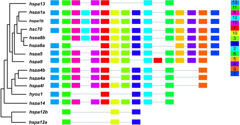
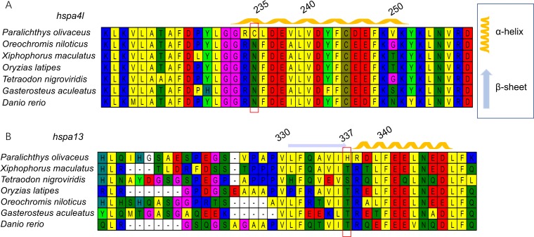
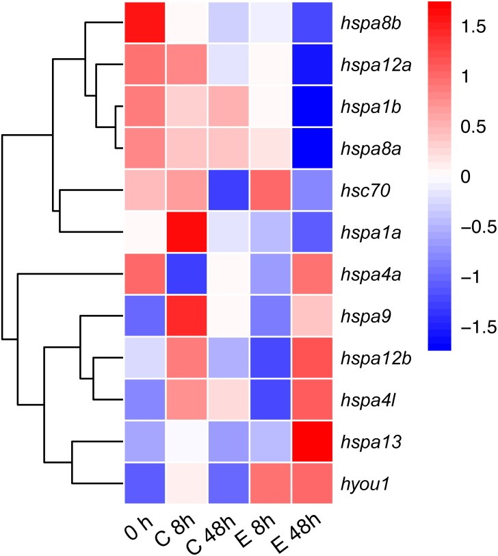
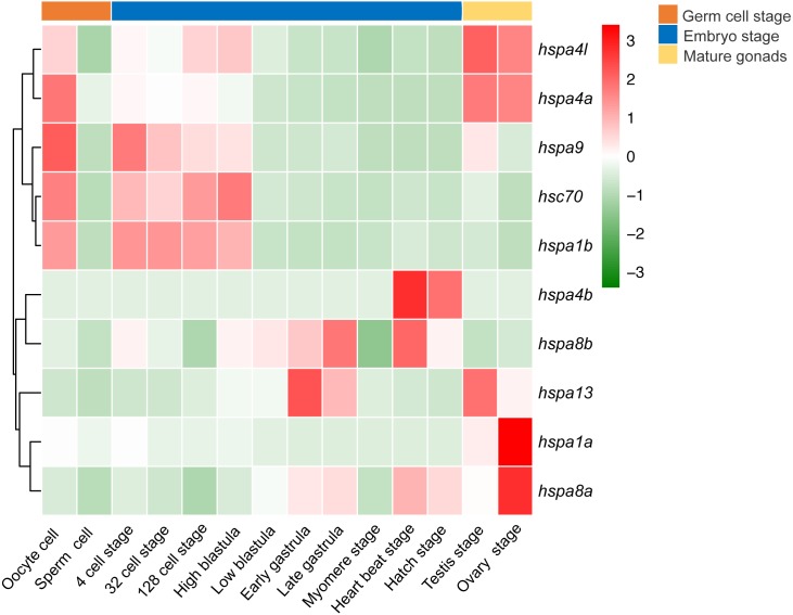
Flatfish undergo extreme morphological development and settle to a benthic in the adult stage, and are likely to be more susceptible to environmental stress. Heat shock proteins 70 ( hsp70 ) are involved in embryonic development and stress response in metazoan animals. However, the evolutionary history and functions of hsp70 in flatfish are poorly understood. Here, we identified 15 hsp70 genes in the genome of Japanese flounder ( Paralichthys olivaceus ), a flatfish endemic to northwestern Pacific Ocean. Gene structure and motifs of the Japanese flounder hsp70 were conserved, and there were few structure variants compared to other fish species. We constructed a maximum likelihood tree to understand the evolutionary relationship of the hsp70 genes among surveyed fish. Selection pressure analysis suggested that four genes, hspa4l , hspa9 , hspa13, and hyou1 , showed signs of positive selection. We then extracted transcriptome data on the Japanese flounder with Edwardsiella tarda to induce stress, and found that hspa9 , hspa12b, hspa4l, hspa13 , and hyou1 were highly expressed, likely to protect cells from stress. Interestingly, expression patterns of hsp70 genes were divergent in different developmental stages of the Japanese flounder. We found that at least one hsp70 gene was always highly expressed at various stages of embryonic development of the Japanese flounder, thereby indicating that hsp70 genes were constitutively expressed in the Japanese flounder. Our findings provide basic and useful resources to better understand hsp70 genes in flatfish.
Overview
The claims and statistics of this paper haven’t been checked yet.
Run the check — free, no account needed. It reads the open-access text and checks the sources it cites.
Integrity signals
Registry
Cites
Statistics
Figures
Verification
Claims the paper makes
The claims and statistics of this paper haven’t been checked yet.
Run the check — free, no account needed. It reads the open-access text and checks the sources it cites.
Claims other papers make about this one
Each is checked against this paper’s own text: does it say what the citing paper says it does?
In the citing paper
“Because these domains are evolutionarily conserved, they may contain epitopes that are recognized by antibodies across species boundaries”
From Parasites and allergies: a complex bidirectional relationship from evolutionary origins to modern therapeutics (2026) · cites this paper as Liu et al. 2019
In the citing paper
“HSP70 homologs from Schistosoma mansoni and Aspergillus fumigatus share conserved structural and functional domains, including regions involved in ATP binding and substrate interaction.”
From Parasites and allergies: a complex bidirectional relationship from evolutionary origins to modern therapeutics (2026) · cites this paper as Liu et al. 2019
In the citing paper
“Heat shock protein 70 (HSP70) is one example of a highly conserved protein family with potential relevance for cross-recognition between parasite and environmental allergens.”
From Parasites and allergies: a complex bidirectional relationship from evolutionary origins to modern therapeutics (2026) · cites this paper as Liu et al. 2019
Self-check
Percentages, sums and reported differences re-computed from the paper’s own numbers.
The self-check needs the paper’s full text.
Citations
Cites
Yang, Z., & Nielsen, R. (2002) · Molecular Biology and Evolution · doi:10.1093/oxfordjournals.molbev.a004148 · Cited by 1,374
Research works printed but not linked: 68
- Ackerman, 2001, Physiological and cellular stress responses of juvenile rainbow trout to vibriosis, Journal of Aquatic Animal Health, 13, 173, 10.1577/1548-8667(2001)013<0173:PACSRO>2.0.CO;2 · DOI
- Angelidis, 1991, Constitutive expression of heat-shock protein 70 in mammalian cells confers thermo resistance, European Journal of Biochemistry, 199, 35, 10.1111/j.1432-1033.1991.tb16088 · DOI
- Asea, 2000, Hsp70 stimulates cytokine production through a cd14-dependant pathway, demonstrating its dual role as a chaperone and cytokine, Nature Medicine, 6, 435, 10.1038/74697 · DOI
- Bertotto, 2011, Whole body cortisol and expression of HSP70, IGF-I and MSTN in early development of sea bass subjected to heat shock, General and Comparative Endocrinology, 174, 44, 10.1016/j.ygcen.2011.08.003 · DOI
- Boone, 2002, Constitutive heat shock protein (HSC70) expression in rainbow trout hepatocytes: effect of heat shock and heavy metal exposure, Comparative Biochemistry and Physiology Part C: Pharmacology, Toxicology and Endocrinology, 132, 223, 10.1016/S1532-0456(02)00066-2 · DOI
- Darwin, 1912, On the origin of species
- Das, 2015, Expression analysis of heat shock protein genes during Aeromonas hydrophila infection in rohu, Labeo rohita, with special reference to molecular characterization of Grp78, Cell Stress Chaperones, 20, 73, 10.1007/s12192-014-0527-2 · DOI
- Daugaard, 2007, The heat shock protein 70 family: highly homologous proteins with overlapping and distinct functions, FEBS Letters, 581, 3702, 10.1016/j.febslet.2007.05.039 · DOI
- De Maio, 2014, Extracellular Hsp70: export and function, Current Protein and Peptide Science, 15, 225, 10.2174/1389203715666140331113057 · DOI
- Forsyth, 1997, Stress protein expression in Coho salmon with bacterial kidney disease, Journal of Aquatic Animal Health, 9, 18, 10.1577/1548-8667(1997)009<0018:SPEICS>2.3.CO;2 · DOI
- Fuji, 2006, Identification of a single major genetic locus controlling the resistance to lymphocystis disease in Japanese flounder (Paralichthys olivaceus), Aquaculture, 254, 203, 10.1016/j.aquaculture.2005.11.024 · DOI
- Gething, 1992, Protein folding in the cell, Nature, 355, 33, 10.1038/355033a0 · DOI
- Gornati, 2004, Rearing density influences the expression of stress-related genes in sea bass (Dicentrarchus labrax, L.), Gene, 341, 111, 10.1016/j.gene.2004.06.020 · DOI
- Hahnel, 1986, Expression of the major heat shock protein (hsp70) family during early mouse embryo development, Teratogenesis, Carcinogenesis, and Mutagenesis, 6, 493, 10.1002/tcm.1770060603 · DOI
- Hodgins, 2016, Expression divergence is correlated with sequence evolution but not positive selection in conifers, Molecular Biology and Evolution, 33, 1502, 10.1093/molbev/msw032 · DOI
- Hu, 2015, GSDS 2.0: an upgraded gene feature visualization server, Bioinformatics, 31, 1296, 10.1093/bioinformatics/btu817 · DOI
- Hunt, 1985, Conserved features of eukaryotic hsp70 genes revealed by comparison with the nucleotide sequence of human hsp70, Proceedings of the National Academy of Sciences of the United States of America, 82, 6455, 10.1073/pnas.82.19.6455 · DOI
- Kelley, 2009, Protein structure prediction on the web: a case study using the phyre server, Nature Protocols, 4, 363, 10.1038/nprot.2009.2 · DOI
- Kiang, 1998, Heat shock protein 70 kDa: molecular biology, biochemistry, and physiology, Pharmacology and Therapeutics, 80, 183, 10.1016/S0163-7258(98)00028-X · DOI
- Koester, 2013, Positive selection within a diatom species acts on putative protein interactions and transcriptional regulation, Molecular Biology and Evolution, 30, 422, 10.1093/molbev/mss242 · DOI
- Kregel, 2002, Invited review: heat shock proteins: modifying factors in physiological stress responses and acquired thermotolerance, Journal of Applied Physiology, 92, 2177, 10.1152/japplphysiol.01267.2001 · DOI
- Krone, 1997, Heat shock genes and the heat shock response in zebrafish embryos, Biochemistry and Cell Biology, 75, 487, 10.1139/o97-083 · DOI
- Krone, 1994, HSP 90 alpha and HSP 90 beta genes are present in the zebrafish and are differentially regulated in developing embryos, Biochemical and Biophysical Research Communications, 204, 746, 10.1006/bbrc.1994.2522 · DOI
- Kumar, 2016, MEGA7: molecular evolutionary genetics analysis version 7.0 for bigger datasets, Molecular Biology and Evolution, 33, 1870, 10.1093/molbev/msw054 · DOI
- Li, 2018, Transcriptome profiling based on protein–protein interaction networks provides a core set of genes for understanding blood immune response mechanisms against Edwardsiella tarda infection in Japanese flounder (Paralichthys olivaceus), Developmental and Comparative Immunology, 78, 100, 10.1016/j.dci.2017.09.013 · DOI
- Li, 2017, Oviduct: roles in fertilization and early embryo development, Journal of Endocrinology, 232, R1, 10.1530/JOE-16-0302 · DOI
- Louryan, 2002, Heat shock proteins, embryogenesis and evolution, Bulletin et Memoires de l Academie Royale de Medecine de Belgique, 157, 293
- Luft, 1999, Hsp70 expression and function during embryogenesis, Cell Stress Chaperones, 4, 162, 10.1379/1466-1268(1999)004<0162:HEAFDE>2.3.CO;2 · DOI
- Machanick, 2011, MEME-ChIP: motif analysis of large DNA datasets, Bioinformatics, 27, 1696, 10.1093/bioinformatics/btr189 · DOI
- Matwee, 2001, The effects of antibodies to heat shock protein 70 in fertilization and embryo development, Molecular Human Reproduction, 7, 829, 10.1093/molehr/7.9.829 · DOI
- Mayer, 2005, Hsp70 chaperones: cellular functions and molecular mechanism, Cellular and Molecular Life Science, 62, 670, 10.1007/s00018-004-4464-6 · DOI
- Minami, 1992, Life-History cycles in flatfish from the Northwestern Pacific, with particular reference to their early life histories, Netherlands Journal of Sea Research, 29, 35, 10.1016/0077-7579(92)90006-Z · DOI
- Morange, 1984, Altered expression of heat shock proteins in embryonal carcinoma and mouse early embryonic cells, Molecular and Cellular Biology, 4, 730, 10.1128/MCB.4.4.730 · DOI
- Morimoto, 1997, The heat-shock response: regulation and function of heat-shock proteins and molecular chaperones, Essays in Biochemistry, 32, 17
- Morimoto, 1990, “The stress response, function of the protein, and perspectives” in stress proteins in biology and medicine
- Munro, 1987, A C-terminal signal prevents secretion of luminal ER proteins, Cell, 48, 899, 10.1016/0092-8674(87)90086-9 · DOI · Paper page
- Neuer, 1998, Monoclonal antibodies to mammalian heat shock proteins impair mouse embryo development in vitro, Human Reproduction, 13, 987, 10.1093/humrep/13.4.987 · DOI
- Nguyen, 2014, IQ-TREE: a fast and effective stochastic algorithm for estimating maximum-likelihood phylogenies, Molecular Biology and Evolution, 32, 268, 10.1093/molbev/msu300 · DOI · Paper page
- Oliver, 2010, Whole-genome positive selection and habitat-driven evolution in a shallow and a deep-sea urchin, Genome Biology and Evolution, 2, 800, 10.1093/gbe/evq063 · DOI
- Parsell, 1994, Protein disaggregation mediated by heat shock protein Hsp104, Nature, 372, 475, 10.1038/372475a0 · DOI
- Pearson, 1996, Cloning and characterization of a cDNA encoding the collagen-binding stress protein hsp47 in zebrafish, DNA and Cell Biology, 15, 263, 10.1089/dna.1996.15.263 · DOI
- Pockley, 2008, The dual immunoregulatory roles of stress proteins, Trends in Biochemical Sciences, 33, 71, 10.1016/j.tibs.2007.10.005 · DOI
- Poltronieri, 2007, Quantitative rt-pcr analysis and immunohistochemical localization of hsp70 in sea bass dicentrarchus labrax exposed to transport stress, European Journal of Histochemistry, 51, 125
- Pratt, 1993, The role of heat shock proteins in regulating the function, folding and trafficking of the glucocorticoid receptor, Journal of Biological Chemistry, 268, 21455, 10.1016/S0021-9258(20)80556-0 · DOI
- Pratt, 1997, Steroid receptor interactions with heat shock protein and immunophilin chaperones, Endocrine Reviews, 18, 306
- Ritossa, 1962, A new puffing pattern induced by temperature shock and DNP in drosophila, Experientia, 18, 571, 10.1007/BF02172188 · DOI
- Rocha, 2006, The quest for the universals of protein evolution, Trends in Genetics, 22, 412, 10.1016/j.tig.2006.06.004 · DOI
- Rupik, 2011, The expression patterns of heat shock genes and proteins and their role during vertebrate’s development, Comparative Biochemistry and Physiology. A: Comparative Physiology, 159, 349, 10.1016/j.cbpa.2011.04.002 · DOI
- Rupik, 2006, Promoter of the heat shock testis-specific Hsp70.2/Hst70 gene is active in nervous system during embryonic development of mice, Anatomy and Embryology, 211, 631, 10.1007/s00429-006-0125 · DOI
- Schlesinger, 1990, Heat shock proteins, Journal of Biological Chemistry, 21, 12111, 10.1016/S0021-9258(19)38314-0 · DOI
- Schultz, 2000, Smart: a web-based tool for the study of genetically mobile domains, Nucleic Acids Research, 28, 231, 10.1093/nar/28.1.231 · DOI
- Schultz, 1998, SMART, a simple modular architecture research tool: identification of signaling domains, Proceedings of the National Academy of Sciences of the United States of America, 95, 5857, 10.1073/pnas.95.11.5857 · DOI
- Sela, 2015, GUIDANCE2: accurate detection of unreliable alignment regions accounting for the uncertainty of multiple parameters, Nucleic Acids Research, 43, W7, 10.1093/nar/gkv318 · DOI · Paper page
- Shao, 2017, The genome and transcriptome of japanese flounder provide insights into flatfish asymmetry, Nature Genetics, 49, 119, 10.1038/ng.3732 · DOI
- Song, 2015, Genome-wide identification of Hsp70 genes in channel catfish and their regulated expression after bacterial infection, Fish Shellfish Immunol, 49, 154, 10.1016/j.fsi.2015.12.009 · DOI
- Sung, 2009, Feeding Artemia franciscana (Kellogg) larvae with bacterial heat shock protein, protects from Vibrio campbellii infection, Journal of Fish Diseases, 32, 675, 10.1111/j.1365-2761.2009.01046 · DOI
- Suyama, 2006, PAL2NAL: robust conversion of protein sequence alignments into the corresponding codon alignments, Nucleic Acids Research, 34, W609, 10.1093/nar/gkl315 · DOI
- Tsan, 2009, Heat shock proteins and immune system, Journal of Leukocyte Biology, 85, 905, 10.1189/jlb.0109005 · DOI
- Vega, 2006, Short-term hyperthermic treatment of Penaeus monodon increases expression of heat shock protein 70 (HSP70) and reduces replication of gill associated virus (GAV), Aquaculture, 253, 82, 10.1016/j.aquaculture.2005.07.041 · DOI
- Voss, 2000, Baroreflex sensitivity, heart rate, and blood pressure variability in normal pregnancy, American Journal of Hypertension, 13, 1218, 10.1016/S0895-7061(00)01199-7 · DOI
- Wagner, 2007, Rapid detection of positive selection in genes and genomes through variation clusters, Genetics, 176, 2451, 10.1534/genetics.107.074732 · DOI
- Wallin, 2002, Heat-shock proteins as activators of the innate immune system, Trends in Immunology, 23, 342, 10.1016/S1471-4906(02)02249-4 · DOI
- Whitley, 1999, Heat shock proteins: a review of the molecular chaperones, Journal of Vascular Surgery, 29, 748, 10.1016/S0741-5214(99)70329-0 · DOI
- Yang, 1997, PAML: a program package for phylogenetic analysis by maximum likelihood, Bioinformatics, 13, 555, 10.1093/bioinformatics/13.5.555 · DOI
- Yang, 2007, PAML 4: a program package for phylogenetic analysis by maximum likelihood, Molecular Biology and Evolution, 24, 1586, 10.1093/molbev/msm088 · DOI · Paper page
- Yang, 2011, Statistical properties of the branch-site test of positive selection, Molecular Biology and Evolution, 28, 1217, 10.1093/molbev/msq303 · DOI
- Zhang, 2012, EvolView, an online tool for visualizing, annotating and managing phylogenetic trees, Nucleic Acids Research, 40, 569, 10.1093/nar/gks576 · DOI
- Zhang, 2005, Evaluation of an improved branch-site likelihood method for detecting positive selection at the molecular level, Molecular Biology and Evolution, 22, 2472, 10.1093/molbev/msi237 · DOI · Paper page
Cited by
Sorted by citations
Packi, K., Świerczewski, D., Klimczak, S., Ochab, D., Gałęziewska, J., Gromek, P., Kruczkowska, W., Płuciennik, E., Kałuzińska-Kołat, Ż., & Kołat, D. (2026) · Antonie van Leeuwenhoek · doi:10.1007/s10482-026-02425-4
Metadata
- Authors
- Liu, K., Hao, X., Wang, Q., Hou, J., Lai, X., Dong, Z., & Shao, C.
- Venue
- PeerJ
- Publisher
- PeerJ
- Published
- 2019-11-11
- Volume
- 7
- Pages
- e7781
- ISSN
- 2167-8359
- PMC
- PMC6855204
- Cited by
- 22
- References
- 1
- Language
- en
- Funders
- National Key R&D Program of China (2018YFD0900301)
- Qingdao National Laboratory for Marine Science and Technology (2017ASTCP-ES06)
- Taishan Scholar Project Fund of Shandong of China
- National Ten-Thousands Talents Special Support Program
- The International Scientific Partnership Program ISPP at King Saud University for funding this research work through ISPP No. 0050
About these checks
Registry, retraction, Cites and statistics results are computed, not judged, with one exception: a printed reference that no index lists is read once by a model before it counts as a citation error. Claims are extracted by a model and kept only when their quote appears verbatim in the text; each cited source is then checked, with a model’s help, for whether it says what the claim relies on. Those verdicts can be wrong — read the claim’s quote and the cited source before relying on one.
Statistics and arithmetic are recomputed, not judged.
Confidence bands are shown only for fields calibrated on our annotated test set. None is calibrated yet, so no band is shown.
What counts as a claim ›How we measure confidence ›How we check this ›
Are you an author, or do you see a problem with this page? Request a review
