

The DOI matches its Crossref record; 28 of the 29 references it cites were found in a registry or an index; none of the 85 sources its claims cite is checked for support yet.
Tendinous Inscriptions of the Rectus Abdominis: A Comprehensive Review
Open accessAbstract
The rectus abdominis muscles are interrupted by tendinous inscriptions, which typically appear as fibrous bands crossing the muscle. The current literature on tendinous inscriptions is scarce; hence, this review will provide a detailed overview of their anatomical description, including their variation, embryology, comparative anatomy, and clinical application. Understanding the anatomy and function of the tendinous inscription assists in providing clinical relevance and in guiding reconstructive surgeons in their surgical planning and design.
Overview
CoChat Summary
Written by AI from the paper’s full text.
TL;DR
This comprehensive review synthesizes anatomical, embryological, comparative, and clinical literature on tendinous inscriptions of the rectus abdominis, emphasizing their variable number, shape, thickness, and vascularity. It highlights surgical relevance for TRAM flap planning and other reconstructive procedures, while noting scarce and uncertain evidence on their development.
Key findings
- Anson and McVay examined 162 rectus abdominis muscles in 81 cadaveric specimens: 1.2% (2/162) had one inscription, 5.9% (9/162) two, 58.0% (94/162) three, 35.2% (57/162) four, and 82% of specimens had an equal number bilaterally. The review also states the muscle is usually divided by three to five irregular fibrous bands and typically has three to four paired tendinous inscriptions.
- Among 530 inscriptions, 67% (356/530) extended completely from lateral to medial muscle borders, 24% (128/530) did not extend across more than half the muscle width, and 9% (46/530) were fragments less than half the width. Milloy et al. classified 484 complete or almost complete inscriptions: 109 (22.5%) straight, 57 (12%) biphasic with apices pointing caudally, 186 (38%) biphasic with apices pointing cephalad, and 132 (27.5%) multiphasic.
- Broyles et al. assessed 168 tendinous inscriptions in 32 fresh human cadavers; 18% (30/168) showed full thickness during their anterior–posterior course without intervening neurovascular structures. Among nine hemiabdominal specimens with 55 inscriptions, 42% (23/55) demonstrated at least one full-thickness tendinous inscription.
- Whetzel and Huang dissected 14 cadavers and found the arterial supply arises from transverse arcades from the superior or inferior epigastric arteries, with the greatest number of perforators within the tendinous inscriptions; the periumbilical inferior tendinous inscription region had the highest density (1.47 per sq cm), while the superior periumbilical muscular segment had the highest concentration (0.123 per sq cm). Embryologically, the rectus abdominis lacks tendinous inscriptions until the 17th week of gestation, with inscriptions appearing between the late 17th and 20th weeks; Rizk and Adieb studied 60 rat embryos and 10 postnatal rat specimens and concluded tendinous intersections were only observed postnatally and attached exclusively to the ventral, not dorsal, rectus sheath.
Methods
- Design: comprehensive narrative review of the literature on tendinous inscriptions of the rectus abdominis.
- Data: existing anatomical, embryological, comparative anatomy, and clinical studies, including cadaveric samples such as 81 cadavers/162 muscles, 32 fresh cadavers, 14 cadavers, and 46 cadavers, plus animal embryological work in 60 rat embryos and 10 postnatal rats.
- Analysis: narrative synthesis of anatomical description, variation, embryology, comparative anatomy, and clinical applications; no systematic search strategy or meta-analysis is described.
Limitations
- Stated in the paper: the current literature on tendinous inscriptions is scarce, and the embryological origins of tendinous inscriptions are uncertain.
- Evident from the text: as a narrative review, it does not report a systematic search strategy, inclusion/exclusion criteria, or quality appraisal, and it synthesizes heterogeneous cadaveric, animal, and clinical studies without presenting primary data.
Model-written summary. Check it against the paper before relying on it.
Integrity signals
Registry
Cites
Statistics
Figures
Verification
Claims the paper makes
70 Not checked yet · 5 Own interpretation · 3 No full text of the sources · 1 Check pending · 1 No basis given
None of the sources cited for this claim have been checked yet.
In the paper
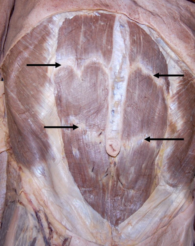
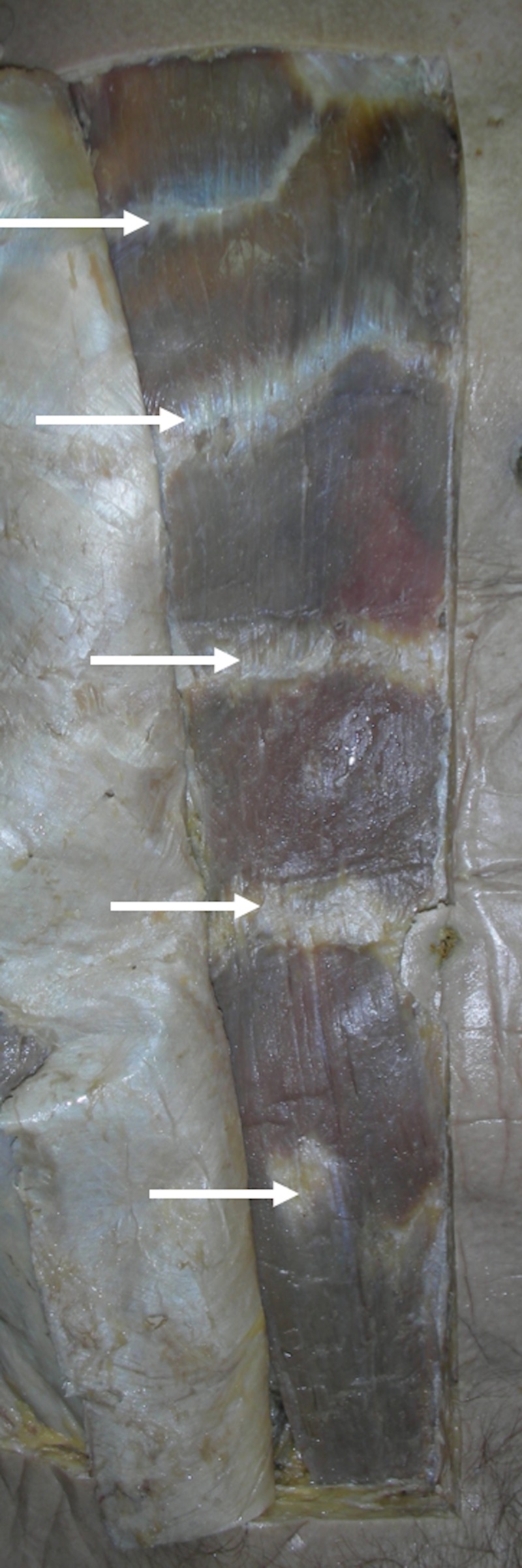
“The tendinous inscriptions may divide the RA into three or four muscular segments: at the opposite free end of the xiphoid process, umbilicus, midway between these two structures, and sporadically below the umbilicus [1,3].”
§ Tendinous Inscriptions of the Rectus Abdominis: A Comprehensive Review > Review
What the sources say
- Not checked yetcites [1] Defining the anatomy of the tendinous intersections of the rectus abdominis muscle and their clinical implications in functional muscle neurotization Ann Plas Surg Broyles JM Schuenke MD Patel SR Vail CM Broyles HV Dellon AL 1 4 0 2017 https://www.ncbi.nlm.nih.gov/pubmed/28671887 28671887
- Not checked yetcites [3] The tendinous intersections of rectus abdominis muscle J MGIMS Meenakshi S Manjunath KY 34 39 13 2008 http://dro.dur.ac.uk/9507/1/9507.pdf?DDD45+zspp51+cqjd36
hedgedstates a possible cause- Asserted: passes — The authors put it forward as true.
- Checkable: passes — It can be checked against evidence.
- Load-bearing: unclear — It is unclear whether the paper’s conclusions depend on it.
- Based: passes — The paper gives a reason why it holds.
In the paper
“24% (128/530) did not completely extend across more than half the width of the muscle”
§ Tendinous Inscriptions of the Rectus Abdominis: A Comprehensive Review > Review
What the sources say
- Not checked yetcites [2] Abdominal Wall. In Surgical Anatomy Anson BJ McVay CB Philadelphia WB Saunders Co 1 1971
plain- Asserted: passes — The authors put it forward as true.
- Checkable: passes — It can be checked against evidence.
- Load-bearing: unclear — It is unclear whether the paper’s conclusions depend on it.
- Based: passes — The paper gives a reason why it holds.
A check of a cited source is scheduled, or we don’t hold its full text yet; the claim is checked once it runs.
In the paper
“The rectus abdominis muscles are interrupted by tendinous inscriptions, which typically appear as fibrous bands crossing the muscle.”
- Also stated in: § Review · plain
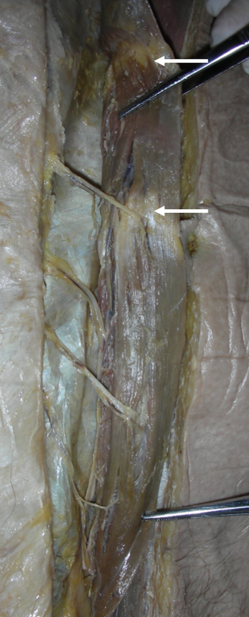
“The RA is divided, in part, by tendinous inscriptions that are usually three to five irregular fibrous bands (Figures 1-2) that cross the RA and adhere mainly to the anterior surface of the sheath [2].”
§ Tendinous Inscriptions of the Rectus Abdominis: A Comprehensive Review > Abstract
What the sources say
- Not checked yetcites [2] Abdominal Wall. In Surgical Anatomy Anson BJ McVay CB Philadelphia WB Saunders Co 1 1971
A check is scheduled.
plain- Asserted: passes — The authors put it forward as true.
- Checkable: passes — It can be checked against evidence.
- Load-bearing: passes — The paper’s conclusions depend on it.
- Based: passes — The paper gives a reason why it holds.
- Also stated in: § Review · plain
In the paper
“Anson and McVay [2] found that out of the 530 inscriptions studied, only 67% (356/530) completely extended without interruption from the lateral to the medial muscle borders”
§ Tendinous Inscriptions of the Rectus Abdominis: A Comprehensive Review > Review
What the sources say
- Not checked yetcites [2] Abdominal Wall. In Surgical Anatomy Anson BJ McVay CB Philadelphia WB Saunders Co 1 1971
plain- Asserted: passes — The authors put it forward as true.
- Checkable: passes — It can be checked against evidence.
- Load-bearing: unclear — It is unclear whether the paper’s conclusions depend on it.
- Based: passes — The paper gives a reason why it holds.
None of the sources cited for this claim have been checked yet.
In the paper
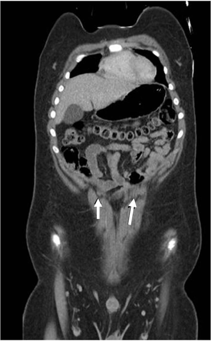
“The tendinous inscriptions limit fluid collection beneath the anterior rectus sheath, prevent muscle rupture, and aid in the biomechanics of the RA [1-2].”
§ Tendinous Inscriptions of the Rectus Abdominis: A Comprehensive Review > Review
What the sources say
- Not checked yetcites [1] Defining the anatomy of the tendinous intersections of the rectus abdominis muscle and their clinical implications in functional muscle neurotization Ann Plas Surg Broyles JM Schuenke MD Patel SR Vail CM Broyles HV Dellon AL 1 4 0 2017 https://www.ncbi.nlm.nih.gov/pubmed/28671887 28671887
- Not checked yetcites [2] Abdominal Wall. In Surgical Anatomy Anson BJ McVay CB Philadelphia WB Saunders Co 1 1971
plainstates a cause- Asserted: passes — The authors put it forward as true.
- Checkable: passes — It can be checked against evidence.
- Load-bearing: passes — The paper’s conclusions depend on it.
- Based: passes — The paper gives a reason why it holds.
- It may state two separate claims as one. 27
- Probably not a substantial claim. 33
Create a free account to see these claims.
Create a free account to see these assumptions.
28 more claims
Create a free account to see all 80 claims and to verify the 82 references still open.
Self-check
Percentages, sums and reported differences re-computed from the paper’s own numbers.
Each contradiction between two passages listed here was confirmed by an independent check.
Rated minor: it would not touch a result — a value no result rests on, a p-value that stays on the same side of the significance level, or a conflict that is clerical, resolved elsewhere in the paper, or outside its own findings.
In the paper
“5.9% (9/162) two inscriptions”
§ Tendinous Inscriptions of the Rectus Abdominis: A Comprehensive Review > Review
Reported 5.9 · recomputed 5.555556
Rated minor: it would not touch a result — a value no result rests on, a p-value that stays on the same side of the significance level, or a conflict that is clerical, resolved elsewhere in the paper, or outside its own findings.
In the paper
§ Tendinous Inscriptions of the Rectus Abdominis: A Comprehensive Review > Review
“which are only present on the anterior surface of the RA and fuse with the anterior layer of the rectus sheath.”
§ Tendinous Inscriptions of the Rectus Abdominis: A Comprehensive Review > Review
“18% (30/168) of these inscriptions showed full thickness during their anterior–posterior course”
Rated minor: it would not touch a result — a value no result rests on, a p-value that stays on the same side of the significance level, or a conflict that is clerical, resolved elsewhere in the paper, or outside its own findings.
In the paper
§ Tendinous Inscriptions of the Rectus Abdominis: A Comprehensive Review > Review
“the absence of the tendinous inscriptions as reported in a case by Ikiz et al. [13] may contribute to resisting abdominal wall herniation”
§ Tendinous Inscriptions of the Rectus Abdominis: A Comprehensive Review > Review
“the absence of these inscriptions may weaken the anterior abdominal wall.”
Citations
Cites
Myofascial pain and dysfunction : the trigger point manual
Travell, J. G., & Simons, D. G. (1982) · Medical Entomology and Zoology · Cited by 2,735
The Vascular Territories of the Superior Epigastric and the Deep Inferior Epigastric Systems
Boyd, J. B., Taylor, G. I., & Corlett, R. J. (1984) · Plastic & Reconstructive Surgery · doi:10.1097/00006534-198401000-00001 · Cited by 532
Kacey, D. J. (2009) · JAMA · doi:10.1001/jama.2009.1467 · Cited by 284
The fasciacytes: A new cell devoted to fascial gliding regulation
Stecco, C., Fede, C., Macchi, V., Porzionato, A., Petrelli, L., Biz, C., Stern, R., & De, R. (2018) · Clinical Anatomy · doi:10.1002/ca.23072 · Cited by 115
The rectus abdominis muscle and the epigastric arteries.
Milloy, F. J., Anson, B. J., & Dk, M. (1960) · PubMed · Cited by 80
The Rectus Abdominis Free Flap in Head and Neck Reconstruction
Urken, M. L., Turk, J. B., Weinberg, H., Vickery, C., Biller, H. F., & Schramm, V. L. (1991) · Archives of Otolaryngology - Head and Neck Surgery · doi:10.1001/archotol.1991.01870210103021 · Cited by 73
The Rectus Abdominis Free Flap in Head and Neck Reconstruction
Urken, M. L., Turk, J. B., Weinberg, H., Vickery, C., & Biller, H. F. (1991) · Archives of Otolaryngology - Head and Neck Surgery · doi:10.1001/archotol.1991.01870200051007 · Cited by 69
Do cutaneous nerves cross the midline?
Capek, S., Tubbs, R. S., & Spinner, R. J. (2014) · Clinical Anatomy · doi:10.1002/ca.22427 · Cited by 35
Self-citationFunctional Morphology of Serially Linked Skeletal Muscle Fibers
Hijikata, T., & Ishikawa, H. (1997) · Cells Tissues Organs · doi:10.1159/000147972 · Cited by 35
The Clinical anatomy of the physical examination of the abdomen: A comprehensive review
Bilal, M., Voin, V., Topale, N., Iwanaga, J., Loukas, M., & Tubbs, R. S. (2017) · Clinical Anatomy · doi:10.1002/ca.22832 · Cited by 31
Self-citationA reappraisal of adult thoracic and abdominal surface anatomy via CT scan in Chinese population
Shen, X., Su, B.-Y., Liu, J., Zhang, G., Xue, H., Jin, Z., Mirjalili, S. A., & Ma, C. (2015) · Clinical Anatomy · doi:10.1002/ca.22556 · Cited by 30
Function of the digastric muscle in carnivores
Scapino, R. P. (1976) · Journal of Morphology · doi:10.1002/jmor.1051500405 · Cited by 28
The Vascular Territories of the Superior Epigastric and the Deep Inferior Epigastric Systems
Boyd, J. B., Taylor, G. I., McCraw, J. B., & McCraw, J. B. (1984) · Plastic & Reconstructive Surgery · doi:10.1097/00006534-198401000-00002 · Cited by 28
The Vascular Anatomy of the Tendinous Intersections of the Rectus Abdominis Muscle
Whetzel, T. P., & Huang, V. (1996) · Plastic & Reconstructive Surgery · doi:10.1097/00006534-199607000-00013 · Cited by 25
Brown, S. H. M., & McGill, S. M. (2008) · European Journal of Applied Physiology · doi:10.1007/s00421-008-0858-8 · Cited by 23
Broyles, J. M., Schuenke, M. D., Patel, S., Vail, C. M., Broyles, H. V., & Dellon, A. L. (2017) · Annals of Plastic Surgery · doi:10.1097/sap.0000000000001193 · Cited by 17
In vivo and in vitro Examination of the Tendinous Inscription of the Human Semitendinosus Muscle
Kellis, E., Galanis, N., Natsis, K. I., & Kapetanos, G. A. (2011) · Cells Tissues Organs · doi:10.1159/000327574 · Cited by 16
A reappraisal of pediatric abdominal surface anatomy utilizing in vivo cross‐sectional imaging
Subramaniam, H., Taghavi, K., & Mirjalili, S. A. (2015) · Clinical Anatomy · doi:10.1002/ca.22674 · Cited by 12
Ogami, K., Murata, H., Sakai, A., Sato, S., Saiki, K., Okamoto, K., Manabe, Y., Hara, T., & Tsurumoto, T. (2017) · Clinical Anatomy · doi:10.1002/ca.22852 · Cited by 12
Rizk, N. N., & Adieb, N. (1982) · Europe PMC (PubMed Central) · Cited by 11
Kellis, E., & Balidou, A. (2013) · Journal of Morphology · doi:10.1002/jmor.20196 · Cited by 9
The tendinous intersections of rectus abdominis muscle.
Meenakshi, S., & Manjunath, K. (2008) · Durham Research Online (Durham University) · Cited by 8
Lancaster, W. C., & Henson, O. W. (1995) · Journal of Morphology · doi:10.1002/jmor.1052230109 · Cited by 8
Preliminary observations on the microarchitecture of the human abdominal muscles
Woodley, S. J., Duxson, M. J., & Mercer, S. R. (2007) · Clinical Anatomy · doi:10.1002/ca.20523 · Cited by 8
Muscle development in the abdominal region of Larval Hylidae (Amphibia: Anura)
Manzano, A. S., & Perotti, M. G. (1999) · Journal of Morphology · doi:10.1002/(sici)1097-4687(199909)241:3 · Cited by 4
Bilateral absence of the tendinous intersections of the rectus abdominis muscle
İkiz, Z. A. A. (2009) · Anatomy (International Journal of Experimental and Clinical Anatomy) · doi:10.2399/ana.09.015 · Cited by 4
[On the functional structure of tendinous inscriptions in the human rectus abdominis muscle].
Lange, W. N. (1968) · PubMed · Cited by 2
Clinical importance of unusual patterns of tendinous intersections of rectus abdominis muscle.
Das, S., Suri, R. K., & Kapur, V. K. (2003) · PubMed · Cited by 2
Research works printed but not linked: 1
- [2]Abdominal Wall. In Surgical Anatomy Anson BJ McVay CB Philadelphia WB Saunders Co 1 1971
Cited by
High-Definition Lipoplasty in Male Patients: A Systematic Review of Surgical Techniques and Outcomes
Escandón, J. M., Vyas, K. S., & Manrique, O. J. (2021) · Aesthetic Surgery Journal · doi:10.1093/asj/sjab300 · Cited by 18
Dafkou, K., Kellis, E., Ellinoudis, A., & Sahinis, C. (2020) · Europe PMC (PubMed Central) · Cited by 15
Umbilicus and the rectus sheath: a study using human fetuses
Xu, D., Jin, Z. W., Kim, J. H., Rodríguez‐Vázquez, J. F., Murakami, G., & Hayashi, S. (2020) · Surgical and Radiologic Anatomy · doi:10.1007/s00276-019-02398-2 · Cited by 9
Sato, T., Kim, J. H., Cho, K. H., Hayashi, S., Rodríguez‐Vázquez, J. F., & Murakami, G. (2021) · Surgical and Radiologic Anatomy · doi:10.1007/s00276-021-02759-w · Cited by 5
Park, C., Cho, H.-Y., & Kang, C. (2022) · International Journal of Environmental Research and Public Health · doi:10.3390/ijerph192114525 · Cited by 4
Metadata
- Authors
- Rai, R., Azih, L. C., Iwanaga, J., Loukas, M., Mortazavi, M., Oskouian, R. J., & Tubbs, R. S.
- Affiliations
- St. George's University (GD)
- Corresponding author
- Iwanaga, Joe
- Venue
- Cureus
- Published
- 2018-08-04
- Volume
- 10
- Issue
- 8
- Pages
- e3100-e3100
- ISSN
- 2168-8184 (ISSN-L)
- PMC
- PMC6173272
- OpenAlex
- W2886318833
- Cited by
- 12
- References
- 29
- Citations by year
- 2025: 2 · 2024: 1 · 2023: 1 · 2022: 3 · 2021: 3 · 2020: 2
- FWCI
- 3.45
- Type
- journal article
- Language
- en
- Open access
- diamond
- doi.org
- Licence
- cc-by
- Locations
- Cureus (publishedVersion, cc-by)
- Europe PMC (PubMed Central) (publishedVersion, cc-by)
- PubMed (publishedVersion)
- PubMed Central (publishedVersion, cc-by)
- Topics
- Health Sciences › Medicine › Anatomy › Classical Anatomy and Histology
- Health Sciences › Medicine › Surgery › Reconstructive Surgery and Microvascular Techniques
- Health Sciences › Medicine › Surgery › Hernia repair and management
- Keywords
- rectus abdominis, abdominal wall anatomy, anatomical variation, embryology, comparative anatomy, reconstructive surgery, surgical planning
About these checks
Registry, retraction, Cites and statistics results are computed, not judged, with one exception: a printed reference that no index lists is read once by a model before it counts as a citation error. Claims are extracted by a model and kept only when their quote appears verbatim in the text; each cited source is then checked, with a model’s help, for whether it says what the claim relies on. Those verdicts can be wrong — read the claim’s quote and the cited source before relying on one.
A language model drew the claims from the paper on 2026-11-11, under version 3 of the claim definition (model deepseek-v4.1-flash, prompt extract-v3).
A second model labelled each claim on 2026-11-11 (model jev-1.13-20260917, definition revision gold-guideline-v0.2).
Statistics and arithmetic are recomputed, not judged.
Confidence bands are shown only for fields calibrated on our annotated test set. None is calibrated yet, so no band is shown.
What counts as a claim ›How we measure confidence ›How we check this ›
Are you an author, or do you see a problem with this page? Request a review
