Cited by 423Neuroinflammation and Neurodegeneration MechanismsNeuroscience and Neuropharmacology ResearchNeurological Disease Mechanisms and Treatments

The DOI matches its Crossref record; 68 of the 68 references it cites were found in a registry or an index.

Dark microglia: A new phenotype predominantly associated with pathological states

Open access

Abstract

The past decade has witnessed a revolution in our understanding of microglia. These immune cells were shown to actively remodel neuronal circuits, leading to propose new pathogenic mechanisms. To study microglial implication in the loss of synapses, the best pathological correlate of cognitive decline across chronic stress, aging, and diseases, we recently conducted ultrastructural analyses. Our work uncovered the existence of a new microglial phenotype that is rarely present under steady state conditions, in hippocampus, cerebral cortex, amygdala, and hypothalamus, but becomes abundant during chronic stress, aging, fractalkine signaling deficiency (CX 3 CR1 knockout mice), and Alzheimer's disease pathology (APP‐PS1 mice). Even though these cells display ultrastructural features of microglia, they are strikingly distinct from the other phenotypes described so far at the ultrastructural level. They exhibit several signs of oxidative stress, including a condensed, electron‐dense cytoplasm and nucleoplasm making them as “dark” as mitochondria, accompanied by a pronounced remodeling of their nuclear chromatin. Dark microglia appear to be much more active than the normal microglia, reaching for synaptic clefts, while extensively encircling axon terminals and dendritic spines with their highly ramified and thin processes. They stain for the myeloid cell markers IBA1 and GFP (in CX 3 CR1‐GFP mice), and strongly express CD11b and microglia‐specific 4D4 in their processes encircling synaptic elements, and TREM2 when they associate with amyloid plaques. Overall, these findings suggest that dark microglia, a new phenotype that we identified based on their unique properties, could play a significant role in the pathological remodeling of neuronal circuits, especially at synapses. GLIA 2016;64:826–839

Overview

The claims and statistics of this paper haven’t been checked yet.

Run the check — free, no account needed. It reads the open-access text and checks the sources it cites.

A few free checks a day · papers already verified open instantly · results are public

Integrity signals

Registry

Crossref · 10.1002/glia.22966

Cites

68 of 68 references verified0 citation errors · 0 not checkable

Statistics

Figures

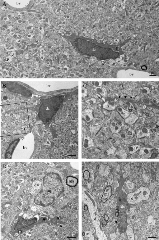
Figure 1 Ultrastructural features of the dark microglia. A–E: Examples of dark microglial cells (dc) encountered in the CA1 region of the hippocampus (stratum lacunosum‐moleculare) of stressed CX3CR1 knockout mice (A–D) or in the median eminence of the hypothalamus in a nontransgenic control mouse (E). In addition to their ultrastructural features of microglia, for instance their frequent long stretches of endoplasmic reticulum (arrowheads in C), these cells are recognized by their various signs of oxidative stress: their condensed, electron‐dense cytoplasm and nucleoplasm, accompanied by cytoplasmic shrinkage, Golgi apparatus (g) and endoplasmic reticulum (er) dilation, and mitochondrial alteration (arrowheads in D, E). Examples of endoplasmic reticulum dilation in cell bodies and a process are, respectively, provided in (B), (D), and (C). The dark microglia contain lipofuscin granules (g) in (D) and (E). Direct contacts with blood vessels (bv), dendrites (d), a neuronal perikaryon (np), axon terminals (t) and dendritic spines (s), and synapses between axon terminals and dendritic spines are also shown. ma = myelinated axon. Scale bars = 1 μm.
Figure 1 Ultrastructural features of the dark microglia. A–E: Examples of dark microglial cells (dc) encountered in the CA1 region of the hippocampus (stratum lacunosum‐moleculare) of stressed CX3CR1 knockout mice (A–D) or in the median eminence of the hypothalamus in a nontransgenic control mouse (E). In addition to their ultrastructural features of microglia, for instance their frequent long stretches of endoplasmic reticulum (arrowheads in C), these cells are recognized by their various signs of oxidative stress: their condensed, electron‐dense cytoplasm and nucleoplasm, accompanied by cytoplasmic shrinkage, Golgi apparatus (g) and endoplasmic reticulum (er) dilation, and mitochondrial alteration (arrowheads in D, E). Examples of endoplasmic reticulum dilation in cell bodies and a process are, respectively, provided in (B), (D), and (C). The dark microglia contain lipofuscin granules (g) in (D) and (E). Direct contacts with blood vessels (bv), dendrites (d), a neuronal perikaryon (np), axon terminals (t) and dendritic spines (s), and synapses between axon terminals and dendritic spines are also shown. ma = myelinated axon. Scale bars = 1 μm.CC BY · PubMed Central
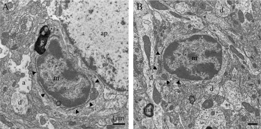
Figure 2 Examples of normal microglia, observed in the median eminence of the hypothalamus in a nontransgenic control mouse. A, B: Unstained microglia (m) generally display a lighter cytoplasm and nucleoplasm with a clearly defined chromatin pattern, compared with the dark microglial cells. They also share with the dark microglia a small elongated nucleus delineated by a narrow nuclear cistern, associated pockets of extracellular space (asterisks), distinctive long stretches of endoplasmic reticulum (arrowheads), frequent endosomes, lipofuscin granules (g), and cellular inclusions. a = astrocytic process, ap = astrocytic perikaryon, d = dendrites, and ma = myelinated axon. Scale bars = 1 μm.
Figure 2 Examples of normal microglia, observed in the median eminence of the hypothalamus in a nontransgenic control mouse. A, B: Unstained microglia (m) generally display a lighter cytoplasm and nucleoplasm with a clearly defined chromatin pattern, compared with the dark microglial cells. They also share with the dark microglia a small elongated nucleus delineated by a narrow nuclear cistern, associated pockets of extracellular space (asterisks), distinctive long stretches of endoplasmic reticulum (arrowheads), frequent endosomes, lipofuscin granules (g), and cellular inclusions. a = astrocytic process, ap = astrocytic perikaryon, d = dendrites, and ma = myelinated axon. Scale bars = 1 μm.CC BY · PubMed Central
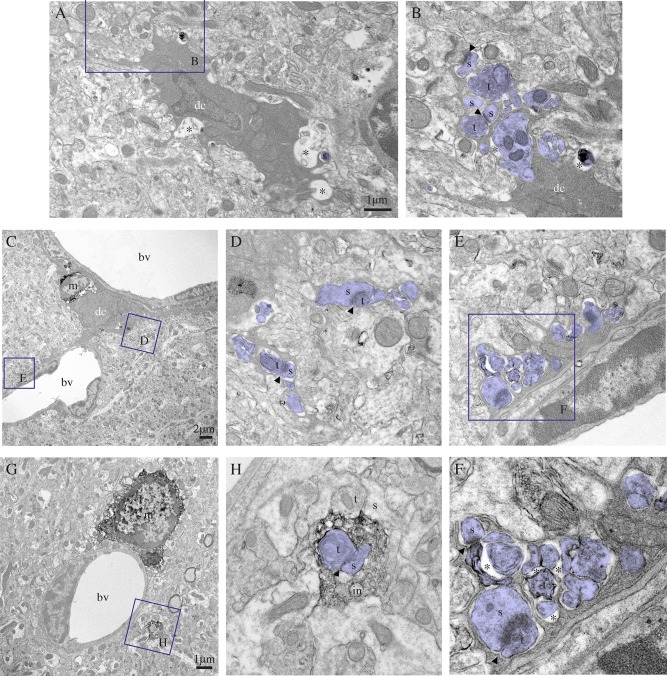
Figure 3 Dark microglia's interactions with synapses. A–F: Examples of dark microglial cells (dc) typically contacting synaptic elements (colored in purple) with their profusion of highly ramified and extremely thin processes, reaching for synaptic clefts (arrowheads), while encircling axon terminals (t) and dendritic spines (s), in the CA1 lacunosum‐moleculare of stressed CX3CR1 knockout mice. In (C), the dark microglia is simultaneously contacting two blood vessels (bv) and a normal microglia (m) that is stained for IBA1. Its processes are extensively encircling various types of synaptic elements, including shrunk axon terminals surrounded by extracellular space (asterisks) in the process of being digested and an entire synapse (see the inset in F). By comparison, an example of IBA1‐stained microglia (m) that is extending a single process, discontinuous from its cell body in ultrathin section, is shown in (G). Contrary to the dark microglia processes, it is bulkier and showing obtuse (instead of acute) angles. It nevertheless contains several phagocytic inclusions (in), among which a synapse between an axon terminal (t) and a dendritic spine (s), in addition to making focal contacts (instead of encircling) synaptic elements. Scale bars = 1 μm for (A) and (G) and 2 μm for (C). [Color figure can be viewed in the online issue, which is available at wileyonlinelibrary.com.]
Figure 3 Dark microglia's interactions with synapses. A–F: Examples of dark microglial cells (dc) typically contacting synaptic elements (colored in purple) with their profusion of highly ramified and extremely thin processes, reaching for synaptic clefts (arrowheads), while encircling axon terminals (t) and dendritic spines (s), in the CA1 lacunosum‐moleculare of stressed CX3CR1 knockout mice. In (C), the dark microglia is simultaneously contacting two blood vessels (bv) and a normal microglia (m) that is stained for IBA1. Its processes are extensively encircling various types of synaptic elements, including shrunk axon terminals surrounded by extracellular space (asterisks) in the process of being digested and an entire synapse (see the inset in F). By comparison, an example of IBA1‐stained microglia (m) that is extending a single process, discontinuous from its cell body in ultrathin section, is shown in (G). Contrary to the dark microglia processes, it is bulkier and showing obtuse (instead of acute) angles. It nevertheless contains several phagocytic inclusions (in), among which a synapse between an axon terminal (t) and a dendritic spine (s), in addition to making focal contacts (instead of encircling) synaptic elements. Scale bars = 1 μm for (A) and (G) and 2 μm for (C). [Color figure can be viewed in the online issue, which is available at wileyonlinelibrary.com.]CC BY · PubMed Central
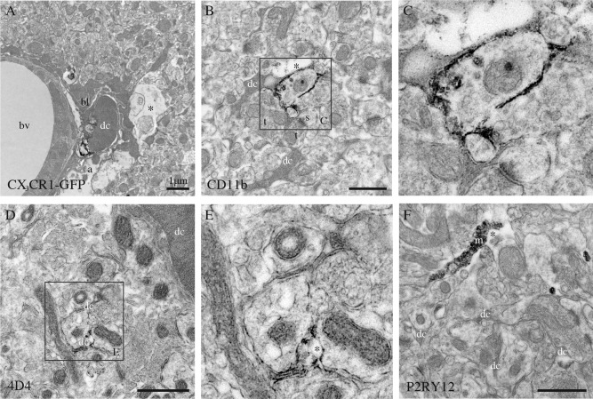
Figure 4 Phenotypic characterization of the dark microglia, using immunoperoxidase staining in the CA1 lacunosum‐moleculare of stressed CX3CR1 knockout mice (A–C, F), or a nontransgenic control mouse (D, E). A: Focal staining for GFP in a dark microglial cell (dc) from a CX3CR1‐GFP mouse. In contrast, normal microglia display strong and diffuse immunoreactivity for IBA1 throughout their cytoplasm. B, C: Examples of dark microglia staining for the myeloid cell marker CD11b, which forms CR3 involved in phagocytosis, strongly expressed at the plasma membrane of their processes encircling synaptic elements. D, E: Dark microglia's staining for 4D4, a recently discovered marker of homeostatic microglia, at the extremity of their ramified processes. In contrast, the dark microglia do not stain for P2RY12 (F), another marker of homeostatic microglia that is abundant in microglial processes (m). a = astrocytic process, bl = basal lamina, bv = blood vessel, s = dendritic spine, and t = axon terminals. Asterisks show the extracellular space. Scale bars = 1 μm.
Figure 4 Phenotypic characterization of the dark microglia, using immunoperoxidase staining in the CA1 lacunosum‐moleculare of stressed CX3CR1 knockout mice (A–C, F), or a nontransgenic control mouse (D, E). A: Focal staining for GFP in a dark microglial cell (dc) from a CX3CR1‐GFP mouse. In contrast, normal microglia display strong and diffuse immunoreactivity for IBA1 throughout their cytoplasm. B, C: Examples of dark microglia staining for the myeloid cell marker CD11b, which forms CR3 involved in phagocytosis, strongly expressed at the plasma membrane of their processes encircling synaptic elements. D, E: Dark microglia's staining for 4D4, a recently discovered marker of homeostatic microglia, at the extremity of their ramified processes. In contrast, the dark microglia do not stain for P2RY12 (F), another marker of homeostatic microglia that is abundant in microglial processes (m). a = astrocytic process, bl = basal lamina, bv = blood vessel, s = dendritic spine, and t = axon terminals. Asterisks show the extracellular space. Scale bars = 1 μm.CC BY · PubMed Central
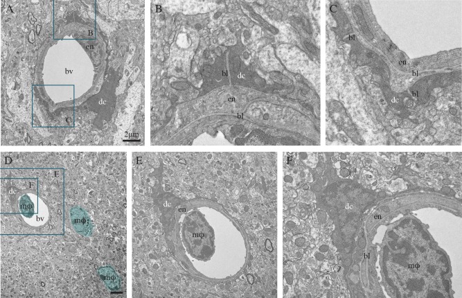
Figure 5 Dark microglia's interactions with the vasculature, in the hypothalamus median eminence of a nontransgenic mouse (A–C) and CA1 radiatum of an APP‐PS1 6‐month‐old mouse (D–F). In these two examples, the dark microglial cell (dc) bodies are directly juxtaposing the blood vessels' (bv) basal lamina (bl), and their processes are extending all around the vessels, as well as ensheathing the basal lamina. In (D–F), the blood vessel contains a circulating, bone marrow‐derived monocyte or macrophage (mφ; colored in blue). Additional examples of bone marrow‐derived macrophages can also be seen in the brain parenchyma (colored in blue). It can be noted that the dark microglial cell's contact with the blood vessel is occurring at the site of docking, raising the intriguing possibility of dark cells functional interactions with endothelial cells, as well as circulating immune cells. Scale bars = 2 μm. [Color figure can be viewed in the online issue, which is available at wileyonlinelibrary.com.]
Figure 5 Dark microglia's interactions with the vasculature, in the hypothalamus median eminence of a nontransgenic mouse (A–C) and CA1 radiatum of an APP‐PS1 6‐month‐old mouse (D–F). In these two examples, the dark microglial cell (dc) bodies are directly juxtaposing the blood vessels' (bv) basal lamina (bl), and their processes are extending all around the vessels, as well as ensheathing the basal lamina. In (D–F), the blood vessel contains a circulating, bone marrow‐derived monocyte or macrophage (mφ; colored in blue). Additional examples of bone marrow‐derived macrophages can also be seen in the brain parenchyma (colored in blue). It can be noted that the dark microglial cell's contact with the blood vessel is occurring at the site of docking, raising the intriguing possibility of dark cells functional interactions with endothelial cells, as well as circulating immune cells. Scale bars = 2 μm. [Color figure can be viewed in the online issue, which is available at wileyonlinelibrary.com.]CC BY · PubMed Central
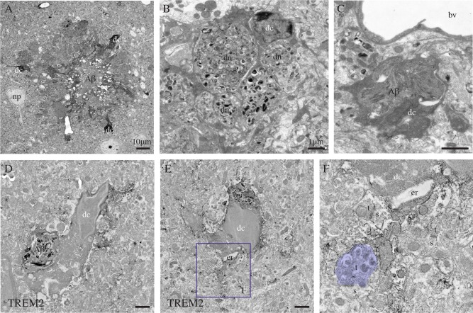
Figure 6 Dark microglia's association with the plaques of amyloid β, examples from the prefrontal cortex subgranular layers (A–C) and CA1 lacunosum‐moleculare (D–F) of 6‐ and 21‐month‐old APP‐PS1 mice, respectively. In (A), three dark microglial cells (dc) showing several signs of cellular stress (i.e., extreme condensation, darkening of their cytoplasm and nucleoplasm) are observed in the close proximity of a plaque of amyloid β (Αβ), which is identified by its ultrastructural features. In (B), farther from the plaque, a dark microglial cell process is encircling several dystrophic neurites (dn) containing autophagic vacuoles. In (C) and (D), a dark cell body (D) and process (C) are containing Αβ deposits. D–F: Examples of dark microglial cells that exhibit immunostaining for TREM2, as frequently observed nearby the plaques of Αβ. The remodeling of their nuclear contents can be noted in D and E, together with the pronounced dilation of their endoplasmic reticulum (er) in (E). In addition, the dark microglial cell in (E) is contacting an axon terminal (colored in purple, t) with an accumulation of autophagic vacuoles that makes a synapse on a healthy looking dendritic spine (s). bv = blood vessel and np = neuronal perikaryon. Scale bars = 10 μm for (A), and 1 μm for (B–E). [Color figure can be viewed in the online issue, which is available at wileyonlinelibrary.com.]
Figure 6 Dark microglia's association with the plaques of amyloid β, examples from the prefrontal cortex subgranular layers (A–C) and CA1 lacunosum‐moleculare (D–F) of 6‐ and 21‐month‐old APP‐PS1 mice, respectively. In (A), three dark microglial cells (dc) showing several signs of cellular stress (i.e., extreme condensation, darkening of their cytoplasm and nucleoplasm) are observed in the close proximity of a plaque of amyloid β (Αβ), which is identified by its ultrastructural features. In (B), farther from the plaque, a dark microglial cell process is encircling several dystrophic neurites (dn) containing autophagic vacuoles. In (C) and (D), a dark cell body (D) and process (C) are containing Αβ deposits. D–F: Examples of dark microglial cells that exhibit immunostaining for TREM2, as frequently observed nearby the plaques of Αβ. The remodeling of their nuclear contents can be noted in D and E, together with the pronounced dilation of their endoplasmic reticulum (er) in (E). In addition, the dark microglial cell in (E) is contacting an axon terminal (colored in purple, t) with an accumulation of autophagic vacuoles that makes a synapse on a healthy looking dendritic spine (s). bv = blood vessel and np = neuronal perikaryon. Scale bars = 10 μm for (A), and 1 μm for (B–E). [Color figure can be viewed in the online issue, which is available at wileyonlinelibrary.com.]CC BY · PubMed Central

Verification

Claims the paper makes

The claims and statistics of this paper haven’t been checked yet.

Run the check — free, no account needed. It reads the open-access text and checks the sources it cites.

A few free checks a day · papers already verified open instantly · results are public

Claims other papers make about this one

Each is checked against this paper’s own text: does it say what the citing paper says it does?

Self-check

Percentages, sums and reported differences re-computed from the paper’s own numbers.

The self-check needs the paper’s full text.

Citations

Cites

1 retracted

Show all 68 references

Cited by

Sorted by citations

See all 423

Metadata

Authors
Bisht, K., Sharma, K. P., Lecours, C., Gabriela Sánchez, M., El Hajj, H., Milior, G., Olmos‐Alonso, A., Gómez‐Nicola, D., Luheshi, G., Vallières, L., Branchi, I., Maggi, L., Limatola, C., Butovsky, O., & Tremblay, M.
Affiliations
  • Centre de recherche du CHU de Québec-Université Laval (CA)
  • Axe Neurosciences Centre De Recherche Du CHU De Québec Québec Québec Canada
  • Istituto Pasteur (IT)
  • University of Southampton (GB)
  • Douglas Mental Health University Institute (CA)
  • Istituto Superiore di Sanità (IT)
  • Brigham and Women's Hospital (US)
Corresponding author
Tremblay, Marie‐Ève
Venue
Glia
Publisher
Wiley
Published
2016-02-05
Volume
64
Issue
5
Pages
826-839
ISSN
  • 0894-1491 (ISSN-L)
  • 1098-1136
OpenAlex
W2256955303
Cited by
421
References
68
Citations by year
2026: 25 · 2025: 36 · 2024: 52 · 2023: 48 · 2022: 44 · 2021: 64 · 2020: 42 · 2019: 42 · 2018: 30 · 2017: 26 · 2016: 12
FWCI
20.09
Type
journal article
Language
en
Open access
Locations
  • Glia (publishedVersion, cc-by)
  • Europe PMC (PubMed Central) (publishedVersion, cc-by)
  • PubMed Central (publishedVersion, cc-by)
  • PubMed (publishedVersion)
  • ePrints Soton (University of Southampton) (acceptedVersion)
  • IRIS Research product catalog (Sapienza University of Rome) (submittedVersion, cc-by)
  • Digital Access to Scholarship at Harvard (DASH) (Harvard University) (submittedVersion, cc-by)
Funders
  • Natural Sciences and Engineering Research Council of Canada (NSERC) (RGPIN-2014-05308)
  • Medical Research Council (MR/K022687/1)
  • The Banting Research Foundation
  • The Scottish Rite Charitable Foundation of Canada
Topics
  • Life Sciences › Neuroscience › Neurology › Neuroinflammation and Neurodegeneration Mechanisms
  • Life Sciences › Neuroscience › Cellular and Molecular Neuroscience › Neuroscience and Neuropharmacology Research
  • Life Sciences › Neuroscience › Neurology › Neurological Disease Mechanisms and Treatments
Keywords
microglial phenotype, oxidative stress, chronic stress, aging, CX3CR1, Alzheimer's disease, APP/PS1 mice, amyloid plaques, TREM2, synaptic loss, dendritic spines, axon terminals, hippocampus, cerebral cortex, amygdala, hypothalamus
MeSH terms
CX3C Chemokine Receptor 1, Formyltetrahydrofolate Dehydrogenase, Aging, Aldehyde Dehydrogenase, Alzheimer Disease, Oxidoreductases Acting on CH-NH Group Donors, Animals, Cerebral Cortex, Disease Models, Animal, Mice, Inbred C57BL, Mice, Transgenic, Nerve Tissue Proteins, Phenotype, Stress, Psychological, Antigens, CD, Amyloid beta-Protein Precursor, Microglia, Receptors, Chemokine, Green Fluorescent Proteins, Mice, Presenilin-1

About these checks

Registry, retraction, Cites and statistics results are computed, not judged, with one exception: a printed reference that no index lists is read once by a model before it counts as a citation error. Claims are extracted by a model and kept only when their quote appears verbatim in the text; each cited source is then checked, with a model’s help, for whether it says what the claim relies on. Those verdicts can be wrong — read the claim’s quote and the cited source before relying on one.

Statistics and arithmetic are recomputed, not judged.

Confidence bands are shown only for fields calibrated on our annotated test set. None is calibrated yet, so no band is shown.

What counts as a claim ›How we measure confidence ›How we check this ›

Are you an author, or do you see a problem with this page? Request a review