

The DOI matches its Crossref record; 65 of the 65 references it cites were found in a registry or an index.
Myricanol rescues dexamethasone-induced muscle dysfunction Via a sirtuin 1-dependent mechanism
Open accessAbstract
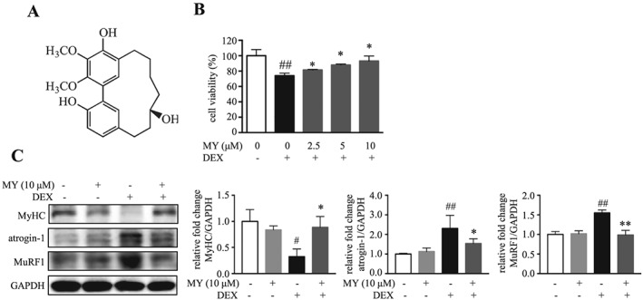
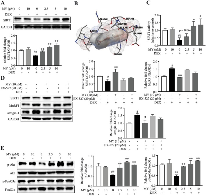
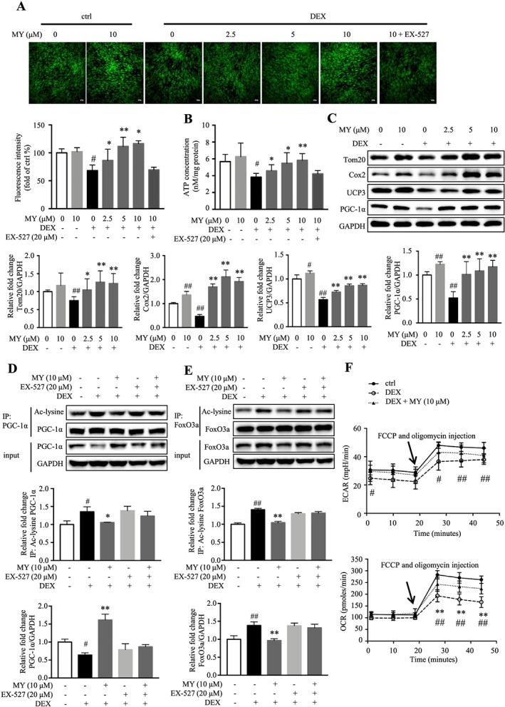
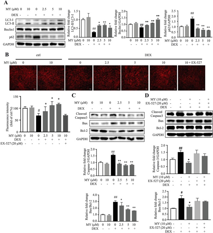
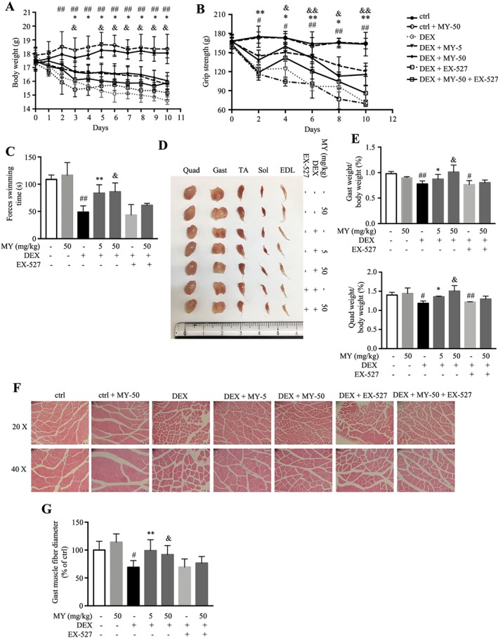
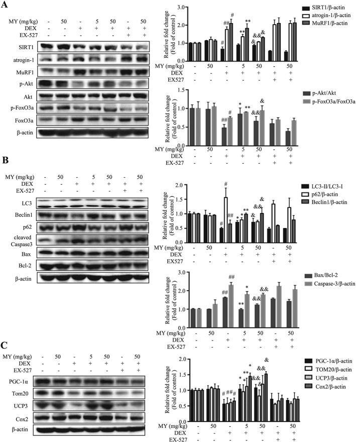
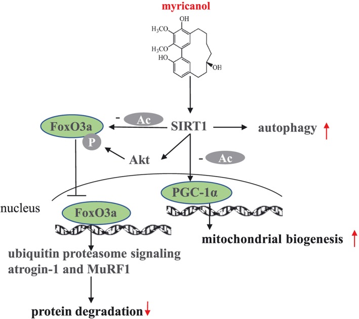
BACKGROUND: Muscle atrophy and weakness are adverse effects of high dose or the sustained usage of glucocorticoids. Loss of mitochondria and degradation of protein are highly correlated with muscle dysfunction. The deacetylase sirtuin 1 (SIRT1) plays a vital role in muscle remodelling. The current study was designed to identify myricanol as a SIRT1 activator, which could protect skeletal muscle against dexamethasone-induced wasting. METHODS: The dexamethasone-induced atrophy in C2C12 myotubes was evaluated by expression of myosin heavy chain, muscle atrophy F-box (atrogin-1), and muscle ring finger 1 (MuRF1), using western blots. The mitochondrial content and oxygen consumption were assessed by MitoTracker staining and extracellular flux analysis, respectively. Muscle dysfunction was established in male C57BL/6 mice (8-10 weeks old, n = 6) treated with a relatively high dose of dexamethasone (25 mg/kg body weight, i.p., 10 days). Body weight, grip strength, forced swimming capacity, muscle weight, and muscle histology were assessed. The expression of proteolysis-related, autophagy-related, apoptosis-related, and mitochondria-related proteins was analysed by western blots or immunoprecipitation. RESULTS: Myricanol (10 μM) was found to rescue dexamethasone-induced muscle atrophy and dysfunction in C2C12 myotubes, indicated by increased expression of myosin heavy chain (0.33 ± 0.14 vs. 0.89 ± 0.21, *P < 0.05), decreased expression of atrogin-1 (2.31 ± 0.67 vs. 1.53 ± 0.25, *P < 0.05) and MuRF1 (1.55 ± 0.08 vs. 0.99 ± 0.12, **P < 0.01), and elevated ATP production (3.83 ± 0.46 vs. 5.84 ± 0.79 nM/mg protein, **P < 0.01), mitochondrial content (68.12 ± 10.07% vs. 116.38 ± 5.12%, *P < 0.05), and mitochondrial oxygen consumption (166.59 ± 22.89 vs. 223.77 ± 22.59 pmol/min, **P < 0.01). Myricanol directly binds and activates SIRT1, with binding energy of -5.87 kcal/mol. Through activating SIRT1 deacetylation, myricanol inhibits forkhead box O 3a transcriptional activity to reduce protein degradation, induces autophagy to enhance degraded protein clearance, and increases peroxisome proliferator-activated receptor γ coactivator-1α activity to promote mitochondrial biogenesis. In dexamethasone-induced muscle wasting C57BL/6 mice, 5 mg/kg myricanol treatment reduces the loss of muscle mass; the percentages of quadriceps and gastrocnemius muscle in myricanol-treated mice are 1.36 ± 0.02% and 0.87 ± 0.08%, respectively (cf. 1.18 ± 0.06% and 0.78 ± 0.05% in dexamethasone-treated mice, respectively). Myricanol also rescues dexamethasone-induced muscle weakness, indicated by improved grip strength (70.90 ± 4.59 vs. 120.58 ± 7.93 g, **P < 0.01) and prolonged swimming exhaustive time (48.80 ± 11.43 vs. 83.75 ± 15.19 s, **P < 0.01). Myricanol prevents dexamethasone-induced muscle atrophy and weakness by activating SIRT1, to reduce muscle protein degradation, enhance autophagy, and promote mitochondrial biogenesis and function in mice. CONCLUSIONS: Myricanol ameliorates dexamethasone-induced skeletal muscle wasting by activating SIRT1, which might be developed as a therapeutic agent for treatment of muscle atrophy and weakness.
Overview
The claims and statistics of this paper haven’t been checked yet.
Run the check — free, no account needed. It reads the open-access text and checks the sources it cites.
Integrity signals
Registry
Cites
Statistics
Figures
Verification
Claims the paper makes
The claims and statistics of this paper haven’t been checked yet.
Run the check — free, no account needed. It reads the open-access text and checks the sources it cites.
Claims other papers make about this one
Each is checked against this paper’s own text: does it say what the citing paper says it does?
In the citing paper
“Additionally, it has been found that SIRT1 serves as a potential target that is activated by myricanol to increase PGC-1α activity to ameliorate dexamethasone-induced skeletal muscle wasting (77).”
From From mitochondria to sarcopenia: role of 17β-estradiol and testosterone (2023) · cites this paper as [77]
Self-check
Percentages, sums and reported differences re-computed from the paper’s own numbers.
The self-check needs the paper’s full text.
Citations
Cites
AutoDock4 and AutoDockTools4: Automated docking with selective receptor flexibility
Morris, G. M., Huey, R., Lindstrom, W., Sanner, M. F., Belew, R. K., Goodsell, D. S., & Olson, A. J. (2009) · Journal of Computational Chemistry · doi:10.1002/jcc.21256 · Cited by 26,294
Wu, Z., Puigserver, P., Andersson, U., Zhang, C., Adelmant, G., Mootha, V., Troy, A., Cinti, S., Lowell, B., Scarpulla, R. C., et al. (1999) · Cell · doi:10.1016/s0092-8674(00)80611-x · Cited by 4,139
Lagouge, M., Argmann, C., Gerhart-Hines, Z., Meziane, H., Lerin, C., Daussin, F., Messadeq, N., Milne, J., Lambert, P., Elliott, P., et al. (2006) · Cell · doi:10.1016/j.cell.2006.11.013 · Cited by 4,057
Identification of ubiquitin ligases required for skeletal muscle atrophy.
Bodine, S. C., Latres, E., Baumhueter, S., Lai, V. K.-M., Nunez, L., Clarke, B. A., Poueymirou, W. T., Panaro, F. J., Na, E., Dharmarajan, K., et al. (2001) · Science · doi:10.1126/science.1065874 · Cited by 3,521
AMPK regulates energy expenditure by modulating NAD+ metabolism and SIRT1 activity
Cantó, C., Gerhart-Hines, Z., Feige, J. N., Lagouge, M., Noriega, L., Milne, J. C., Elliott, P. J., Puigserver, P., & Auwerx, J. (2009) · Nature · doi:10.1038/nature07813 · Cited by 3,300
Antiinflammatory Action of Glucocorticoids — New Mechanisms for Old Drugs
Rhen, T., & Cidlowski, J. A. (2005) · New England Journal of Medicine · doi:10.1056/nejmra050541 · Cited by 3,149
Python: a programming language for software integration and development.
Sanner, M. F. (1999) · PubMed · Cited by 3,093
AMP-activated protein kinase (AMPK) action in skeletal muscle via direct phosphorylation of PGC-1α
Jäger, S., Handschin, C., St.-Pierre, J., & Spiegelman, B. M. (2007) · Proceedings of the National Academy of Sciences · doi:10.1073/pnas.0705070104 · Cited by 2,491
Autophagy is required to maintain muscle mass.
Masiero, E., Agatea, L., Mammucari, C., Blaauw, B., Loro, E., Komatsu, M., Metzger, D., Reggiani, C., Schiaffino, S., & Sandri, M. (2009) · Cell Metabolism · doi:10.1016/j.cmet.2009.10.008 · Cited by 1,282
Sandri, M., Lin, J., Handschin, C., Yang, W., Arany, Z. P., Lecker, S. H., Goldberg, A. L., & Spiegelman, B. M. (2006) · Proceedings of the National Academy of Sciences · doi:10.1073/pnas.0607795103 · Cited by 974
Fulco, M., Cen, Y., Zhao, P., Hoffman, E. P., McBurney, M. W., Sauve, A. A., & Sartorelli, V. (2008) · Developmental Cell · doi:10.1016/j.devcel.2008.02.004 · Cited by 843
Milan, G., Romanello, V., Pescatore, F., Armani, A., Paik, J.-H., Frasson, L., Seydel, A., Zhao, J., Abraham, R., Goldberg, A. L., et al. (2015) · Nature Communications · doi:10.1038/ncomms7670 · Cited by 709
The E3 Ligase MuRF1 Degrades Myosin Heavy Chain Protein in Dexamethasone-Treated Skeletal Muscle
Clarke, B. A., Drujan, D., Willis, M. S., Murphy, L. O., Corpina, R. A., Burova, E., Rakhilin, S. V., Stitt, T. N., Patterson, C., Latres, E., et al. (2007) · Cell Metabolism · doi:10.1016/j.cmet.2007.09.009 · Cited by 683
Lenk, K., Schuler, G. C., & Adams, V. (2010) · Journal of Cachexia Sarcopenia and Muscle · doi:10.1007/s13539-010-0007-1 · Cited by 498
Quercetin increases brain and muscle mitochondrial biogenesis and exercise tolerance
Davis, J. M., Murphy, E. A., Carmichael, M. D., & Davis, B. (2009) · American Journal of Physiology-Regulatory, Integrative and Comparative Physiology · doi:10.1152/ajpregu.90925.2008 · Cited by 486
Corticosteroids Contribute to Muscle Weakness in Chronic Airflow Obstruction
Decramer, M., Lacquet, L. M., Fagard, R., & Rogiers, P. (1994) · American Journal of Respiratory and Critical Care Medicine · doi:10.1164/ajrccm.150.1.8025735 · Cited by 481
Sandri, M. (2010) · FEBS Letters · doi:10.1016/j.febslet.2010.01.056 · Cited by 451
Bowen, T. S., Schuler, G. C., & Adams, V. (2015) · Journal of Cachexia Sarcopenia and Muscle · doi:10.1002/jcsm.12043 · Cited by 445
Mechanisms for fiber-type specificity of skeletal muscle atrophy
Wang, Y., & Pessin, J. E. (2013) · Current Opinion in Clinical Nutrition & Metabolic Care · doi:10.1097/mco.0b013e328360272d · Cited by 432
Molecular and cellular mechanisms of skeletal muscle atrophy: an update
Fanzani, A., Conraads, V. M., Penna, F., & Martinet, W. (2012) · Journal of Cachexia Sarcopenia and Muscle · doi:10.1007/s13539-012-0074-6 · Cited by 386
Ethical guidelines for publishing in the journal of cachexia, sarcopenia and muscle: update 2017
von Haehling, S., Morley, J. E., Coats, A. J. S., & Anker, S. D. (2017) · Journal of Cachexia Sarcopenia and Muscle · doi:10.1002/jcsm.12261 · Cited by 351
Ou, X., Lee, M. R., Huang, X., Messina‐Graham, S., & Broxmeyer, H. E. (2014) · Stem Cells · doi:10.1002/stem.1641 · Cited by 338
Ex-527 inhibits Sirtuins by exploiting their unique NAD + -dependent deacetylation mechanism
Gertz, M., Fischer, F. F., Nguyen, G. T. T., Lakshminarasimhan, M., Schutkowski, M., Weyand, M., & Steegborn, C. (2013) · Proceedings of the National Academy of Sciences · doi:10.1073/pnas.1303628110 · Cited by 325
Regulation of Muscle Protein Degradation
Lee, S. W., Dai, G., Hu, Z., Wang, X., Du, J., & Mitch, W. E. (2004) · Journal of the American Society of Nephrology · doi:10.1097/01.asn.0000127211.86206.e1 · Cited by 324
Self-citationRegulation of SIRT1 in aging: Roles in mitochondrial function and biogenesis
Yuan, Y., Cruzat, V. F., Newsholme, P., Cheng, J., Chen, Y., & Lu, Y. (2016) · Mechanisms of Ageing and Development · doi:10.1016/j.mad.2016.02.003 · Cited by 306
The regulation of muscle mass by endogenous glucocorticoids
Braun, T. P., & Marks, D. L. (2015) · Frontiers in Physiology · doi:10.3389/fphys.2015.00012 · Cited by 273
Wang, H., Kubica, N., Ellisen, L. W., Jefferson, L. S., & Kimball, S. R. (2006) · Journal of Biological Chemistry · doi:10.1074/jbc.m610023200 · Cited by 262
Altun, M., Besche, H. C., Overkleeft, H. S., Piccirillo, R., Edelmann, M. J., Kessler, B. M., Goldberg, A. L., & Ulfhake, B. (2010) · Journal of Biological Chemistry · doi:10.1074/jbc.m110.129718 · Cited by 226
AMPK Activation Stimulates Autophagy and Ameliorates Muscular Dystrophy in the mdx Mouse Diaphragm
Pauly, M., Daussin, F., Burelle, Y., Li, T., Godin, R., Fauconnier, J., Koechlin-Ramonatxo, C., Hugon, G., Lacampagne, A., Coisy-Quivy, M., et al. (2012) · The American Journal of Pathology · doi:10.1016/j.ajpath.2012.04.004 · Cited by 225 · cited 1×
“Notably, AMPK activator, such as AICAR (5-aminoimidazole-4-carboxamide ribonucleotide), has been reported to clean the damaged mitochondria through activating autophagy, resulting in improved dystrophic phenotype in diaphragm atrophy.(57) Interestingly, MY increased the phosphorylated AMPK level in C2C12 myotubes, and…”
BackgroundCrystallographic structure of a small molecule SIRT1 activator-enzyme complex
Dai, H., Case, A. W., Riera, T. V., Considine, T., Lee, J. E., Hamuro, Y., Zhao, H., Jiang, Y., Sweitzer, S. M., Pietrak, B., et al. (2015) · Nature Communications · doi:10.1038/ncomms8645 · Cited by 221
Lee, D., & Goldberg, A. L. (2013) · Journal of Biological Chemistry · doi:10.1074/jbc.m113.489716 · Cited by 211
Scaling of muscle architecture and fiber types in the rat hindlimb
Eng, C. M., Smallwood, L. H., Rainiero, M. P., Lahey, M., Ward, S. R., & Lieber, R. L. (2008) · Journal of Experimental Biology · doi:10.1242/jeb.017640 · Cited by 208
FOXO signaling is required for disuse muscle atrophy and is directly regulated by Hsp70
Judge, S. M., DODD, S. L., & Judge, A. R. (2009) · American Journal of Physiology-Cell Physiology · doi:10.1152/ajpcell.00315.2009 · Cited by 202
Zhao, H., Chen, S., Gao, K., Zhou, Z., Wang, C., Shen, Z., Guo, Y., Li, Z., Wan, Z., Liu, C., et al. (2017) · Neuroscience · doi:10.1016/j.neuroscience.2017.02.027 · Cited by 202
Gurd, B. J. (2011) · Applied Physiology, Nutrition, and Metabolism · doi:10.1139/h11-070 · Cited by 171
Antioxidant and Anti-Fatigue Constituents of Okra
Xia, F., Zhong, Y., Li, M., Chang, Q., Liao, Y., Liu, X., & Pan, R. (2015) · Nutrients · doi:10.3390/nu7105435 · Cited by 164
Self-citationLiu, J., Li, D., Zhang, T., Tong, Q., Ye, R. D., & Lin, L. (2017) · Cell Death and Disease · doi:10.1038/cddis.2017.564 · Cited by 161
Self-citationDexamethasone-induced autophagy mediates muscle atrophy through mitochondrial clearance
Troncoso, R., Paredes, F., Parra, V., Gatica, D., Vásquez-Trincado, C., Quiroga, C., Bravo, R., López‐Crisosto, C., Rodriguez, A. E., Oyarzún, A. P., et al. (2014) · Cell Cycle · doi:10.4161/cc.29272 · Cited by 113
Bowen, T. S., Adams, V., Werner, S., Fischer, T., Vinke, P., Brögger, M. N., Mangner, N., Linke, A. H.-P., Sehr, P. A., Lewis, J., et al. (2017) · Journal of Cachexia Sarcopenia and Muscle · doi:10.1002/jcsm.12233 · Cited by 109
The physiological roles of Sirt1 in skeletal muscle
Pardo, P. S., & Boriek, A. M. (2011) · Aging · doi:10.18632/aging.100312 · Cited by 105
Endocrine determinants of incident sarcopenia in middle-aged and elderly European men
Gielen, E., O’Neill, T. W., Pye, S. R., Adams, J. E., Wu, F. C. W., Laurent, M. R., Claessens, F., Ward, K. A., Boonen, S., Bouillon, R., et al. (2015) · Journal of Cachexia Sarcopenia and Muscle · doi:10.1002/jcsm.12030 · Cited by 102
Update on clinical trials of growth factors and anabolic steroids in cachexia and wasting
Gullett, N. P., Hebbar, G., & Ziegler, T. R. (2010) · American Journal of Clinical Nutrition · doi:10.3945/ajcn.2010.28608e · Cited by 100
Alamdari, N., Aversa, Z., Castillero, E., Gurav, A., Petkova, V., Tizio, S., & Hasselgren, P.-O. J. (2011) · Biochemical and Biophysical Research Communications · doi:10.1016/j.bbrc.2011.11.154 · Cited by 99
Drescher, C., Konishi, M., Ebner, N., & Springer, J. (2015) · Journal of Cachexia Sarcopenia and Muscle · doi:10.1002/jcsm.12082 · Cited by 98
Shen, S., Liao, Q., Zhang, T., Pan, R., & Lin, L. (2019) · British Journal of Pharmacology · doi:10.1111/bph.14802 · Cited by 86
Self-citationKou, X., Li, J., Liu, X., Yang, X., Fan, J., & Chen, N. (2017) · Biomedicine & Pharmacotherapy · doi:10.1016/j.biopha.2017.03.070 · Cited by 81
Tao, J., Morikawa, T., Toguchida, I., Ando, S., Matsuda, H., & Yoshikawa, M. (2002) · Bioorganic & Medicinal Chemistry · doi:10.1016/s0968-0896(02)00314-0 · Cited by 79
Jones, J. R., Lebar, M. D., Jinwal, U. K., Abisambra, J. F., Koren, J., Blair, L. J., O’Leary, J. C., Davey, Z. S., Trotter, J. H., Johnson, A. G., et al. (2010) · Journal of Natural Products · doi:10.1021/np100572z · Cited by 76
Mukai, R., Matsui, N., Fujikura, Y., Matsumoto, N., Hou, D., Kanzaki, N., Shibata, H., Horikawa, M., Iwasa, K., Hirasaka, K., et al. (2016) · The Journal of Nutritional Biochemistry · doi:10.1016/j.jnutbio.2016.02.001 · Cited by 76
Yu, R., Chen, J., Xu, J., Cao, J., Wang, Y., Thomas, S. S., & Hu, Z. (2016) · Journal of Cachexia Sarcopenia and Muscle · doi:10.1002/jcsm.12162 · Cited by 73
Mitochondrial biogenesis and fragmentation as regulators of protein degradation in striated muscles
Romanello, V., & Sandri, M. (2012) · Journal of Molecular and Cellular Cardiology · doi:10.1016/j.yjmcc.2012.08.001 · Cited by 70
Myricetin improves endurance capacity and mitochondrial density by activating SIRT1 and PGC-1α
Jung, H., Lee, D.-Y., Ryu, H. G., Choi, B.-H., Go, Y., Lee, N., Lee, D., Son, H. G., Jeon, J., Kim, S., et al. (2017) · Scientific Reports · doi:10.1038/s41598-017-05303-2 · Cited by 67
Self-citationMusolino, V., Palus, S., Tschirner, A., Drescher, C., Gliozzi, M., Carresi, C., Vitale, C., Muscoli, C., Doehner, W., von Haehling, S., et al. (2016) · Journal of Cachexia Sarcopenia and Muscle · doi:10.1002/jcsm.12116 · Cited by 62
Li, D., Liu, Q., Sun, W. C., Chen, X., Wang, Y., Sun, Y., & Lin, L. (2018) · British Journal of Pharmacology · doi:10.1111/bph.14162 · Cited by 59
Self-citationLow-dose dexamethasone affects osteoblast viability by inducing autophagy via intracellular ROS
Zhang, S., Liu, Y., & Liang, Q. (2018) · Molecular Medicine Reports · doi:10.3892/mmr.2018.8461 · Cited by 57
Drescher, C., Konishi, M., Ebner, N., & Springer, J. (2015) · International Journal of Cardiology · doi:10.1016/j.ijcard.2015.10.033 · Cited by 55
Cho, J. E., Fournier, M., Da, X., & Lewis, M. I. (2009) · Journal of Applied Physiology · doi:10.1152/japplphysiol.00704.2009 · Cited by 50
Rahnert, J. A., Zheng, B., Hudson, M. B., Woodworth‐Hobbs, M. E., & Price, S. R. (2016) · PLoS ONE · doi:10.1371/journal.pone.0159181 · Cited by 38
Cid-Diaz, T., Santos‐Zas, I., González‐Sánchez, J., Gurriarán‐Rodríguez, U., Mosteiro, C. S., Casabiell, X. A., García‐Caballero, T., Mouly, V., Pazos, Y., & Camiña, J. P. (2017) · Journal of Cachexia Sarcopenia and Muscle · doi:10.1002/jcsm.12222 · Cited by 35
Time-course changes of catabolic proteins following muscle atrophy induced by dexamethasone
Macedo, A. G., Krug, A. L. O., Souza, L. M., Martuscelli, A. M., Constantino, P. B., Zago, A. S., Rush, J. W. E., Santos, C. F., & Amaral, S. L. (2015) · Steroids · doi:10.1016/j.steroids.2015.12.016 · Cited by 33
Dai, G., Meng, G., Tong, Y., Chen, X., Ren, Z., Wang, K., & Yang, F. (2014) · Phytomedicine · doi:10.1016/j.phymed.2014.04.025 · Cited by 32
Martin, M. D., Calcul, L., Smith, C., Jinwal, U. K., Fontaine, S. N., Darling, A. L., Seeley, K. W., Wojtas, Ł., Narayan, M., Gestwicki, J. E., et al. (2015) · ACS Chemical Biology · doi:10.1021/cb501013w · Cited by 25
Wendowski, O., Redshaw, Z., & Mutungi, G. (2016) · Journal of Cachexia Sarcopenia and Muscle · doi:10.1002/jcsm.12122 · Cited by 25
Breakthrough in cachexia treatment through a novel selective androgen receptor modulator?!
Thum, T., & Springer, J. (2011) · Journal of Cachexia Sarcopenia and Muscle · doi:10.1007/s13539-011-0040-8 · Cited by 14
[A new cyclic diarylheptanoid from the bark of Myrica rubra].
Shen, S.-N., Xia, F., Li, H., Liu, Y., & Pan, R. (2015) · PubMed · Cited by 3
Self-citation
Cited by
Sorted by citations
Mitochondrial dysfunction: roles in skeletal muscle atrophy
Chen, X., Ji, Y., Liu, R., Zhu, X., Wang, K., Yang, X., Liu, B., Gao, Z., Huang, Y. H., Shen, Y., et al. (2023) · Journal of Translational Medicine · doi:10.1186/s12967-023-04369-z · Cited by 353
SIRT3 promotes lipophagy and chaperon-mediated autophagy to protect hepatocytes against lipotoxicity
Zhang, T., Liu, J., Shen, S., Tong, Q., Ma, X., & Lin, L. (2019) · Cell Death and Differentiation · doi:10.1038/s41418-019-0356-z · Cited by 187
Self-citationExperimental Models of Sarcopenia: Bridging Molecular Mechanism and Therapeutic Strategy
Mankhong, S., Kim, S., Moon, S., Kwak, H., Park, D., & Kang, J. (2020) · Cells · doi:10.3390/cells9061385 · Cited by 150
Wang, L., Jiao, X.-F., Wu, C., Li, X., Sun, H., Shen, X.-Y., Zhang, K., Zhao, C., Liu, L., Wang, M., et al. (2021) · Cell Death Discovery · doi:10.1038/s41420-021-00648-0 · Cited by 114
Shen, S., Liao, Q., Zhang, T., Pan, R., & Lin, L. (2019) · British Journal of Pharmacology · doi:10.1111/bph.14802 · Cited by 86
Self-citation
Metadata
- Authors
- Shen, S., Liao, Q., Liu, J., Pan, R., Lee, S. M., & Lin, L.
- Affiliations
- University of Macau (MO)
- Chinese Academy of Medical Sciences & Peking Union Medical College (CN)
- Corresponding author
- Lin, Ligen
- Venue
- Journal of Cachexia Sarcopenia and Muscle
- Publisher
- Wiley
- Published
- 2019-02-21
- Volume
- 10
- Issue
- 2
- Pages
- 429-444
- ISSN
- 2190-5991 (ISSN-L)
- 2190-6009
- PMC
- PMC6463464
- OpenAlex
- W2915142269
- Cited by
- 142
- References
- 64
- Citations by year
- 2026: 15 · 2025: 22 · 2024: 34 · 2023: 19 · 2022: 19 · 2021: 15 · 2020: 14 · 2019: 4
- FWCI
- 4.30
- Type
- journal article
- Language
- en
- Open access
- gold
- doi.org
- Licence
- Locations
- Journal of Cachexia Sarcopenia and Muscle (publishedVersion, cc-by-nc)
- Europe PMC (PubMed Central) (publishedVersion, cc-by-nc)
- PubMed Central (publishedVersion, cc-by-nc)
- PubMed (publishedVersion)
- DOAJ (DOAJ: Directory of Open Access Journals) (submittedVersion, cc-by-sa)
- Funders
- National Natural Science Foundation of China (81872754)
- Topics
- Life Sciences › Biochemistry, Genetics and Molecular Biology › Molecular Biology › Muscle Physiology and Disorders
- Health Sciences › Medicine › Physiology (medical) › Nutrition and Health in Aging
- Health Sciences › Medicine › Geriatrics and Gerontology › Sirtuins and Resveratrol in Medicine
- Keywords
- dexamethasone, glucocorticoid-induced muscle atrophy, skeletal muscle dysfunction, SIRT1, mitochondrial function, mitochondrial oxygen consumption, atrogin-1, MuRF1, ubiquitin ligase, muscle protein degradation, autophagy, apoptosis, myosin heavy chain, muscle strength
- MeSH terms
- Animals, Autophagy, Organelle Biogenesis, Cell Line, Dexamethasone, Disease Models, Animal, Gene Expression Regulation, Models, Molecular, Molecular Conformation, Muscular Atrophy, Structure-Activity Relationship, Biomarkers, Apoptosis, Muscle, Skeletal, Muscle Fibers, Skeletal, Diarylheptanoids, Mice, Sirtuin 1
About these checks
Registry, retraction, Cites and statistics results are computed, not judged, with one exception: a printed reference that no index lists is read once by a model before it counts as a citation error. Claims are extracted by a model and kept only when their quote appears verbatim in the text; each cited source is then checked, with a model’s help, for whether it says what the claim relies on. Those verdicts can be wrong — read the claim’s quote and the cited source before relying on one.
Statistics and arithmetic are recomputed, not judged.
Confidence bands are shown only for fields calibrated on our annotated test set. None is calibrated yet, so no band is shown.
What counts as a claim ›How we measure confidence ›How we check this ›
Are you an author, or do you see a problem with this page? Request a review
