

The DOI matches its Crossref record; 2 of the 2 references it cites were found in a registry or an index.
Implantation of the clinical-grade human neural stem cell line, CTX0E03 , rescues the behavioral and pathological deficits in the quinolinic acid-lesioned rodent model of Huntington's disease
Abstract
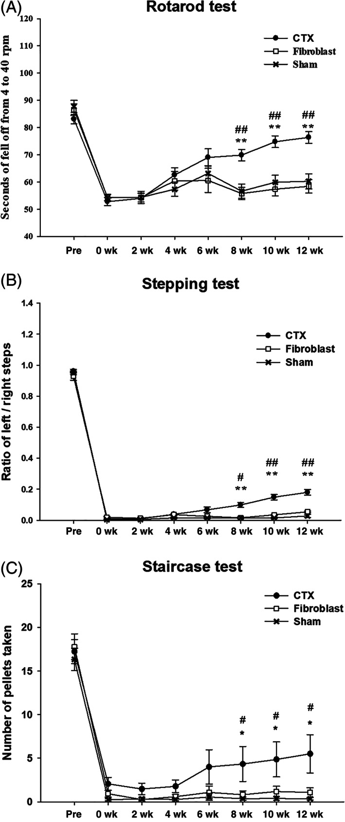
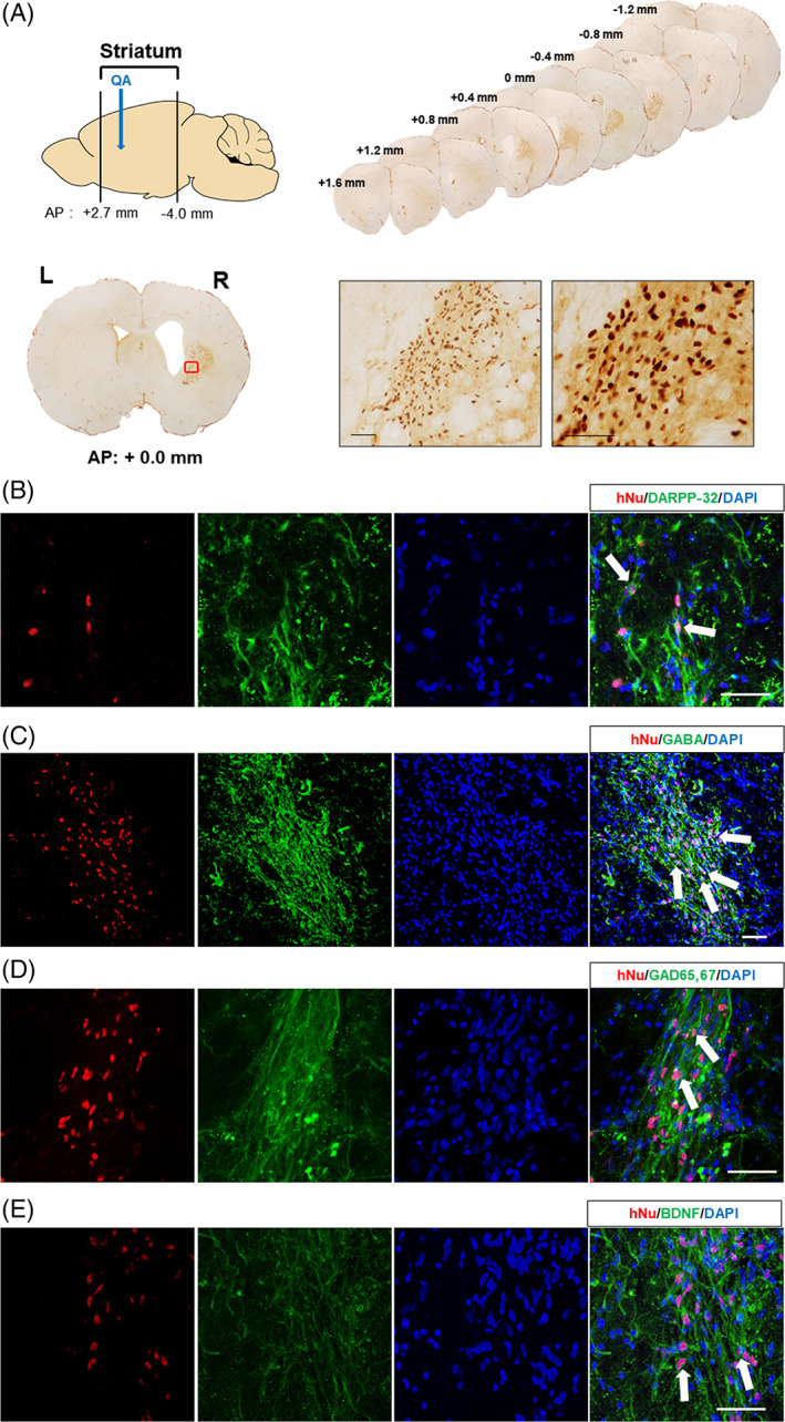
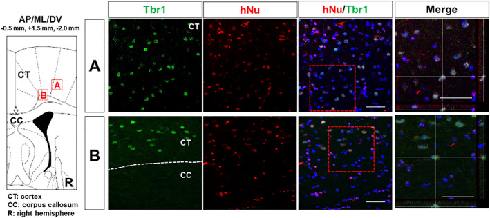
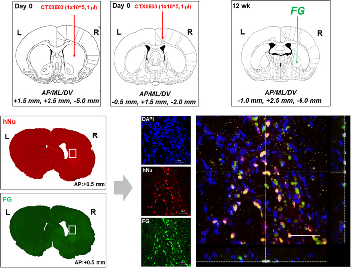
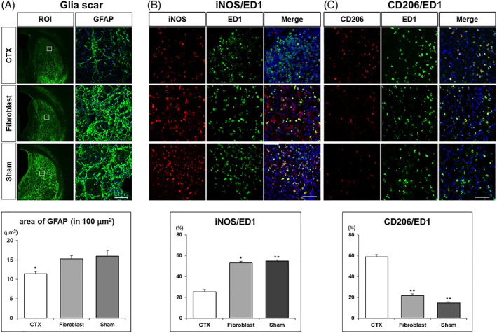
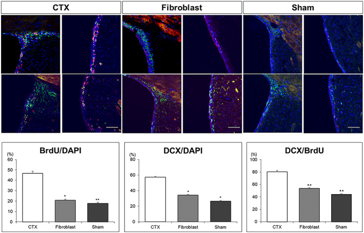
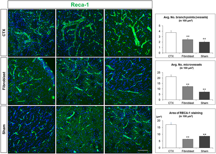
Huntington's disease (HD) is a devastating, autosomal-dominant neurodegenerative disease, for which there are currently no disease-modifying therapies. Clinical trials to replace the damaged striatal medium spiny neurons (MSNs) have been attempted in the past two decades but have met with only limited success. In this study, we investigated whether a clonal, conditionally immortalized neural stem cell line (CTX0E03), which has already shown safety and signals of efficacy in chronic ischemic stroke patients, could rescue deficits seen in an animal model of HD. After CTX0E03 transplantation into the quinolinic acid-lesioned rat model of HD, behavioral changes were measured using the rotarod, stepping, and staircase tests. In vivo differentiation and neuronal connections of the transplanted CTX0E03 cells were evaluated with immunohistochemical staining and retrograde tracing with Fluoro-Gold. We found that transplantation of CTX0E03 gave rise to a significant behavioral improvement compared with the sham- or fibroblast-transplanted group. Transplanted CTX0E03 formed MSNs (DARPP-32) and GABAergic neurons (GABA, GAD65/67) with BDNF expression in the striatum, while cortically transplanted cells formed Tbr1-positive neurons. Using a retrograde label, we also found stable engraftment and connection of the transplanted cells with host brain tissues. CTX0E03 transplantation also reduced glial scar formation and inflammation, as well as increasing endogenous neurogenesis and angiogenesis. Overall, our results demonstrate that CTX0E03, a clinical-grade neural stem cell line, is effective for preclinical test in HD, and, therefore, will be useful for clinical development in the treatment of HD patients.
Overview
The claims and statistics of this paper haven’t been checked yet.
Run the check — free, no account needed. It reads the open-access text and checks the sources it cites.
Integrity signals
Registry
Cites
Statistics
Figures
Verification
Claims the paper makes
The claims and statistics of this paper haven’t been checked yet.
Run the check — free, no account needed. It reads the open-access text and checks the sources it cites.
Claims other papers make about this one
Each is checked against this paper’s own text: does it say what the citing paper says it does?
In the citing paper
“For example, they have used immortalized cell lines as model systems for studying neuronal development and performance recovery in neurological disease models, such as Huntington’s disease [28,29].”
From Cell Immortality: In Vitro Effective Techniques to Achieve and Investigate Its Applications and Challenges (2024) · cites this paper as [29]
Self-check
Percentages, sums and reported differences re-computed from the paper’s own numbers.
The self-check needs the paper’s full text.
Citations
Cites
The choreography of neuroinflammation in Huntington's disease
Crotti, A., & Glass, C. K. (2015) · Trends in Immunology · doi:10.1016/j.it.2015.04.007 · Cited by 276
Human glia can both induce and rescue aspects of disease phenotype in Huntington disease
Benraiss, A., Wang, S., Herrlinger, S., Li, X., Chandler-Militello, D., Mauceri, J., Burm, H. B., Toner, M., Osipovitch, M., Jim Xu, Q., et al. (2016) · Nature Communications · doi:10.1038/ncomms11758 · Cited by 199
Research works printed but not linked: 36
- Pringsheim, 2012, The incidence and prevalence of Huntington's disease: a systematic review and meta-analysis, Mov Disord, 27, 1083, 10.1002/mds.25075 · DOI
- Connor, 2018, Concise review: the use of stem cells for understanding and treating Huntington's disease, Stem Cells, 36, 146, 10.1002/stem.2747 · DOI
- Dunnett, 2004, Cell therapy in Huntington's disease, NeuroRx, 1, 394, 10.1602/neurorx.1.4.394 · DOI
- Kim, 2015, Progress in Stem Cell Transplantation, 77
- Lee, 2005, Intravenous administration of human neural stem cells induces functional recovery in Huntington's disease rat model, Neurosci Res, 52, 243, 10.1016/j.neures.2005.03.016 · DOI
- Ryu, 2004, Proactive transplantation of human neural stem cells prevents degeneration of striatal neurons in a rat model of Huntington disease, Neurobiol Dis, 16, 68, 10.1016/j.nbd.2004.01.016 · DOI
- Johann, 2007, Time of transplantation and cell preparation determine neural stem cell survival in a mouse model of Huntington's disease, Exp Brain Res, 177, 458, 10.1007/s00221-006-0689-y · DOI
- Al-Gharaibeh, 2017, Induced pluripotent stem cell-derived neural stem cell transplantations reduced behavioral deficits and ameliorated neuropathological changes in YAC128 mouse model of Huntington's disease, Front Neurosci, 11, 628, 10.3389/fnins.2017.00628 · DOI
- Stroemer, 2009, The neural stem cell line CTX0E03 promotes behavioral recovery and endogenous neurogenesis after experimental stroke in a dose-dependent fashion, Neurorehabil Neural Repair, 23, 895, 10.1177/1545968309335978 · DOI
- Kalladka, 2016, Human neural stem cells in patients with chronic ischaemic stroke (PISCES): a phase 1, first-in-man study, Lancet, 388, 787, 10.1016/S0140-6736(16)30513-X · DOI
- Hicks, 2013, In vivo and in vitro characterization of the angiogenic effect of CTX0E03 human neural stem cells, Cell Transplant, 22, 1541, 10.3727/096368912X657936 · DOI
- Sinden, 2017, Human neural stem cell therapy for chronic ischemic stroke: charting Progress from laboratory to patients, Stem Cells Dev, 26, 933, 10.1089/scd.2017.0009 · DOI
- Lelos, 1780, Generating Excitotoxic lesion models of Huntington's disease, Methods Mol Biol, 2018, 209
- Pollock, 2006, A conditionally immortal clonal stem cell line from human cortical neuroepithelium for the treatment of ischemic stroke, Exp Neurol, 199, 143, 10.1016/j.expneurol.2005.12.011 · DOI
- Conte-Perales, 2010, Neuroanatomical tracing combined with in situ hybridization: analysis of gene expression patterns within brain circuits of interest, J Neurosci Methods, 194, 28, 10.1016/j.jneumeth.2009.11.021 · DOI
- Wojtowicz, 2006, BrdU assay for neurogenesis in rodents, Nat Protoc, 1, 1399, 10.1038/nprot.2006.224 · DOI
- Hanna, 2007, Treatment of sickle cell anemia mouse model with iPS cells generated from autologous skin, Science, 318, 1920, 10.1126/science.1152092 · DOI · Paper page
- Jeon, 2012, Neuronal properties, in vivo effects, and pathology of a Huntington's disease patient-derived induced pluripotent stem cells, Stem Cells, 30, 2054, 10.1002/stem.1135 · DOI
- Chang, 2013, Therapeutic effect of BDNF-overexpressing human neural stem cells (HB1. F3. BDNF) in a rodent model of middle cerebral artery occlusion, Cell Transplant, 22, 1441, 10.3727/096368912X657323 · DOI
- Takahashi, 2007, Induction of pluripotent stem cells from adult human fibroblasts by defined factors, Cell, 131, 861, 10.1016/j.cell.2007.11.019 · DOI · Paper page
- Schwarcz, 1983, Quinolinic acid: an endogenous metabolite that produces axon-sparing lesions in rat brain, Science, 219, 316, 10.1126/science.6849138 · DOI
- Beal, 1991, Chronic quinolinic acid lesions in rats closely resemble Huntington's disease, J Neurosci, 11, 1649, 10.1523/JNEUROSCI.11-06-01649.1991 · DOI
- Popoli, 1994, Behavioral and electrophysiological correlates of the quinolinic acid rat model of Huntington's disease in rats, Brain Res Bull, 35, 329, 10.1016/0361-9230(94)90109-0 · DOI
- Francis, 2000, Behavior characterization of a model of Huntington's disease in rats, induced by quinolinic acid, Rev Neurol, 30, 1016
- Sanberg, 1989, The quinolinic acid model of Huntington's disease: locomotor abnormalities, Exp Neurol, 105, 45, 10.1016/0014-4886(89)90170-2 · DOI
- McBride, 2004, Human neural stem cell transplants improve motor function in a rat model of Huntington's disease, J Comp Neurol, 475, 211, 10.1002/cne.20176 · DOI
- Rosas, 2008, Cerebral cortex and the clinical expression of Huntington's disease: complexity and heterogeneity, Brain, 131, 1057, 10.1093/brain/awn025 · DOI · Paper page
- Thu, 2010, Cell loss in the motor and cingulate cortex correlates with symptomatology in Huntington's disease, Brain, 133, 1094, 10.1093/brain/awq047 · DOI
- Smith, 2012, Implantation site and lesion topology determine efficacy of a human neural stem cell line in a rat model of chronic stroke, Stem Cells, 30, 785, 10.1002/stem.1024 · DOI
- Amor, 2014, Inflammation in neurodegenerative diseases—an update, Immunology, 142, 151, 10.1111/imm.12233 · DOI · Paper page
- Neumann, 2009, Debris clearance by microglia: an essential link between degeneration and regeneration, Brain, 132, 288, 10.1093/brain/awn109 · DOI · Paper page
- Loov, 2012, Engulfing astrocytes protect neurons from contact-induced apoptosis following injury, PLoS One, 7, 10.1371/journal.pone.0033090 · DOI
- Mosser, 2008, Exploring the full spectrum of macrophage activation, Nat Rev Immunol, 8, 958, 10.1038/nri2448 · DOI · Paper page
- Mills, 2012, M1 and M2 macrophages: oracles of health and disease, Crit Rev Immunol, 32, 463, 10.1615/CritRevImmunol.v32.i6.10 · DOI
- Hassani, 2012, Human neural progenitor cell engraftment increases neurogenesis and microglial recruitment in the brain of rats with stroke, PLoS One, 7, 10.1371/journal.pone.0050444 · DOI
- Reidling, 2018, Human neural stem cell transplantation rescues functional deficits in R6/2 and Q140 Huntington's disease mice, Stem Cell Rep, 10, 58, 10.1016/j.stemcr.2017.11.005 · DOI
Cited by
Practical Use of Immortalized Cells in Medicine: Current Advances and Future Perspectives
Voloshin, N., Tyurin-Kuzmin, P., Karagyaur, M., Akopyan, Z., & Kulebyakin, K. (2023) · International Journal of Molecular Sciences · doi:10.3390/ijms241612716 · Cited by 77
Chalak, M., Hesaraki, M., Mirbahari, S. N., Yeganeh, M., Abdi, S., Rajabi, S., & Hemmatzadeh, F. (2024) · Life · doi:10.3390/life14030417 · Cited by 66
Metadata
- Authors
- Yoon, Y., Kim, H. S., Jeon, I., Noh, J.-E., Park, H. J., Lee, S., Park, I.-H., Stevanato, L., Hicks, C., Corteling, R., Barker, R. A., Sinden, J. D., & Song, J.
- Affiliations
- 1 Department of Biomedical Science, CHA Stem Cell Institute, CHA University, Seongnam-si, Gyeonggi-do, Republic of Korea
- 2 Department of Neurology, CHA Bundang Medical Center, CHA University, Seongnam-si, Gyeonggi-do, Republic of Korea
- 3 Department of Genetics, Yale Stem Cell Center, Yale School of Medicine, New Haven, Connecticut, USA
- 4 ReNeuron, Pencoed Business Park, Bridgend, UK
- 5 John van Geest Cambridge Centre for Brain Repair, Department of Clinical Neuroscience, University of Cambridge, Cambridge, UK
- ORCID
- Song, Jihwan
- Venue
- Stem Cells
- Publisher
- Oxford University Press (OUP)
- Published
- 2020-05-06
- Volume
- 38
- Issue
- 8
- Pages
- 936-947
- ISSN
- 1066-5099
- 1549-4918
- PMC
- PMC7496241
- Cited by
- 34
- References
- 2
- Language
- en
- Licence
- Funders
- National Research Foundation of Korea (NRF-2017M3A9B4061407, NRF‐2017M3A9B4061407)
- iPS Bio, Inc
- ReNeuron, United Kingdom
About these checks
Registry, retraction, Cites and statistics results are computed, not judged, with one exception: a printed reference that no index lists is read once by a model before it counts as a citation error. Claims are extracted by a model and kept only when their quote appears verbatim in the text; each cited source is then checked, with a model’s help, for whether it says what the claim relies on. Those verdicts can be wrong — read the claim’s quote and the cited source before relying on one.
Statistics and arithmetic are recomputed, not judged.
Confidence bands are shown only for fields calibrated on our annotated test set. None is calibrated yet, so no band is shown.
What counts as a claim ›How we measure confidence ›How we check this ›
Are you an author, or do you see a problem with this page? Request a review
