

Aging microglia
Open accessThe DOI matches its Crossref record; 156 of the 156 references it cites were found in a registry or an index.
Abstract
Microglia are the tissue-resident macrophage population of the brain, specialized in supporting the CNS environment and protecting it from endogenous and exogenous insults. Nonetheless, their function declines with age, in ways that remain to be fully elucidated. Given the critical role played by microglia in neurodegenerative diseases, a better understanding of the aging microglia phenotype is an essential prerequisite in designing better preventive and therapeutic strategies. In this review, we discuss the most recent literature on microglia in aging, comparing findings in rodent models and human subjects.
Overview
The claims and statistics of this paper haven’t been checked yet.
Free: it reads the open-access text and checks the first 5 claims that cite a source we can check. Create a free account to check every claim.
Integrity signals
Registry
Cites
Statistics
Figures
Verification
Claims the paper makes
The claims and statistics of this paper haven’t been checked yet.
Free: it reads the open-access text and checks the first 5 claims that cite a source we can check. Create a free account to check every claim.
Claims other papers make about this one
Each is checked against this paper’s own text: does it say what the citing paper says it does?
The cited source says what the claim attributes to it.
In the citing paper
“The contribution of microglia in disease associated with age is dichotomous: protective homeostatic roles described on the one hand, yet dysregulated function and phenotype implicated with greater disease severity or progression on the other hand.32,33,34”
From Characterization of the ocular inflammatory response to AAV reveals divergence by sex and age (2025) · cites this paper as [32]
Self-check
Percentages, sums and reported differences re-computed from the paper’s own numbers.
The self-check needs the paper’s full text.
Citations
Cites
López-Otín, C., Blasco, M. A., Partridge, L., Serrano, M., & Kroemer, G. (2013) · Cell · doi:10.1016/j.cell.2013.05.039 · Cited by 14,982
Neurotoxic reactive astrocytes are induced by activated microglia
Liddelow, S. A., Guttenplan, K. A., Clarke, L. E., Bennett, F. C., Bohlen, C. J., Schirmer, L., Bennett, M. L., Münch, A. E., Chung, W.-S., Peterson, T. C., et al. (2017) · Nature · doi:10.1038/nature21029 · Cited by 7,971
A Unique Microglia Type Associated with Restricting Development of Alzheimer’s Disease
Keren-Shaul, H., Spinrad, A., Weiner, A., Matcovitch-Natan, O., Dvir-Szternfeld, R., Ulland, T. K., David, E., Baruch, K., Lara-Astaiso, D., Toth, B., et al. (2017) · Cell · doi:10.1016/j.cell.2017.05.018 · Cited by 5,493 · cited 5×
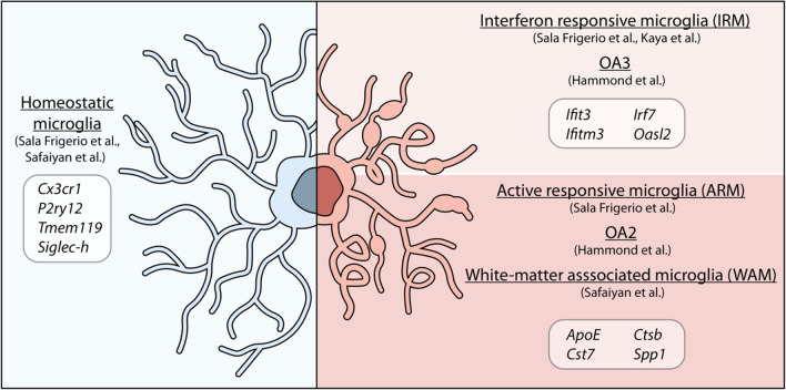
“At transcriptional level, WAM were defined by upregulation of lipid metabolism and phagosome-related genes (Apoe, Cst7, Bm2, Lyz2, Cd63 and Clec7a), cathepsins (Cstb, Ctss, and Ctsz) and MHC II-related genes, further confirming their similarity to the DAM phenotype [59], of which they appear to be a precursory stage […”
BackgroundResultInfluentialThe senescence-associated secretory phenotype: the dark side of tumor suppression
Coppé, J.-P., Desprez, P.-Y., Krtolica, A., & Campisi, J. (2010) · Annual Review of Pathology: Mechanisms of Disease · doi:10.1146/annurev-pathol-121808-102144 · Cited by 5,206 · cited 1×
“Senescence has been linked to organismal aging, as a factor contributing to age-related dysfunctions, nonetheless, it can also be induced in younger tissues by stressful insults, leading to distinctive secretory phenotypes [77].”
BackgroundFate Mapping Analysis Reveals That Adult Microglia Derive from Primitive Macrophages
Ginhoux, F., Greter, M., Leboeuf, M., Nandi, S., See, P., Gokhan, S., Mehler, M. F., Conway, S. J., Ng, L. G., Stanley, E. R., et al. (2010) · Science · doi:10.1126/science.1194637 · Cited by 5,175 · cited 1×
“Microglia have a mesodermal origin and originate from yolk-sac progenitors during embryogenesis; after their early migration and proliferation, they colonize the CNS and selfrenew throughout the lifespan [1, 2], with no or perhaps little contribution from bone marrow-derived monocytes [3], as previously hypothesized […”
BackgroundStructural and functional features of central nervous system lymphatic vessels
Louveau, A., Smirnov, I., Keyes, T. J., Eccles, J. D., Rouhani, S. J., Peske, J. D., Derecki, N. C., Castle, D., Mandell, J. W., Lee, K. S., et al. (2015) · Nature · doi:10.1038/nature14432 · Cited by 4,413 · cited 1×
“The discovery of meningeal lymphatics revealed new concepts of lymphatic drainage in the brain, which plays a central role in draining protein aggregates and, therefore, is particularly important in aging and neurodegeneration [98, 99].”
BackgroundMicroglia Sculpt Postnatal Neural Circuits in an Activity and Complement-Dependent Manner
Schafer, D. P., Lehrman, E. K., Kautzman, A. G., Koyama, R., Mardinly, A. R., Yamasaki, R., Ransohoff, R. M., Greenberg, M. E., Barres, B. A., & Stevens, B. (2012) · Neuron · doi:10.1016/j.neuron.2012.03.026 · Cited by 4,089
Interaction between microbiota and immunity in health and disease
Zheng, D., Liwinski, T., & Elinav, E. (2020) · Cell Research · doi:10.1038/s41422-020-0332-7 · Cited by 3,947 · cited 1×
“It is known that the microbiome influences immune cells [125] and more recent studies have demonstrated that also microglia are sensitive to factors produced by the gut microbiota, which can alter microglial numbers, size, transcriptome, and surveillance functions.”
BackgroundClearance of p16Ink4a-positive senescent cells delays ageing-associated disorders
Baker, D. J., Wijshake, T., Tchkonia, T., LeBrasseur, N. K., Childs, B. G., van de Sluis, B., Kirkland, J. L., & van Deursen, J. M. (2011) · Nature · doi:10.1038/nature10600 · Cited by 3,670 · cited 1×
“, microglia) but affects the whole body, cell-specific targeting strategies, in combination with or without senescence-targeting transgenic mouse models [79, 89, 90], would help clarify the specific role of senescent microglia.”
BackgroundGut microbiota regulate motor deficits and neuroinflammation in a model of Parkinson's disease
Sampson, T. R., Debelius, J. W., Thron, T., Janssen, S., Shastri, G. G., Ilhan, Z. E., Challis, C., Schretter, C. E., Rocha, S., Gradinaru, V., et al. (2016) · Cell · doi:10.1016/j.cell.2016.11.018 · Cited by 3,622 · cited 1×
“Nonetheless, subsequent studies have shown that SCFAs play a detrimental role on microglia in the context of neurodegenerative diseases, such as Parkinson’s disease [128] and Alzheimer’s disease [128], exacerbating disease progression.”
Background
Cited by
Emerging role of senescent microglia in brain aging-related neurodegenerative diseases
Rim, C., You, M., Nahm, M., & Kwon, M. (2024) · Translational Neurodegeneration · doi:10.1186/s40035-024-00402-3 · Cited by 169
“Recent studies propose that senescent microglia, rather than reactive microglia, could be a novel therapeutic target in neurodegenerative diseases [5–7].”
BackgroundLabarta-Bajo, L., & Allen, N. J. (2025) · Neuron · doi:10.1016/j.neuron.2024.12.010 · Cited by 69
The ageing central nervous system in multiple sclerosis: the imaging perspective
Filippi, M., Preziosa, P., Barkhof, F., Ciccarelli, O., Cossarizza, A., Stefano, N. D., Gasperini, C., Geraldes, R., Granziera, C., Haider, L., et al. (2024) · Brain · doi:10.1093/brain/awae251 · Cited by 56
Shin, H. J., Kim, I. S., Choi, S. G., Lee, K., Park, H., Shin, J., Kim, D., Beom, J., Yi, Y. Y., Gupta, D. P., et al. (2024) · Molecular Neurodegeneration · doi:10.1186/s13024-024-00715-x · Cited by 55
“Further confirmation of these findings over the years has corroborated the idea that aged microglia have a priming phenotype that tends to respond more strongly to stimuli, rather than a more inflammatory state in normal condition [48].”
BackgroundHe, C., Chen, B., Yang, H., & Zhou, X. (2025) · Frontiers in Aging Neuroscience · doi:10.3389/fnagi.2025.1554398 · Cited by 48
Metadata
- Authors
- Antignano, I., Liu, Y., Offermann, N., & Capasso, M.
- Affiliations
- German Center for Neurodegenerative Diseases (DE)
- Corresponding author
- Capasso, Melania
- Venue
- Cellular and Molecular Life Sciences
- Published
- 2023-04-21
- Volume
- 80
- Issue
- 5
- Pages
- 126-126
- ISSN
- 1420-682X (ISSN-L)
- 1420-9071
- PMC
- PMC10119228
- OpenAlex
- W4366601967
- Cited by
- 117
- References
- 155
- Citations by year
- 2026: 38 · 2025: 32 · 2024: 42 · 2023: 5
- FWCI
- 3.16
- Type
- journal article
- Language
- en
- Open access
- hybrid
- doi.org
- Licence
- cc-by
- creativecommons.org/licenses/by/4.0 (tdm)
- creativecommons.org/licenses/by/4.0 (vor)
- Locations
- Cellular and Molecular Life Sciences (publishedVersion, cc-by)
- PubMed Central (publishedVersion, cc-by)
- PubMed (publishedVersion)
- DZNE Pub (submittedVersion, cc-by)
- Funders
- Deutsches Zentrum für Neurodegenerative Erkrankungen
- Deutsches Zentrum für Neurodegenerative Erkrankungen e.V. (DZNE) in der Helmholtz-Gemeinschaft
- Topics
- Life Sciences › Neuroscience › Neurology › Neuroinflammation and Neurodegeneration Mechanisms
- Life Sciences › Neuroscience › Neurology › Neurological Disease Mechanisms and Treatments
- Life Sciences › Biochemistry, Genetics and Molecular Biology › Aging › Genetics, Aging, and Longevity in Model Organisms
- Keywords
- microglia, brain aging, neuroinflammation, neurodegenerative diseases, microglial phenotype, tissue-resident macrophages, rodent models, human aging
- MeSH terms
- Brain-Gut Axis, Aging, Animals, Humans, Monocytes, Signal Transduction, Cellular Senescence, Microglia, Oxidative Stress
About these checks
Registry, retraction, Cites and statistics results are computed, not judged, with one exception: a printed reference that no index lists is read once by a model before it counts as a citation error. Claims are extracted by a model and kept only when their quote appears verbatim in the text; each cited source is then checked, with a model’s help, for whether it says what the claim relies on. Those verdicts can be wrong — read the claim’s quote and the cited source before relying on one.
Statistics and arithmetic are recomputed, not judged.
Confidence bands are shown only for fields calibrated on our annotated test set. None is calibrated yet, so no band is shown.
What counts as a claim ›How we measure confidence ›How we check this ›
Are you an author, or do you see a problem with this page? Request a review
How to cite
Cite CoChat for the checks on this page, and the paper for its own findings.
- This page
- CoChat. (2026). Citation check: Aging microglia. https://app.cochat.ai/papers/10.1007/s00018-023-04775-y
- The paper
- Antignano, I., Liu, Y., Offermann, N., & Capasso, M. (2023). Aging microglia. Cellular and Molecular Life Sciences. https://doi.org/10.1007/s00018-023-04775-y
Data sources
Bibliographic data: Semantic Scholar, OpenAlex, Crossref.
Contains information from the Semantic Scholar Academic Graph Datasets, made available under the ODC Attribution License (ODC-BY 1.0).
Semantic Scholar’s data is described in: Kinney, R., et al. (2023). The Semantic Scholar Open Data Platform. arXiv:2301.10140.
