

The DOI matches its Crossref record; 219 of the 220 references it cites were found in a registry or an index.
Quantification of amyloid PET for future clinical use: a state-of-the-art review
Open accessAbstract
Amyloid-β (Aβ) pathology is one of the earliest detectable brain changes in Alzheimer’s disease (AD) pathogenesis. The overall load and spatial distribution of brain Aβ can be determined in vivo using positron emission tomography (PET), for which three fluorine-18 labelled radiotracers have been approved for clinical use. In clinical practice, trained readers will categorise scans as either Aβ positive or negative, based on visual inspection. Diagnostic decisions are often based on these reads and patient selection for clinical trials is increasingly guided by amyloid status. However, tracer deposition in the grey matter as a function of amyloid load is an inherently continuous process, which is not sufficiently appreciated through binary cut-offs alone. State-of-the-art methods for amyloid PET quantification can generate tracer-independent measures of Aβ burden. Recent research has shown the ability of these quantitative measures to highlight pathological changes at the earliest stages of the AD continuum and generate more sensitive thresholds, as well as improving diagnostic confidence around established binary cut-offs. With the recent FDA approval of aducanumab and more candidate drugs on the horizon, early identification of amyloid burden using quantitative measures is critical for enrolling appropriate subjects to help establish the optimal window for therapeutic intervention and secondary prevention. In addition, quantitative amyloid measurements are used for treatment response monitoring in clinical trials. In clinical settings, large multi-centre studies have shown that amyloid PET results change both diagnosis and patient management and that quantification can accurately predict rates of cognitive decline. Whether these changes in management reflect an improvement in clinical outcomes is yet to be determined and further validation work is required to establish the utility of quantification for supporting treatment endpoint decisions. In this state-of-the-art review, several tools and measures available for amyloid PET quantification are summarised and discussed. Use of these methods is growing both clinically and in the research domain. Concurrently, there is a duty of care to the wider dementia community to increase visibility and understanding of these methods.
Overview
The claims and statistics of this paper haven’t been checked yet.
Run the check — free, no account needed. It reads the open-access text and checks the sources it cites.
Integrity signals
Registry
Cites
Statistics
Figures
Verification
Claims the paper makes
The claims and statistics of this paper haven’t been checked yet.
Run the check — free, no account needed. It reads the open-access text and checks the sources it cites.
Claims other papers make about this one
Each is checked against this paper’s own text: does it say what the citing paper says it does?
The source backs part of the claim, not all of it — or backs it for a different population, condition or comparison.
In the citing paper
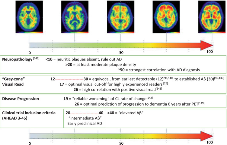
“Moreover, the range from 12 Centiloids to 30 Centiloids has been proposed to reflect the ‘gray zone’ of Aβ deposition24.”
From Plasma p-tau231 and p-tau217 as state markers of amyloid-β pathology in preclinical Alzheimer’s disease (2022) · cites this paper as [24]
Experimental — accuracy not yet measured
Self-check
Percentages, sums and reported differences re-computed from the paper’s own numbers.
The self-check needs the paper’s full text.
Citations
Cites
NIA‐AA Research Framework: Toward a biological definition of Alzheimer's disease
Jack, C. R., Bennett, D. A., Blennow, K., Carrillo, M. C., Dunn, B., Haeberlein, S. B., Holtzman, D. M., Jagust, W., Jessen, F., Karlawish, J., et al. (2018) · Alzheimer's & Dementia · doi:10.1016/j.jalz.2018.02.018 · Cited by 10,537
Alzheimer's disease: the amyloid cascade hypothesis
Hardy, J. A., & Higgins, G. A. (1992) · Science · doi:10.1126/science.1566067 · Cited by 7,484
Scheltens, P., De Strooper, B., Kivipelto, M., Holstege, H., Chételat, G., Teunissen, C. E., Cummings, J., & van der Flier, W. M. (2021) · The Lancet · doi:10.1016/s0140-6736(20)32205-4 · Cited by 5,465
Imaging brain amyloid in Alzheimer's disease with Pittsburgh Compound‐B
Klunk, W. E., Engler, H., Nordberg, A., Wang, Y., Blomqvist, G., Holt, D. P., Bergström, M., Savitcheva, I., Huang, G., Estrada, S., et al. (2004) · Annals of Neurology · doi:10.1002/ana.20009 · Cited by 4,496
Research criteria for the diagnosis of Alzheimer's disease: revising the NINCDS–ADRDA criteria
Dubois, B., Feldman, H. H., Jacova, C., DeKosky, S. T., Barberger-Gateau, P., Cummings, J., Delacourte, A., Galasko, D., Gauthier, S., Jicha, G., et al. (2007) · The Lancet Neurology · doi:10.1016/s1474-4422(07)70178-3 · Cited by 4,411
Jack, C. R., Knopman, D. S., Jagust, W. J., Petersen, R. C., Weiner, M. W., Aisen, P. S., Shaw, L. M., Vemuri, P., Wiste, H. J., Weigand, S. D., et al. (2013) · The Lancet Neurology · doi:10.1016/s1474-4422(12)70291-0 · Cited by 4,396
Phases of Aβ-deposition in the human brain and its relevance for the development of AD
Thal, D. R., Rüb, U., Orantes, M., & Braak, H. (2002) · Neurology · doi:10.1212/wnl.58.12.1791 · Cited by 3,490
Advancing research diagnostic criteria for Alzheimer's disease: the IWG-2 criteria
Dubois, B., Feldman, H. H., Jacova, C., Hampel, H., Molinuevo, J. L., Blennow, K., DeKosky, S. T., Gauthier, S., Selkoe, D., Bateman, R., et al. (2014) · The Lancet Neurology · doi:10.1016/s1474-4422(14)70090-0 · Cited by 3,321
The antibody aducanumab reduces Aβ plaques in Alzheimer’s disease
Sevigny, J., Chiao, P., Bussière, T., Weinreb, P. H., Williams, L., Maier, M., Dunstan, R., Salloway, S., Chen, T., Ling, Y., et al. (2016) · Nature · doi:10.1038/nature19323 · Cited by 3,059
Amyloid deposition as the central event in the aetiology of Alzheimer’s disease
Hardy, J., & Allsop, D. (1991) · Trends in Pharmacological Sciences · doi:10.1016/0165-6147(91)90609-v · Cited by 2,538
Cited by
Sorted by citations
Items 101–200 of 226
Strohl, J. J., Carrión, J., & Huerta, P. T. (2024) · Scientific Reports · doi:10.1038/s41598-024-79259-5 · Cited by 4
Giarratana, A. O. (2025) · Frontiers in Neurology · doi:10.3389/fneur.2025.1637243 · Cited by 4
Collij, L. E., Bischof, G. N., Altomare, D., Bader, I., Battle, M. R., García, D. V., Alves, I. L., Wolz, R., Gismondi, R., Stephens, A. W., et al. (2024) · medRxiv · doi:10.1101/2024.05.22.24307653 · Cited by 4
Self-citationDeep learning-based amyloid PET harmonization to predict cognitive decline in non-demented elderly
Choi, Y. S., Ngam, P. I., Lee, J. R., Hwang, D., Tan, E., Study, F. T. H. A. B., Aging, F. T. A. I. B. A. L. S. O., Initiative, F. T. J. A. D. N., & Initiative, A. D. N. (2024) · Radiology Advances · doi:10.1093/radadv/umae019 · Cited by 3
Polygenic risk discriminates Lewy body dementia from Alzheimer's disease
McKeever, A., Swann, P., Malpetti, M., Donaghy, P. C., Thomas, A. J., Mak, E., Carter, S. F., Tan, J. H. K., Hong, Y. T., Fryer, T. D., et al. (2025) · Alzheimer s & Dementia · doi:10.1002/alz.14381 · Cited by 4
Nasrallah, I. M., Kuo, P. H., Nordberg, A. K., Bohnen, N. I., & Ponisio, M. R. (2025) · American Journal of Roentgenology · doi:10.2214/ajr.24.32325 · Cited by 4
A Question of Origins: Non-neuronal Sources of Amyloid-β
Sasmita, A. O., & Depp, C. (2026) · Neuroscience Bulletin · doi:10.1007/s12264-025-01572-4 · Cited by 4
Centiloids in Amyloid PET: A Practical Guide to Quantitation Interpretation
Grabher, B. J. (2026) · Journal of Nuclear Medicine Technology · doi:10.2967/jnmt.125.271823 · Cited by 3
Functional imaging constrained diffusion for brain PET synthesis from structural MRI
Yu, M., Wu, M., Yue, L., Bozoki, A., & Liu, M. (2026) · Medical Image Analysis · doi:10.1016/j.media.2026.104044 · Cited by 11
Kim, D., Choo, K., Lee, S., Kang, S., Yun, M., & Yang, J. (2025) · European Journal of Nuclear Medicine and Molecular Imaging · doi:10.1007/s00259-025-07132-2 · Cited by 3
Yang, Z. X., Sheng, J., Zhang, Q., Chou, J. T. C., Xin, Y., & Wang, L. (2025) · Scientific Reports · doi:10.1038/s41598-025-01927-x · Cited by 3
Transforming Neurology and Psychiatry
Taha, A., Alassi, A., GJEDDE, A. H., & Wong, D. F. (2023) · PET Clinics · doi:10.1016/j.cpet.2023.06.002 · Cited by 3
Subcortical hemorrhage caused by cerebral amyloid angiopathy compared with hypertensive hemorrhage
Yamada, S. M., Tomita, Y., Iwamoto, N., Takeda, R., Nakane, M., Aso, T., & Takahashi, M. (2023) · Clinical Neurology and Neurosurgery · doi:10.1016/j.clineuro.2023.108076 · Cited by 4
Hartz, S. M., Schindler, S. E., Streitz, M. L., Moulder, K. L., Mozersky, J. T., Wang, G., Xiong, C., & Morris, J. C. (2024) · medRxiv · doi:10.1101/2024.07.16.24310511 · Cited by 3
Veljanoska, E., Gomes, S., Szende, Á., Cabra, A., Munter-Young, R., & Gordois, A. (2025) · Value in Health · doi:10.1016/j.jval.2025.02.011 · Cited by 2
Machine Learning Framework of EEG for binary classification of Early on-set Alzheimer’s Disease
Shyam Sundar, S., Chintanpalli, A., & Rajesh Kumar, M. (2025) · International Conference on Biomedical Engineering and Sustainable Healthcare (ICBMESH) · doi:10.1109/icbmesh66209.2025.11182251 · Cited by 2
Ross, D., Chang, F., Edwards, L., Thomas, K. R., Bangen, K. J., & Initiative, A. D. N. (2025) · The Clinical Neuropsychologist · doi:10.1080/13854046.2025.2570301 · Cited by 2
Amyloid PET Z Score Quantification and Correlation with Visual Semiquantitative Grading
Yadav, D., Knight‐Greenfield, A., Moirano, J., Nordvig, A. S., Salgado, M. W., Hamed, M. A., Lin, M., RoyChoudhury, A., Blum, S., Keil, S. A., et al. (2025) · American Journal of Neuroradiology · doi:10.3174/ajnr.a9079 · Cited by 2
Morooka, S., Fushimi, Y., Okuchi, S., Sakata, A., Yamamoto, T., Nakajima, S., Mitsumoto, K., Itagaki, K., Kubota, M., Shima, A., et al. (2025) · Japanese Journal of Radiology · doi:10.1007/s11604-025-01915-1 · Cited by 2
Nai, Y., O’Donnell, J. L., Tanenbaum, A., Soda, A. K., Jiang, H., Moerlein, S. M., Norris, S. A., Perlmutter, J. S., & Tu, Z. (2025) · Nuclear Medicine and Biology · doi:10.1016/j.nucmedbio.2025.109599 · Cited by 2
SupReMix: Supervised contrastive learning for medical imaging regression with mixup
Wu, Y., Dong, Z., Chen, C., Zhou, W., & Zhou, J. H. (2025) · Medical Image Analysis · doi:10.1016/j.media.2025.103909 · Cited by 5
Chen, T., Hutchison, R. M., Rubel, C. E., Murphy, J. M., Xie, J., O'Gorman, J. T., Dent, G. W., Molenberghs, G., Sormani, M. P., Hendrix, S. B., et al. (2026) · The Journal of Prevention of Alzheimer s Disease · doi:10.1016/j.tjpad.2025.100458 · Cited by 3
Factors associated with discordant visual and quantitative amyloid PET results
Guevara, J. E., Matusz, E. F., Wang, W., DeSimone, J. C., Yawer, B. A., Fiala, J. A., Levy, S., Arias, F., Rayaprolu, S., Barker, W. W., et al. (2026) · Alzheimer s & Dementia Diagnosis Assessment & Disease Monitoring · doi:10.1002/dad2.70241 · Cited by 2
Doroudinia, A., York, B., & Colletti, P. M. (2026) · Journal of Nuclear Medicine Technology · doi:10.2967/jnmt.125.271678 · Cited by 2
Tang, C., Sun, X., Tang, A., Ruan, W., Fang, H., Fang, H., Gai, Y., Liang, Z., Su, Y., Wang, X., et al. (2026) · Radiology · doi:10.1148/radiol.252321 · Cited by 2
Xue, H., Bi, S., Wang, M., Zhu, X., Wang, F., Wang, Y., He, Y., Yan, W., Liu, X., Cui, B., et al. (2026) · Alzheimer s Research & Therapy · doi:10.1186/s13195-026-02071-y · Cited by 2
Balaji, N., Balachandar, R., Algodayan, S., Lilburn, D. M. L., Fraioli, F., & Bomanji, J. B. (2023) · Nuclear Medicine Communications · doi:10.1097/mnm.0000000000001688 · Cited by 2
Predicting cognitive decline in older people by structural and molecular imaging
Teipel, S. J., Tang, Y., & Boccardi, M. (2023) · Current Opinion in Neurology · doi:10.1097/wco.0000000000001172 · Cited by 2
Rhodius‐Meester, H. F. M., van Maurik, I. S., Collij, L. E., van Gils, A. M., Koikkalainen, J. R., Tolonen, A. J., Pijnenburg, Y. A. L., Berkhof, J. H., Barkhof, F., van de Giessen, E., et al. (2024) · PLoS ONE · doi:10.1371/journal.pone.0303111 · Cited by 2
Self-citationUltra-fast [18F]florbetapir PET imaging using the uMI Panorama PET/CT system
Yang, X., Wu, M., Liang, M., Zhang, H., Li, B., Mao, C., Dong, L., Wang, Y., Xing, H., Ren, C., et al. (2024) · EJNMMI Physics · doi:10.1186/s40658-024-00712-5 · Cited by 2
Galvin, J. E., Kleiman, M. J., Estes, P. W., Harris, H. M., & Fung, E. (2025) · Journal of Alzheimer s Disease · doi:10.1177/13872877251314117 · Cited by 2
Kim, J., Choe, Y. S., Park, Y., Kim, Y., Kim, J. P., Jang, H., Kim, H. J., Na, D. L., Cho, S., Moon, S. H., et al. (2023) · Frontiers in Aging Neuroscience · doi:10.3389/fnagi.2023.1124445 · Cited by 2
Data-Driven Image-Based Protocol for Brain PET Image Harmonization
Štokelj, E., Simončič, U., & Initiative, A. D. N. (2025) · Sensors · doi:10.3390/s25134230 · Cited by 1
Human subject study of a transmission-aided attenuation correction for precision PET neuroimaging
Lotfimarangloo, S., Rahman, S. T., Park, M., Fei, B., Badawi, R. D., & Bowen, S. (2025) · EJNMMI Physics · doi:10.1186/s40658-025-00803-x · Cited by 1
Geiger, A. R., Guevara, J. E., King, J. B., Hoffman, J. M., & Duff, K. M. (2025) · Journal of Clinical and Experimental Neuropsychology · doi:10.1080/13803395.2025.2578345 · Cited by 1
Updates of nuclear medicine imaging in neurodegenerative disorders
Huang, S., Lin, K., Hung, G., & Hsiao, I. (2025) · Journal of the Formosan Medical Association · doi:10.1016/j.jfma.2025.11.032 · Cited by 1
Badarchin, M., Otomi, Y., Shinya, T., Otsuka, H., Takaoka, Y., Matsushita, T., Matsubara, T., Fujita, K., Tomioka, Y., Nakataki, M., et al. (2025) · Annals of Nuclear Medicine · doi:10.1007/s12149-025-02146-0 · Cited by 1
Sheng, J., Yang, Z. X., Wang, Y., Zhang, Q., Xin, Y., Song, Y., & Wang, L. (2026) · Communications Medicine · doi:10.1038/s43856-025-01172-3 · Cited by 1
Standardized quantification of [18F]Florbetazine amyloid PET with the Centiloid scale
Wu, M., Liang, M., Mao, C., Dong, L., Ge, Q., Li, Y., Wang, J., Ren, C., Zhang, H., Huang, Z., et al. (2026) · NeuroImage · doi:10.1016/j.neuroimage.2026.121746 · Cited by 1
Beyond CT: Attenuation correction for stand-alone brain PET
Jehl, M., Bonta, M. J., Mikhaylova, E., Ahnen, M. L., Cherry, S. R., Sacco, I., & Fischer, J. (2026) · Zeitschrift für Medizinische Physik · doi:10.1016/j.zemedi.2026.02.001 · Cited by 1
Rajalakshmi, R., M., R. C., Naaz, R., Madhunapantula, S. V., Uthaiah, C. A., & Salimath, P. V. (2026) · Frontiers in Aging · doi:10.3389/fragi.2026.1696711 · Cited by 1
Ha, S., Choi, B., Kwon, S. J., Kang, S., Son, J., Jeong, S. Y., Lee, S.-W., Choe, Y. S., Kim, D., & Hong, C. M. (2026) · European Radiology · doi:10.1007/s00330-026-12687-1 · Cited by 1
APOE and amyloid‐tau pathology in cognitively unimpaired older adults
Morillo, C. A., Zheng, L., Ghanbarian, E., Khorsand, B., Glover, C. M., Grill, J. D., Sajjadi, A., Initiative, A. D. N., & Ezzati, A. (2026) · Alzheimer s & Dementia Diagnosis Assessment & Disease Monitoring · doi:10.1002/dad2.70496 · Cited by 1
Associations of Antibodies Against Several Infections With Alzheimer Disease Neuropathology
Felici, C., Green, R., Warren‐Gash, C., Butt, J., Waterboer, T., Hughes, A. D., Chaturvedi, N., Keshavan, A., Coath, W., Schott, J. M., et al. (2026) · Neurology · doi:10.1212/wnl.0000000000218584 · Cited by 1
Campero-García, L. A., Cantoral-Ceballos, J. A., Martínez-Maldonado, A., Luna‐Muñoz, J., Ontiveros‐Torres, M. Á., & Gutierrez-Rodríguez, A. E. (2022) · Biology · doi:10.3390/biology11081131 · Cited by 1
Patterns of amyloid accumulation in amyloid-negative cases
Alchera, N., Garibotto, V., Tomczyk, S., Treyer, V., Höck, C., Gietl, A. F., Lövblad, K., Scheffler, M., Chincarini, A., Frisoni, G. B., et al. (2023) · Neurobiology of Aging · doi:10.1016/j.neurobiolaging.2023.05.006 · Cited by 2
Self-citationAndersen, A. B. A., Lehel, S., Grove, E. K., Langkjær, N., Fuglø, D., & Huynh, T. H. V. (2024) · Pharmaceuticals · doi:10.3390/ph17020217 · Cited by 1
Mathies, F. L., Heeman, F., Visser, P. J., den Braber, A., Yaqub, M. M., Klutmann, S., Schöll, M., van de Giessen, E., Collij, L. E., & Buchert, R. (2024) · Clinical Nuclear Medicine · doi:10.1097/rlu.0000000000005360 · Cited by 1
Self-citationEstablishing cutoff values for visual amyloid positivity in [18F]flutemetamol PET
Park, Y.-J., Moon, S. Y., Yoon, J., Lee, S. J., & An, Y. (2025) · EJNMMI Research · doi:10.1186/s13550-024-01193-4 · Cited by 1
Pérez-Martínez, D. A. (2023) · Revista de Neurología · doi:10.33588/rn.7606.2023055 · Cited by 1
Linking Amyloid Burden to Prognosis
Masri, A. (2024) · JACC. Cardiovascular imaging · doi:10.1016/j.jcmg.2024.06.002 · Cited by 1
Bhargava, V., Wang, M. S., Chen, Y., Luo, J., Weiner, M. W., Landau, S. M., Jagust, W., Sabbagh, M. N., Su, Y., Reiman, E. M., et al. (2025) · medRxiv · doi:10.1101/2025.09.19.25336123 · Cited by 1
Bartels, P. H., Rougé, S. L. S., Scripter, J. D., Zeng, Z., Estrada‐Tobar, Z. M., Price, J., Jacobi, A. A., Berumen, R. A., Ho, S., Avedisyan, A., et al. (2026) · bioRxiv (Cold Spring Harbor Laboratory) · doi:10.64898/2026.04.25.720803 · Cited by 1
Sun, X., Badachhape, A., Chin, J., Annapragada, A., & Tanifum, E. A. (2023) · bioRxiv (Cold Spring Harbor Laboratory) · doi:10.1101/2023.07.25.550555
Suresh, K., Dahal, E., & Badano, A. G. (2024) · bioRxiv (Cold Spring Harbor Laboratory) · doi:10.1101/2024.01.31.578167
Jiménez-Bonilla, J. F., de Arcocha-Torres, M., Lage, C., López‐García, S., Martínez‐Rodríguez, I., Rodríguez‐Rodríguez, E., Sánchez‐Juan, P., & Quirce, R. (2024) · Research Square · doi:10.21203/rs.3.rs-4000629/v1
Nuclear Medicine Division, A. O. –. U. D. P., Ruffini, L., Zilioli, A., Neurology Division, A. O. –. U. D. P., Cervati, V., Nuclear Medicine Division, A. O. –. U. D. P., Lauretani, F., Misirocchi, F., Neurology Division, A. O. –. U. D. P., Maggio, M. G., et al. (2024) · European Journal of Clinical and Experimental Medicine · doi:10.15584/ejcem.2024.1.20 · Cited by 1
Curiel, R. E., Vaillancourt, D. E., Ortega, A., Crocco, E., Carballea, D., Asken, B. M., Armstrong, M. J., Vélez-Uribe, I., Wang, W.-I., Rosselli, M., et al. (2024) · SSRN Electronic Journal · doi:10.2139/ssrn.4825251
Degree of amyloid-β burden could be indicative of the primary etiology underlying dementia
Collij, L. E., Smith, A. P. L., & Buckley, C. J. (2024) · European Journal of Nuclear Medicine and Molecular Imaging · doi:10.1007/s00259-024-06875-8
Self-citationYang, L., Dong, P., Li, L., Lin, L., & Su, M. (2024) · European Journal of Nuclear Medicine and Molecular Imaging · doi:10.1007/s00259-024-06917-1
Radionuclide Imaging of Regional Cerebral Blood Flow
Buchert, R. (2024) · Springer eBooks · doi:10.1007/978-3-031-61846-8_24
Molecular imaging for senescent cells-targeted therapeutics in aging and age-related diseases
Cen, P. (2025) · Advances in pharmacology · doi:10.1016/bs.apha.2025.01.020
Qi, Z., Guo, W., Cui, M., & Lu, J. (2025) · Radiation Research · doi:10.1667/rade-25-00035.1
Amyloid PET Imaging and Quantification of Amyloid Deposition
Matsuda, H., & Yamao, T. (2025) · Neuromethods · doi:10.1007/978-1-0716-4494-2_5
Central Nervous System Nuclear Imaging
Muhleman, M., Brown, R. K. J., Wong, K. K., Al-Dulaimi, R., & Bohnen, N. I. (2025) · Seminars in Roentgenology · doi:10.1053/j.ro.2025.04.006
Mohlapholi, M. S., Doruyter, A., Mix, M. D., Trauernicht, C. J., Ellmann, A. V. Z., Warwick, J., & Dupont, P. J. (2025) · Nuclear Medicine Communications · doi:10.1097/mnm.0000000000001995
Imaging Markers of Amyloid and Tau in Alzheimer Disease
Tarawneh, R. M. (2025) · Springer eBooks · doi:10.1007/978-3-031-80849-4_20
Shekari, M., Escalante, A. G., Milà‐Alomà, M., Falcón, C., López‐Martos, D., Sánchez‐Benavides, G., Brugulat‐Serrat, A., Niñerola‐Baizán, A., Ashton, N. J., Karikari, T. K., et al. (2025) · medRxiv · doi:10.1101/2025.07.09.25331011
Self-citationBiomarker-driven diagnosis of Alzheimer's disease: Toward precision neurology
Altuna, M. (2025) · Medicina Clínica · doi:10.1016/j.medcli.2025.107105
Li, J., Barinas‐Mitchell, E., Chang, Y., Snitz, B. E., Lopresti, B. J., DelBene, A., Sekikawa, A. M., & López, O. L. (2025) · Journal of Alzheimer s Disease Reports · doi:10.1177/25424823251358278
Mehadji, B., Abdelhafez, Y. G., Alavi, A., Chung, K. J., Karakatsanis, N. A., Omidvari, N., & Nardo, L. (2025) · PET Clinics · doi:10.1016/j.cpet.2025.07.009
Ou, Z., Jiang, C., Liu, Y., Sun, K., & Shen, D. G. (2025) · Lecture notes in computer science · doi:10.1007/978-3-032-05472-2_23
Boiaryna, L., Pieri, L., Chalon, S., Sérrière, S., Bodard, S., Chicheri, G., Chenaf, E., Suzenet, F., Melki, R., Buron, F., et al. (2025) · Pharmaceuticals · doi:10.3390/ph18111638
Biomarker-driven diagnosis of Alzheimer's disease: Toward precision neurology
Altuna, M. (2025) · Medicina Clínica (English Edition) · doi:10.1016/j.medcle.2025.107105
Kamitaka, Y., Sakata, M., Oda, K., Katsuki, A., Kawakami, H., Wagatsuma, K., Kobayashi, M., & Ishii, K. (2025) · EJNMMI Physics · doi:10.1186/s40658-025-00820-w
Additional file 1 of Non-invasive quantification of 18F-florbetaben with total-body EXPLORER PET
Holy, E. N., Li, E. J., Bhattarai, A., Fletcher, E., Alfaro, E. R., Harvey, D. J., Spencer, B. A., Cherry, S. R., DeCarli, C. S., & Fan, A. P. (2024) · Figshare · doi:10.6084/m9.figshare.26707781
Quenon, L., Collij, L. E., García, D. V., Lopes Alves, I., Gérard, T., Malotaux, V., Huyghe, L., Gispert, J. D., Jessen, F., Visser, P. J., et al. (2024) · Figshare · doi:10.6084/m9.figshare.26734833
Self-citationShekari, M., Verwer, E. E., Yaqub, M. M., Daamen, M., Buckley, C. J., Frisoni, G. B., Visser, P. J., Farrar, G., Barkhof, F., Gispert, J. D., et al. (2023) · Figshare · doi:10.6084/m9.figshare.24472521
Self-citationAdditional file 2 of Non-invasive quantification of 18F-florbetaben with total-body EXPLORER PET
Holy, E. N., Li, E. J., Bhattarai, A., Fletcher, E., Alfaro, E. R., Harvey, D. J., Spencer, B. A., Cherry, S. R., DeCarli, C. S., & Fan, A. P. (2024) · Figshare · doi:10.6084/m9.figshare.26707784
Additional file 3 of Non-invasive quantification of 18F-florbetaben with total-body EXPLORER PET
Holy, E. N., Li, E. J., Bhattarai, A., Fletcher, E., Alfaro, E. R., Harvey, D. J., Spencer, B. A., Cherry, S. R., DeCarli, C. S., & Fan, A. P. (2024) · Figshare · doi:10.6084/m9.figshare.26707787
Shekari, M., Verwer, E. E., Yaqub, M. M., Daamen, M., Buckley, C. J., Frisoni, G. B., Visser, P. J., Farrar, G., Barkhof, F., Gispert, J. D., et al. (2024) · Figshare · doi:10.6084/m9.figshare.24472578
Self-citationAdditional file 4 of Non-invasive quantification of 18F-florbetaben with total-body EXPLORER PET
Holy, E. N., Li, E. J., Bhattarai, A., Fletcher, E., Alfaro, E. R., Harvey, D. J., Spencer, B. A., Cherry, S. R., DeCarli, C. S., & Fan, A. P. (2024) · Figshare · doi:10.6084/m9.figshare.26707790
Quenon, L., Collij, L. E., García, D. V., Lopes Alves, I., Gérard, T., Malotaux, V., Huyghe, L., Gispert, J. D., Jessen, F., Visser, P. J., et al. (2024) · Figshare · doi:10.6084/m9.figshare.26734836
Self-citationNuclear Medicine Imaging in the Major Neurodegenerative Disorders
Kim, S., & Seok, J. W. (2025) · Korean Journal of Geriatrics & Gerontology · doi:10.15656/kjgg.2025.26.3.103
Matsuda, H. (2026) · Springer eBooks · doi:10.1007/978-981-95-5222-1_3
Tagmazian, A. A., Schwarz, C., Lange, C., Pitkänen, E., Vuoksimaa, E., & Initiative, T. F. L. D. N. (2026) · bioRxiv (Cold Spring Harbor Laboratory) · doi:10.64898/2026.02.02.703218
Wachi, K., Yamao, T., Miwa, K., Miyaji, N., Akiya, N., Horikawa, T., Masubuchi, M., Kimura, S., & Matsuda, H. (2026) · Annals of Nuclear Medicine · doi:10.1007/s12149-026-02184-2
Felici, C., Green, R., Warren‐Gash, C., Butt, J., Waterboer, T., Hughes, A. D., Chaturvedi, N., Keshavan, A., Coath, W., Schott, J. M., et al. (2026) · medRxiv · doi:10.64898/2026.03.04.26347593
Zhu, Z., Ni, M., Lv, X., Peng, J., Gao, F., Pan, B., Shen, Y., Wang, S., & Shi, J. (2026) · European Journal of Nuclear Medicine and Molecular Imaging · doi:10.1007/s00259-026-07798-2
Jali, V., Li, L., Toh, A. Q., Tanaka, T., Hilal, S., Gyanwali, B., Chen, C. L., & Ngam, P. I. (2026) · NeuroImage · doi:10.1016/j.neuroimage.2026.121844
Zhang, T., Nie, B., Liu, H., Mao, G., OuYang, C., Jiang, X., & Shan, B. (2026) · European Journal of Nuclear Medicine and Molecular Imaging · doi:10.1007/s00259-026-07797-3 · Cited by 1
Gerards, M., Sannemann, L., & Jessen, F. (2026) · The Journal of Prevention of Alzheimer s Disease · doi:10.1016/j.tjpad.2026.100563
Zhou, Z., Gao, J., Wang, Q., Wang, Q., Liu, L., Zhang, X., Li, C., Liu, J., Wei, Y., Zhang, S., et al. (2026) · Alzheimer s Research & Therapy · doi:10.1186/s13195-026-02069-6
Q amyloid: an open platform for quantitative Aβ PET imaging
Marin, B., Piva, F., Villa, C. D., Luchetti, L., Demurtas, G., Maccioni, L., Moretto, M., Lorenzini, L., Battaglini, M., & Veronese, M. (2026) · EANM Innovation · doi:10.1016/j.eanmi.2026.100273
Yamakuni, R., Seino, S., Yamaki, A., Shima, A., Ishikawa, H., Abe, M., Takano, H., Sawamoto, N., Sekino, H., Ishii, S., et al. (2026) · Annals of Nuclear Medicine · doi:10.1007/s12149-026-02218-9
Szujewski, C., Shepherd, T. M., Ghesani, M., Ponisio, M. R., Lavely, W. C., Schramm, G., Bollack, A., Ades‐Aron, B., Lemberskiy, G., & Initiative, T. F. L. D. N. (2026) · medRxiv · doi:10.64898/2026.05.14.26353149
Self-citationInvestigating AD and non-AD [18F]flortaucipir distribution patterns in a memory clinic cohort
Piva, F., Boccalini, C., Ashton, N. J., Zetterberg, H., Blennow, K., Frisoni, G. B., Veronese, M., Garibotto, V., & Peretti, D. E. (2026) · European Journal of Nuclear Medicine and Molecular Imaging · doi:10.1007/s00259-026-07995-z
Self-citationDou, X., Hu, D., Wang, J., Zhong, Y., Yu, C., Zhan, H., Chen, Y., Zhou, R., Zhang, X., Zhang, B., et al. (2026) · European Journal of Nuclear Medicine and Molecular Imaging · doi:10.1007/s00259-026-07989-x · Cited by 1
Yuan, Z., Zhang, J., Zhou, Z., Chen, X., Qi, N., Wang, W., Cheng, X., Lin, Y., & Zhao, J. (2026) · European Radiology Experimental · doi:10.1186/s41747-026-00766-3
Adjunctive quantification for more reproducible amyloid PET interpretation
Shiiba, T., & Yamashita, K. (2026) · European Radiology · doi:10.1007/s00330-026-12692-4
Metadata
- Authors
- Pemberton, H. G., Collij, L. E., Heeman, F., Bollack, A., Shekari, M., Salvadó, G., Alves, I. L., Garcia, D. V., Battle, M., Buckley, C., Stephens, A. W., Bullich, S., Garibotto, V., Barkhof, F., Gispert, J. D., Farrar, G., & on behalf of the AMYPAD consortium
- Affiliations
- UCL Queen Square Institute of Neurology (GB)
- Amsterdam University Medical Centers (NL)
- University College London (GB)
- Universitat Pompeu Fabra (ES)
- Lund University (SE)
- University of Geneva (CH)
- Corresponding author
- Pemberton, Hugh G.
- ORCID
- Venue
- European Journal of Nuclear Medicine and Molecular Imaging
- Published
- 2022-04-07
- Volume
- 49
- Issue
- 10
- Pages
- 3508-3528
- ISSN
- 1619-7070 (ISSN-L)
- 1619-7089
- PMC
- PMC9308604
- OpenAlex
- W4225623397
- Cited by
- 226
- References
- 220
- Citations by year
- 2026: 61 · 2025: 71 · 2024: 64 · 2023: 25 · 2022: 5
- FWCI
- 7.10
- Type
- journal article
- Language
- en
- Open access
- hybrid
- doi.org
- Locations
- European Journal of Nuclear Medicine and Molecular Imaging (publishedVersion, cc-by)
- PubMed Central (publishedVersion, cc-by)
- PubMed (publishedVersion)
- Amsterdam UMC (VU Amsterdam) - Institutional Repository (publishedVersion, other-oa)
- Archive ouverte UNIGE (University of Geneva) (publishedVersion, other-oa)
- Pure Amsterdam UMC (submittedVersion, cc-by)
- UCL Discovery (University College London) (submittedVersion)
- Lund University Publications (Lund University) (submittedVersion)
- Repositori digital de la UPF (Universitat Pompeu Fabra) (submittedVersion, cc-by)
- Data Archiving and Networked Services (DANS) (submittedVersion, other-oa)
- Funders
- Innovative Medicines Initiative (115952)
- Topics
- Health Sciences › Medicine › Psychiatry and Mental health › Dementia and Cognitive Impairment Research
- Life Sciences › Neuroscience › Cellular and Molecular Neuroscience › Alzheimer's disease research and treatments
- Health Sciences › Medicine › Radiology, Nuclear Medicine and Imaging › Medical Imaging Techniques and Applications
- Keywords
- amyloid PET quantification, amyloid-beta, Alzheimer's disease, fluorine-18 radiotracer, aducanumab, treatment response monitoring, cognitive decline prediction, patient selection for clinical trials, amyloid imaging
- MeSH terms
- Alzheimer Disease, Amyloid, Amyloidosis, Brain, Humans, Amyloid beta-Peptides, Positron-Emission Tomography, Cognitive Dysfunction
About these checks
Registry, retraction, Cites and statistics results are computed, not judged, with one exception: a printed reference that no index lists is read once by a model before it counts as a citation error. Claims are extracted by a model and kept only when their quote appears verbatim in the text; each cited source is then checked, with a model’s help, for whether it says what the claim relies on. Those verdicts can be wrong — read the claim’s quote and the cited source before relying on one.
Statistics and arithmetic are recomputed, not judged.
Confidence bands are shown only for fields calibrated on our annotated test set. None is calibrated yet, so no band is shown.
What counts as a claim ›How we measure confidence ›How we check this ›
Are you an author, or do you see a problem with this page? Request a review
