

The DOI matches its Crossref record; 219 of the 220 references it cites were found in a registry or an index.
Quantification of amyloid PET for future clinical use: a state-of-the-art review
Open accessAbstract
Amyloid-β (Aβ) pathology is one of the earliest detectable brain changes in Alzheimer’s disease (AD) pathogenesis. The overall load and spatial distribution of brain Aβ can be determined in vivo using positron emission tomography (PET), for which three fluorine-18 labelled radiotracers have been approved for clinical use. In clinical practice, trained readers will categorise scans as either Aβ positive or negative, based on visual inspection. Diagnostic decisions are often based on these reads and patient selection for clinical trials is increasingly guided by amyloid status. However, tracer deposition in the grey matter as a function of amyloid load is an inherently continuous process, which is not sufficiently appreciated through binary cut-offs alone. State-of-the-art methods for amyloid PET quantification can generate tracer-independent measures of Aβ burden. Recent research has shown the ability of these quantitative measures to highlight pathological changes at the earliest stages of the AD continuum and generate more sensitive thresholds, as well as improving diagnostic confidence around established binary cut-offs. With the recent FDA approval of aducanumab and more candidate drugs on the horizon, early identification of amyloid burden using quantitative measures is critical for enrolling appropriate subjects to help establish the optimal window for therapeutic intervention and secondary prevention. In addition, quantitative amyloid measurements are used for treatment response monitoring in clinical trials. In clinical settings, large multi-centre studies have shown that amyloid PET results change both diagnosis and patient management and that quantification can accurately predict rates of cognitive decline. Whether these changes in management reflect an improvement in clinical outcomes is yet to be determined and further validation work is required to establish the utility of quantification for supporting treatment endpoint decisions. In this state-of-the-art review, several tools and measures available for amyloid PET quantification are summarised and discussed. Use of these methods is growing both clinically and in the research domain. Concurrently, there is a duty of care to the wider dementia community to increase visibility and understanding of these methods.
Overview
The claims and statistics of this paper haven’t been checked yet.
Run the check — free, no account needed. It reads the open-access text and checks the sources it cites.
Integrity signals
Registry
Cites
Statistics
Figures
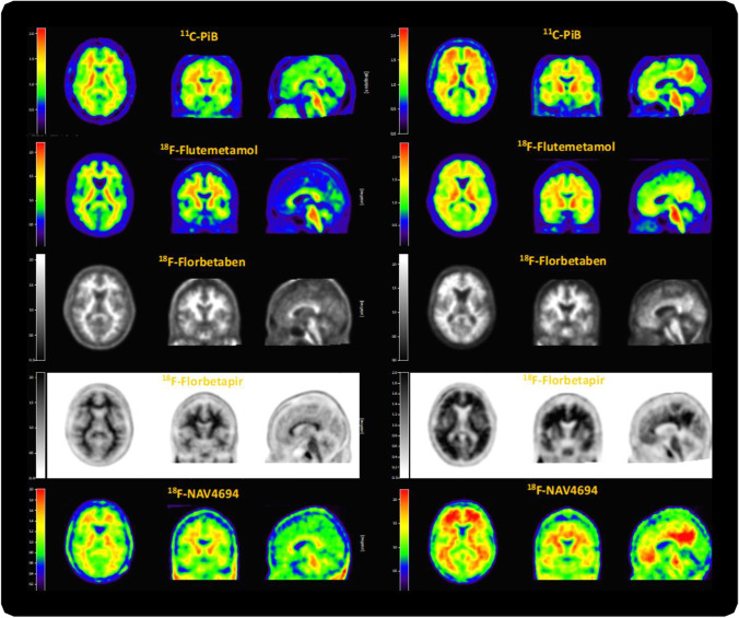
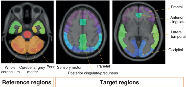
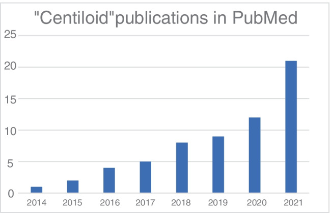
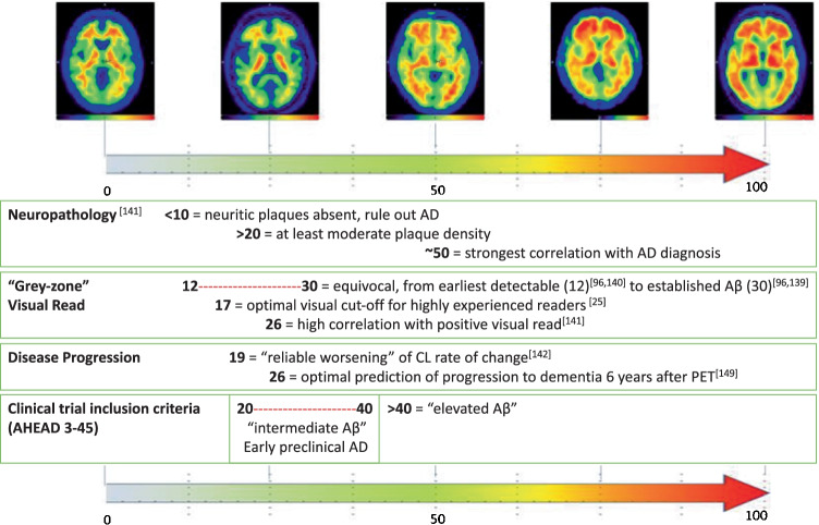
Verification
Claims the paper makes
The claims and statistics of this paper haven’t been checked yet.
Run the check — free, no account needed. It reads the open-access text and checks the sources it cites.
Claims other papers make about this one
Each is checked against this paper’s own text: does it say what the citing paper says it does?
The source backs part of the claim, not all of it — or backs it for a different population, condition or comparison.
In the citing paper
“Moreover, the range from 12 Centiloids to 30 Centiloids has been proposed to reflect the ‘gray zone’ of Aβ deposition24.”
From Plasma p-tau231 and p-tau217 as state markers of amyloid-β pathology in preclinical Alzheimer’s disease (2022) · cites this paper as [24]
Experimental — accuracy not yet measured
Self-check
Percentages, sums and reported differences re-computed from the paper’s own numbers.
The self-check needs the paper’s full text.
Citations
Cites
NIA‐AA Research Framework: Toward a biological definition of Alzheimer's disease
Jack, C. R., Bennett, D. A., Blennow, K., Carrillo, M. C., Dunn, B., Haeberlein, S. B., Holtzman, D. M., Jagust, W., Jessen, F., Karlawish, J., et al. (2018) · Alzheimer's & Dementia · doi:10.1016/j.jalz.2018.02.018 · Cited by 10,537
Alzheimer's disease: the amyloid cascade hypothesis
Hardy, J. A., & Higgins, G. A. (1992) · Science · doi:10.1126/science.1566067 · Cited by 7,484
Scheltens, P., De Strooper, B., Kivipelto, M., Holstege, H., Chételat, G., Teunissen, C. E., Cummings, J., & van der Flier, W. M. (2021) · The Lancet · doi:10.1016/s0140-6736(20)32205-4 · Cited by 5,465
Imaging brain amyloid in Alzheimer's disease with Pittsburgh Compound‐B
Klunk, W. E., Engler, H., Nordberg, A., Wang, Y., Blomqvist, G., Holt, D. P., Bergström, M., Savitcheva, I., Huang, G., Estrada, S., et al. (2004) · Annals of Neurology · doi:10.1002/ana.20009 · Cited by 4,496
Research criteria for the diagnosis of Alzheimer's disease: revising the NINCDS–ADRDA criteria
Dubois, B., Feldman, H. H., Jacova, C., DeKosky, S. T., Barberger-Gateau, P., Cummings, J., Delacourte, A., Galasko, D., Gauthier, S., Jicha, G., et al. (2007) · The Lancet Neurology · doi:10.1016/s1474-4422(07)70178-3 · Cited by 4,411
Jack, C. R., Knopman, D. S., Jagust, W. J., Petersen, R. C., Weiner, M. W., Aisen, P. S., Shaw, L. M., Vemuri, P., Wiste, H. J., Weigand, S. D., et al. (2013) · The Lancet Neurology · doi:10.1016/s1474-4422(12)70291-0 · Cited by 4,396
Phases of Aβ-deposition in the human brain and its relevance for the development of AD
Thal, D. R., Rüb, U., Orantes, M., & Braak, H. (2002) · Neurology · doi:10.1212/wnl.58.12.1791 · Cited by 3,490
Advancing research diagnostic criteria for Alzheimer's disease: the IWG-2 criteria
Dubois, B., Feldman, H. H., Jacova, C., Hampel, H., Molinuevo, J. L., Blennow, K., DeKosky, S. T., Gauthier, S., Selkoe, D., Bateman, R., et al. (2014) · The Lancet Neurology · doi:10.1016/s1474-4422(14)70090-0 · Cited by 3,321
The antibody aducanumab reduces Aβ plaques in Alzheimer’s disease
Sevigny, J., Chiao, P., Bussière, T., Weinreb, P. H., Williams, L., Maier, M., Dunstan, R., Salloway, S., Chen, T., Ling, Y., et al. (2016) · Nature · doi:10.1038/nature19323 · Cited by 3,059
Amyloid deposition as the central event in the aetiology of Alzheimer’s disease
Hardy, J., & Allsop, D. (1991) · Trends in Pharmacological Sciences · doi:10.1016/0165-6147(91)90609-v · Cited by 2,538
Simplified Reference Tissue Model for PET Receptor Studies
Lammertsma, A. A., & Hume, S. P. (1996) · NeuroImage · doi:10.1006/nimg.1996.0066 · Cited by 2,025
Two Phase 3 Trials of Bapineuzumab in Mild-to-Moderate Alzheimer's Disease
Salloway, S., Sperling, R., Fox, N. C., Blennow, K., Klunk, W., Raskind, M., Sabbagh, M., Honig, L. S., Porsteinsson, A. P., Ferris, S., et al. (2014) · New England Journal of Medicine · doi:10.1056/nejmoa1304839 · Cited by 1,960
A/T/N: An unbiased descriptive classification scheme for Alzheimer disease biomarkers
Jack, C. R., Bennett, D. A., Blennow, K., Carrillo, M. C., Feldman, H. H., Frisoni, G. B., Hampel, H., Jagust, W. J., Johnson, K. A., Knopman, D. S., et al. (2016) · Neurology · doi:10.1212/wnl.0000000000002923 · Cited by 1,864
Donanemab in Early Alzheimer’s Disease
Mintun, M. A., Lo, A. C., Duggan Evans, C., Wessels, A. M., Ardayfio, P. A., Andersen, S. W., Shcherbinin, S., Sparks, J., Sims, J. R., Brys, M., et al. (2021) · New England Journal of Medicine · doi:10.1056/nejmoa2100708 · Cited by 1,728 · cited 1×
“Studies so far have demonstrated the added value of this information for both diseasemodifying therapies [112] and in clinical use, especially during the earliest phases of amyloid accumulation where cognitive symptoms are subtle [38, 110, 204–206].”
BackgroundPhase 3 Trials of Solanezumab for Mild-to-Moderate Alzheimer's Disease
Doody, R. S., Thomas, R. G., Farlow, M., Iwatsubo, T., Vellas, B., Joffe, S., Kieburtz, K., Raman, R., Sun, X., Aisen, P. S., et al. (2014) · New England Journal of Medicine · doi:10.1056/nejmoa1312889 · Cited by 1,668
The Centiloid Project: standardizing quantitative amyloid plaque estimation by PET
Klunk, W. E., Koeppe, R. A., Price, J. C., Benzinger, T. L., Devous, M. D., Jagust, W. J., Johnson, K. A., Mathis, C. A., Minhas, D., Pontecorvo, M. J., et al. (2015) · Alzheimer's & Dementia · doi:10.1016/j.jalz.2014.07.003 · Cited by 1,348
Mueller, S. G., Weiner, M. W., Thal, L. J., Petersen, R. C., Jack, C. R., Jagust, W., Trojanowski, J. Q., Toga, A. W., & Beckett, L. (2005) · Alzheimer's & Dementia · doi:10.1016/j.jalz.2005.06.003 · Cited by 1,233
A Phase 3 Trial of Semagacestat for Treatment of Alzheimer's Disease
Doody, R. S., Raman, R., Farlow, M., Iwatsubo, T., Vellas, B., Joffe, S., Kieburtz, K., He, F., Sun, X., Thomas, R. G., et al. (2013) · New England Journal of Medicine · doi:10.1056/nejmoa1210951 · Cited by 1,224
Clinical diagnosis of Alzheimer’s disease: Recommendations of the International Working Group
Dubois, B., Villain, N., Frisoni, G. B., Rabinovici, G. D., Sabbagh, M., Cappa, S., Bejanin, A., Bombois, S., Epelbaum, S., Teichmann, M., et al. (2021) · The Lancet Neurology · doi:10.1016/s1474-4422(21)00066-1 · Cited by 1,051 · cited 1×
“Updated in 2021 [7], the guidelines now further highlight amyloid’s central role in the AD diagnostic process.”
BackgroundTrial of Solanezumab for Mild Dementia Due to Alzheimer’s Disease
Honig, L. S., Vellas, B., Woodward, M., Boada, M., Bullock, R., Borrie, M., Hager, K., Andreasen, N., Scarpini, E., Liu-Seifert, H., et al. (2018) · New England Journal of Medicine · doi:10.1056/nejmoa1705971 · Cited by 1,049
Ellis, K. A., Bush, A. I., Darby, D., Fazio, D. D., Foster, J. K., Hudson, P. J., Lautenschlager, N. T., Lenzo, N. P., Martins, R. N., Maruff, P. T., et al. (2009) · International Psychogeriatrics · doi:10.1017/s1041610209009405 · Cited by 1,005
Defining imaging biomarker cut points for brain aging and Alzheimer's disease
Jack, C. R., Wiste, H. J., Weigand, S. D., Therneau, T. M., Lowe, V. J., Knopman, D. S., Gunter, J. L., Senjem, M. L., Jones, D. T., Kantarci, K., et al. (2017) · Alzheimer's & Dementia · doi:10.1016/j.jalz.2016.08.005 · Cited by 931
Palmqvist, S., Schöll, M., Strandberg, O., Mattsson, N., Stomrud, E., Zetterberg, H., Blennow, K., Landau, S., Jagust, W., & Hansson, O. (2017) · Nature Communications · doi:10.1038/s41467-017-01150-x · Cited by 911
Clark, C. M., Pontecorvo, M. J., Beach, T. G., Bedell, B. J., Coleman, R. E., Doraiswamy, P. M., Fleisher, A. S., Reiman, E. M., Sabbagh, M. N., Sadowsky, C. H., et al. (2012) · The Lancet Neurology · doi:10.1016/s1474-4422(12)70142-4 · Cited by 855
Episodic memory loss is related to hippocampal-mediated -amyloid deposition in elderly subjects
Mormino, E. C., Kluth, J. T., Madison, C. M., Rabinovici, G. D., Baker, S. L., Miller, B. L., Koeppe, R. A., Mathis, C. A., Weiner, M. W., Jagust, W. J., et al. (2009) · Brain · doi:10.1093/brain/awn320 · Cited by 684
Prevalence of Amyloid PET Positivity in Dementia Syndromes: A Meta-analysis
Ossenkoppele, R., Jansen, W. J., Rabinovici, G. D., Knol, D. L., van der Flier, W. M., van Berckel, B. N. M., Scheltens, P., Visser, P. J., Verfaillie, S. C. J., Zwan, M. D., et al. (2015) · JAMA · doi:10.1001/jama.2015.4669 · Cited by 677
Self-citationRabinovici, G. D., Gatsonis, C., Apgar, C., Chaudhary, K., Gareen, I., Hanna, L., Hendrix, J., Hillner, B. E., Olson, C., Lesman-Segev, O. H., et al. (2019) · JAMA · doi:10.1001/jama.2019.2000 · Cited by 674
Vandenberghe, R. R., Laere, K. V., Ivanoiu, A., Salmon, É., Bastin, C., Triau, E., Hasselbalch, S. G., Law, I. G., Andersen, A., Kørner, A., et al. (2010) · Annals of Neurology · doi:10.1002/ana.22068 · Cited by 664
Self-citationKinahan, P. E., & Fletcher, J. W. (2010) · Seminars in Ultrasound, CT and MRI · doi:10.1053/j.sult.2010.10.001 · Cited by 631
Johnson, K. A., Minoshima, S., Bohnen, N. I., Donohoe, K. J., Foster, N. L., Herscovitch, P., Karlawish, J. H., Rowe, C. C., Carrillo, M. C., Hartley, D. M., et al. (2013) · Alzheimer's & Dementia · doi:10.1016/j.jalz.2013.01.002 · Cited by 603
Florbetaben PET imaging to detect amyloid beta plaques in Alzheimer's disease: Phase 3 study
Sabri, O., Sabbagh, M. N., Seibyl, J., Barthel, H., Akatsu, H., Ouchi, Y., Senda, K., Murayama, S., Ishii, K., Takao, M., et al. (2015) · Alzheimer's & Dementia · doi:10.1016/j.jalz.2015.02.004 · Cited by 602
Self-citationA phase III randomized trial of gantenerumab in prodromal Alzheimer’s disease
for the SCarlet RoAD Investigators, Ostrowitzki, S., Lasser, R. A., Dorflinger, E., Scheltens, P., Barkhof, F., Nikolcheva, T., Ashford, E., Retout, S., Hofmann, C., et al. (2017) · Alzheimer's Research & Therapy · doi:10.1186/s13195-017-0318-y · Cited by 567
Self-citationThe Alzheimer's Disease Neuroimaging Initiative positron emission tomography core
Jagust, W. J., Bandy, D., Chen, K., Foster, N. L., Landau, S. M., Mathis, C. A., Price, J. C., Reiman, E. M., Skovronsky, D., Koeppe, R. A., et al. (2010) · Alzheimer s & Dementia · doi:10.1016/j.jalz.2010.03.003 · Cited by 542
Lopresti, B. J., Klunk, W. E., Mathis, C. A., Hoge, J. A., Ziolko, S. K., Lu, X., MELTZER, C. C., Schimmel, K., Tsopelas, N. D., DeKosky, S. T., et al. (2005) · PubMed · Cited by 536
Chételat, G., Arbizu, J., Barthel, H., Garibotto, V., Law, I., Morbelli, S., van de Giessen, E., Agosta, F., Barkhof, F., Brooks, D. J., et al. (2020) · The Lancet Neurology · doi:10.1016/s1474-4422(20)30314-8 · Cited by 526
Self-citationWeiner, M. W., Veitch, D. P., Aisen, P. S. S., Beckett, L. A., Cairns, N. J., Green, R. C., Harvey, D. J., Jack, C. R., Jagust, W. J., Morris, J. C., et al. (2016) · Alzheimer s & Dementia · doi:10.1016/j.jalz.2016.10.006 · Cited by 523
In vivo cortical spreading pattern of tau and amyloid in the Alzheimer disease spectrum
Cho, H., Choi, J. Y., Hwang, M. S., Kim, Y. J., Lee, H. M., Lee, H. S., Lee, J. H., Ryu, Y. H., Lee, M. S., & Lyoo, C. H. (2016) · Annals of Neurology · doi:10.1002/ana.24711 · Cited by 499
Imaging amyloid deposition in Lewy body diseases
Gomperts, S. N., Rentz, D. M., Moran, E., Becker, J. A., Locascio, J. J., Klunk, W. E., Mathis, C. A., Elmaleh, D. R., Shoup, T., Fischman, A. J., et al. (2008) · Neurology · doi:10.1212/01.wnl.0000326146.60732.d6 · Cited by 443
In vivo staging of regional amyloid deposition
Grothe, M. J., Barthel, H., Sepulcre, J., Dyrba, M., Sabri, O., Teipel, S. J., Initiative, F. T. A. D. N., Initiative, A. D. N., Weiner, M., Aisen, P., et al. (2017) · Neurology · doi:10.1212/wnl.0000000000004643 · Cited by 440
Joshi, A. D., Pontecorvo, M. J., Clark, C. M., Carpenter, A. P., Jennings, D. L., Sadowsky, C., Adler, L. P., Kovnat, K. D., Seibyl, J. P., Arora, A., et al. (2012) · Journal of Nuclear Medicine · doi:10.2967/jnumed.111.090340 · Cited by 401
Amsterdam Dementia Cohort: Performing Research to Optimize Care
van der Flier, W. M., & Scheltens, P. (2018) · Journal of Alzheimer s Disease · doi:10.3233/jad-170850 · Cited by 388
Fleisher, A. S. (2011) · Archives of Neurology · doi:10.1001/archneurol.2011.150 · Cited by 364
Landau, S. M., Fero, A., Baker, S. L., Koeppe, R., Mintun, M., Chen, K., Reiman, E. M., & Jagust, W. J. (2015) · Journal of Nuclear Medicine · doi:10.2967/jnumed.114.148981 · Cited by 364
A trial of gantenerumab or solanezumab in dominantly inherited Alzheimer’s disease
Salloway, S., Farlow, M., McDade, E., Clifford, D. B., Wang, G., Llibre-Guerra, J. J., Hitchcock, J. M., Mills, S. L., Santacruz, A. M., Aschenbrenner, A. J., et al. (2021) · Nature Medicine · doi:10.1038/s41591-021-01369-8 · Cited by 359
Jack, C. R., Wiste, H. J., Weigand, S. D., Therneau, T. M., Knopman, D. S., Lowe, V., Vemuri, P., Mielke, M. M., Roberts, R. O., Machulda, M. M., et al. (2017) · The Lancet Neurology · doi:10.1016/s1474-4422(17)30077-7 · Cited by 333
La Joie, R., Ayakta, N., Seeley, W. W., Borys, E., Boxer, A. L., DeCarli, C., Doré, V., Grinberg, L. T., Huang, E., Hwang, J., et al. (2019) · Alzheimer's & Dementia · doi:10.1016/j.jalz.2018.09.001 · Cited by 317
Ossenkoppele, R., Smith, R., Mattsson-Carlgren, N., Groot, C., Leuzy, A., Strandberg, O., Palmqvist, S., Olsson, T., Jögi, J., Stormrud, E., et al. (2021) · JAMA Neurology · doi:10.1001/jamaneurol.2021.1858 · Cited by 311
Amyloid PET imaging in Alzheimer’s disease: a comparison of three radiotracers
Initiative, T. A. D. N., Landau, S. M., Thomas, B. A., Thurfjell, L., Schmidt, M. E., Margolin, R. A., Mintun, M. A., Pontecorvo, M. J., Baker, S. L., & Jagust, W. J. (2014) · European Journal of Nuclear Medicine and Molecular Imaging · doi:10.1007/s00259-014-2753-3 · Cited by 306
The Alzheimer's Disease Neuroimaging Initiative 2 PET Core: 2015
Jagust, W. J., Landau, S. M., Koeppe, R. A., Reiman, E. M., Chen, K., Mathis, C. A., Price, J. C., Foster, N. L., & Wang, A. Y. (2015) · Alzheimer s & Dementia · doi:10.1016/j.jalz.2015.05.001 · Cited by 302
Jack, C. R., Therneau, T. M., Weigand, S. D., Wiste, H. J., Knopman, D. S., Vemuri, P., Lowe, V. J., Mielke, M. M., Roberts, R. O., Machulda, M. M., et al. (2019) · JAMA Neurology · doi:10.1001/jamaneurol.2019.1971 · Cited by 300
In vivo staging of regional amyloid deposition
Grothe, M. J., Barthel, H., Sepulcre, J., Dyrba, M., Sabri, O., Teipel, S. J., Stern, Y., & Initiative, A. D. N. (2022) · DZNE Pub · doi:10.7916/0vj1-4c02 · Cited by 294
Prevalence Estimates of Amyloid Abnormality Across the Alzheimer Disease Clinical Spectrum
Jansen, W. J., Janssen, O., Tijms, B. M., Vos, S. J. B., Ossenkoppele, R., Visser, P. J., Amyloid Biomarker Study Group, Aarsland, D., Alcolea, D., Altomare, D., et al. (2022) · JAMA Neurology · doi:10.1001/jamaneurol.2021.5216 · Cited by 290
Using PET with 18F-AV-45 (florbetapir) to quantify brain amyloid load in a clinical environment
Camus, V., Payoux, P., Barré, L., Desgranges, B., Voisin, T., Tauber, C., Joie, R. L., Tafani, M., Hommet, C., Chételat, G., et al. (2012) · European Journal of Nuclear Medicine and Molecular Imaging · doi:10.1007/s00259-011-2021-8 · Cited by 257
Partial volume correction in quantitative amyloid imaging
Su, Y., Blazey, T. M., Snyder, A. Z., Raichle, M. E., Marcus, D. S., Ances, B. M., Bateman, R. J., Cairns, N. J., Aldea, P., Cash, L., et al. (2015) · NeuroImage · doi:10.1016/j.neuroimage.2014.11.058 · Cited by 247
Validation of amyloid PET positivity thresholds in centiloids: a multisite PET study approach
Initiative, F. T. A. D. N., Royse, S. K., Minhas, D. S., Lopresti, B. J., Murphy, A., Ward, T., Koeppe, R. A., Bullich, S., DeSanti, S., Jagust, W. J., et al. (2021) · Alzheimer's Research & Therapy · doi:10.1186/s13195-021-00836-1 · Cited by 237
Self-citationStaging β -Amyloid Pathology With Amyloid Positron Emission Tomography
Mattsson, N., Palmqvist, S., Stomrud, E., Vogel, J., & Hansson, O. (2019) · JAMA Neurology · doi:10.1001/jamaneurol.2019.2214 · Cited by 237
Navitsky, M., Joshi, A. D., Kennedy, I., Klunk, W. E., Rowe, C. C., Wong, D. F., Pontecorvo, M. J., Mintun, M. A., & Devous, M. D. (2018) · Alzheimer's & Dementia · doi:10.1016/j.jalz.2018.06.1353 · Cited by 229
Thurfjell, L., Lilja, J., Lundqvist, R., Buckley, C. J., Smith, A. P. L., Vandenberghe, R. R., & Sherwin, P. F. (2014) · Journal of Nuclear Medicine · doi:10.2967/jnumed.114.142109 · Cited by 225
Self-citationAducanumab produced a clinically meaningful benefit in association with amyloid lowering
Cummings, J., Aisen, P., Lemere, C., Atri, A., Sabbagh, M., & Salloway, S. (2021) · Alzheimer's Research & Therapy · doi:10.1186/s13195-021-00838-z · Cited by 224
Klein, G., Delmar, P., Voyle, N., Rehal, S., Hofmann, C., Abi-Saab, D., Andjelkovic, M., Ristic, S., Wang, G., Bateman, R., et al. (2019) · Alzheimer's Research & Therapy · doi:10.1186/s13195-019-0559-z · Cited by 212
Fleisher, A. S., Chen, K., Liu, X., Ayutyanont, N., Roontiva, A., Thiyyagura, P., Protas, H., Joshi, A. D., Sabbagh, M., Sadowsky, C. H., et al. (2013) · Neurobiology of Aging · doi:10.1016/j.neurobiolaging.2012.04.017 · Cited by 202
Comparison of 11C-PiB and 18F-florbetaben for Aβ imaging in ageing and Alzheimer’s disease
Villemagne, V. L., Mulligan, R. S., Pejoska, S., Ong, K. T., Jones, G. R., O'Keefe, G. J., Chan, J. G., Young, K., Tochon‐Danguy, H., Masters, C. L., et al. (2012) · European Journal of Nuclear Medicine and Molecular Imaging · doi:10.1007/s00259-012-2088-x · Cited by 197
Rowe, C. C., Pejoska, S., Mulligan, R. S., Jones, G. R., Chan, J. G., Svensson, S., Cselényi, Z., Masters, C. L., & Villemagne, V. L. (2013) · Journal of Nuclear Medicine · doi:10.2967/jnumed.112.114785 · Cited by 195
Centiloid cut-off values for optimal agreement between PET and CSF core AD biomarkers
for the Alzheimer’s Disease Neuroimaging Initiative, F. T. A. S., Salvadó, G., Molinuevo, J. L., Brugulat-Serrat, A., Falcon, C., Grau-Rivera, O., Suárez-Calvet, M., Pavia, J., Niñerola-Baizán, A., Perissinotti, A., et al. (2019) · Alzheimer's Research & Therapy · doi:10.1186/s13195-019-0478-z · Cited by 195
Self-citationAmadoru, S., Doré, V., McLean, C. A., Hinton, F., Shepherd, C. E., Halliday, G. M., Leyton, C. E., Yates, P. A., Hodges, J. R., Masters, C. L., et al. (2020) · Alzheimer's Research & Therapy · doi:10.1186/s13195-020-00587-5 · Cited by 181
18F-Florbetaben PET beta-amyloid binding expressed in Centiloids
Rowe, C. C., Doré, V., Jones, G. R., Baxendale, D., Mulligan, R. S., Bullich, S., Stephens, A. W., Santi, S. D., Masters, C. L., Dinkelborg, L. M., et al. (2017) · European Journal of Nuclear Medicine and Molecular Imaging · doi:10.1007/s00259-017-3749-6 · Cited by 178
Self-citationForward to the Past: The Case for Quantitative PET Imaging
Lammertsma, A. A. (2017) · Journal of Nuclear Medicine · doi:10.2967/jnumed.116.188029 · Cited by 177
Implementing the centiloid transformation for 11C-PiB and β-amyloid 18F-PET tracers using CapAIBL
Bourgeat, P., Doré, V., Fripp, J., Ames, D. J., Masters, C. L., Salvado, O., Villemagne, V. L., & Rowe, C. C. (2018) · NeuroImage · doi:10.1016/j.neuroimage.2018.08.044 · Cited by 158
A phase 3 trial of IV immunoglobulin for Alzheimer disease
Relkin, N. R., Thomas, R. G., Rissman, R. A., Brewer, J. B., Rafii, M. S., van Dyck, C. H., Jack, C. R., Sano, M., Knopman, D. S., Raman, R., et al. (2017) · Neurology · doi:10.1212/wnl.0000000000003904 · Cited by 157
de Wilde, A., van der Flier, W. M., Pelkmans, W., Bouwman, F. H., Verwer, J. H., Groot, C., van Buchem, M. M., Zwan, M. D., Ossenkoppele, R., Yaqub, M. M., et al. (2018) · JAMA Neurology · doi:10.1001/jamaneurol.2018.1346 · Cited by 154
Self-citationAmyloid-β 11 C-PiB-PET imaging results from 2 randomized bapineuzumab phase 3 AD trials
Liu, E., Schmidt, M. E., Margolin, R., Sperling, R., Koeppe, R., Mason, N. S., Klunk, W. E., Mathis, C. A., Salloway, S., Fox, N. C., et al. (2015) · Neurology · doi:10.1212/wnl.0000000000001877 · Cited by 151
Roberts, B. R., Lind, M., Wagen, A. Z., Rembach, A., Frugier, T., Li, Q.-X., Ryan, T. M., McLean, C. A., Doecke, J. D., Rowe, C. C., et al. (2017) · Brain · doi:10.1093/brain/awx057 · Cited by 143
Comparison of Pittsburgh compound B and florbetapir in cross‐sectional and longitudinal studies
Su, Y., Flores, S. E., Wang, G., Hornbeck, R. C., Speidel, B. A., Joseph‐Mathurin, N., Vlassenko, A. G., Gordon, B. A., Koeppe, R. A., Klunk, W. E., et al. (2019) · Alzheimer s & Dementia Diagnosis Assessment & Disease Monitoring · doi:10.1016/j.dadm.2018.12.008 · Cited by 138
Standardized Expression of 18F-NAV4694 and 11C-PiB β-Amyloid PET Results with the Centiloid Scale
Rowe, C. C., Jones, G. R., Doré, V., Pejoska, S., Margison, L., Mulligan, R. S., Chan, J. G., Young, K., & Villemagne, V. L. (2016) · Journal of Nuclear Medicine · doi:10.2967/jnumed.115.171595 · Cited by 137
Leuzy, A., Chiotis, K., Hasselbalch, S. G., Rinne, J. O., de Mendonça, A., Otto, M., Lleó, A., Castelo-Branco, M. S., Santana, I. J., Johansson, J. J., et al. (2016) · Brain · doi:10.1093/brain/aww160 · Cited by 135
The Health & Aging Brain among Latino Elders (HABLE) study methods and participant characteristics
O’Bryant, S. E., Johnson, L. A., Barber, R. C., Braskie, M. N., Christian, B. T., Hall, J. R., Hazra, N., King, K. S., Kothapalli, D., Large, S., et al. (2021) · Alzheimer s & Dementia Diagnosis Assessment & Disease Monitoring · doi:10.1002/dad2.12202 · Cited by 134
Farrell, M. E., Jiang, S., Schultz, A. P., Properzi, M. J., Price, J. C., Becker, J. A., Jacobs, H. I. L., Hanseeuw, B. J., Rentz, D. M., Villemagne, V. L., et al. (2020) · Neurology · doi:10.1212/wnl.0000000000011214 · Cited by 132
Regional amyloid accumulation and cognitive decline in initially amyloid-negative adults
Farrell, M. E., Chen, X., Rundle, M. M., Chan, M. Y., Wig, G. S., & Park, D. C. (2018) · Neurology · doi:10.1212/wnl.0000000000006469 · Cited by 130
Battle, M. R., Pillay, L. C., Lowe, V. J., Knopman, D. S., Kemp, B. J., Rowe, C. C., Doré, V., Villemagne, V. L., & Buckley, C. J. (2018) · EJNMMI Research · doi:10.1186/s13550-018-0456-7 · Cited by 130
Self-citationUtilizing the Centiloid scale in cross-sectional and longitudinal PiB PET studies
Su, Y., Flores, S., Hornbeck, R. C., Speidel, B., Vlassenko, A. G., Gordon, B. A., Koeppe, R. A., Klunk, W. E., Xiong, C., Morris, J. C., et al. (2018) · NeuroImage: Clinical · doi:10.1016/j.nicl.2018.04.022 · Cited by 126
Schmidt, M. E., Chiao, P., Klein, G., Matthews, D. C., Thurfjell, L., Cole, P. E., Margolin, R. A., Landau, S. M., Foster, N. L., Mason, N. S., et al. (2014) · Alzheimer s & Dementia · doi:10.1016/j.jalz.2014.09.004 · Cited by 125
Bullich, S., Seibyl, J. P., Catafau, A. M., Jovalekic, A., Koglin, N., Barthel, H., Sabri, O., & Santi, S. D. (2017) · NeuroImage Clinical · doi:10.1016/j.nicl.2017.04.025 · Cited by 123
Self-citationJ-ADNI Study Group, Yamane, T., Ishii, K., Sakata, M., Ikari, Y., Nishio, T., Ishii, K., Kato, T., Ito, K., & Senda, M. (2016) · European Journal of Nuclear Medicine and Molecular Imaging · doi:10.1007/s00259-016-3591-2 · Cited by 121
Diagnostic impact of [18F]flutemetamol PET in early-onset dementia
Zwan, M. D., Bouwman, F. H., Konijnenberg, E., van der Flier, W. M., Lammertsma, A. A., Verhey, F. R. J., Aalten, P., van Berckel, B. N. M., & Scheltens, P. (2017) · Alzheimer's Research & Therapy · doi:10.1186/s13195-016-0228-4 · Cited by 118
Is amyloid-β harmful to the brain? Insights from human imaging studies
Jagust, W. (2016) · Brain · doi:10.1093/brain/awv326 · Cited by 110
Diagnostic accuracy of CSF Ab42 and florbetapir PET for Alzheimer's disease
Mattsson, N., Insel, P. S., Landau, S. M., Jagust, W., Donohue, M., Shaw, L. M. J., Trojanowski, J. Q., Zetterberg, H., Blennow, K., Weiner, M. W., et al. (2014) · Annals of Clinical and Translational Neurology · doi:10.1002/acn3.81 · Cited by 108
Seibyl, J. P., Catafau, A. M., Barthel, H., Ishii, K., Rowe, C. C., Leverenz, J. B., Ghetti, B. F., Ironside, J. W., Takao, M., Akatsu, H., et al. (2016) · Journal of Nuclear Medicine · doi:10.2967/jnumed.115.161927 · Cited by 108
Self-citationATN profiles among cognitively normal individuals and longitudinal cognitive outcomes
Soldan, A., Pettigrew, C. A., Fagan, A. M., Schindler, S. E., Moghekar, A., Fowler, C. J., Li, Q.-X., Collins, S. J., Carlsson, C. M., Asthana, S., et al. (2019) · Neurology · doi:10.1212/wnl.0000000000007248 · Cited by 107
Multitracer model for staging cortical amyloid deposition using PET imaging
Collij, L. E., Heeman, F., Salvadó, G., Ingala, S., Altomare, D., de Wilde, A., Konijnenberg, E., van Buchem, M. M., Yaqub, M. M., Markiewicz, P. J., et al. (2020) · Neurology · doi:10.1212/wnl.0000000000010256 · Cited by 103
Self-citationRevolutionizing Alzheimer's disease and clinical trials through biomarkers
Mattsson, N., Carrillo, M. C., Dean, R. A., Devous, M. D., Nikolcheva, T., Pesini, P., Salter, H., Potter, W. Z., Sperling, R. A., Bateman, R. J., et al. (2015) · Alzheimer s & Dementia Diagnosis Assessment & Disease Monitoring · doi:10.1016/j.dadm.2015.09.001 · Cited by 102
Clinical Use and Utility of Amyloid Imaging
Barthel, H., & Sabri, O. (2017) · Journal of Nuclear Medicine · doi:10.2967/jnumed.116.185017 · Cited by 102
Cohen, A. D., Mowrey, W. B., Weissfeld, L. A., Aizenstein, H. J., McDade, E., Mountz, J. M., Nebes, R. D., Saxton, J. A., Snitz, B. E., DeKosky, S. T., et al. (2013) · NeuroImage · doi:10.1016/j.neuroimage.2013.01.015 · Cited by 99
Brooks, D. J. (2009) · Movement Disorders · doi:10.1002/mds.22581 · Cited by 97
Relationship between amyloid and tau levels and its impact on tau spreading
Doré, V., Krishnadas, N., Bourgeat, P., Huang, K., Li, S., Burnham, S. C., Masters, C. L., Fripp, J., Villemagne, V. L., & Rowe, C. C. (2021) · European Journal of Nuclear Medicine and Molecular Imaging · doi:10.1007/s00259-021-05191-9 · Cited by 93
Doré, V., Bullich, S., Rowe, C. C., Bourgeat, P., Konate, S., Sabri, O., Stephens, A. W., Barthel, H., Fripp, J., Masters, C. L., et al. (2019) · Alzheimer's & Dementia · doi:10.1016/j.jalz.2019.02.005 · Cited by 93
Self-citationHanseeuw, B. J., Malotaux, V., Dricot, L., Quenon, L., Sznajer, Y., Cerman, J., Woodard, J. L., Buckley, C. J., Farrar, G., Ivanoiu, A., et al. (2020) · European Journal of Nuclear Medicine and Molecular Imaging · doi:10.1007/s00259-020-04942-4 · Cited by 92
Self-citationIngala, S., de Boer, C., Masselink, L. A., Vergari, I., Lorenzini, L., Blennow, K., Chételat, G., Perri, C. D., Ewers, M., van der Flier, W. M., et al. (2021) · Alzheimer s & Dementia · doi:10.1002/alz.12292 · Cited by 88
Self-citationSalloway, S., Gamez, J. E., Singh, U., Sadowsky, C. H., Villena, T., Sabbagh, M. N., Beach, T. G., Duara, R., Fleisher, A. S., Frey, K. A., et al. (2017) · Alzheimer's & Dementia: Diagnosis, Assessment & Disease Monitoring · doi:10.1016/j.dadm.2017.06.001 · Cited by 87
Self-citationA Comparison of Partial Volume Correction Techniques for Measuring Change in Serial Amyloid PET SUVR
Schwarz, C. G., Gunter, J. L., Lowe, V. J., Weigand, S. D., Vemuri, P., Senjem, M. L., Petersen, R. C., Knopman, D. S., & Jack, C. R. (2018) · Journal of Alzheimer s Disease · doi:10.3233/jad-180749 · Cited by 85
Resection Probability Maps for Quality Assessment of Glioma Surgery without Brain Location Bias
Hamer, P. C. D. W., Hendriks, E. J., Mandonnet, E., Barkhof, F., Zwinderman, A. H., & Duffau, H. (2013) · PLoS ONE · doi:10.1371/journal.pone.0073353 · Cited by 84
Self-citationRinne, J. O., Wong, D. F., Wolk, D. A., Leinonen, V., Arnold, S. E., Buckley, C. J., Smith, A. P. L., McLain, R. W., Sherwin, P. F., Farrar, G., et al. (2012) · Acta Neuropathologica · doi:10.1007/s00401-012-1051-z · Cited by 84
Self-citationvan der Kall, L. M., Truong, T. H., Burnham, S. C., Doré, V., Mulligan, R. S., Bozinovski, S., Lamb, F., Bourgeat, P., Fripp, J., Schultz, S. A., et al. (2020) · Neurology · doi:10.1212/wnl.0000000000011222 · Cited by 84
Haeberlein, S. B., von Hehn, C., Tian, Y., Chalkias, S., Muralidharan, K. K., Chen, T., Wu, S., Skordos, L., Nisenbaum, L., Rajagovindan, R., et al. (2020) · Alzheimer's & Dementia · doi:10.1002/alz.047259 · Cited by 81
Applying the ATN scheme in a memory clinic population
Altomare, D., de Wilde, A., Ossenkoppele, R., Pelkmans, W., Bouwman, F. H., Groot, C., van Maurik, I. S., Zwan, M. D., Yaqub, M. M., Barkhof, F., et al. (2019) · Neurology · doi:10.1212/wnl.0000000000008361 · Cited by 81
Self-citationPemberton, H., Zaki, L. A. M., Goodkin, O., Das, R., Steketee, R. M. E., Barkhof, F., & Vernooij, M. W. (2021) · Neuroradiology · doi:10.1007/s00234-021-02746-3 · Cited by 80
Self-citationSchreiber, S., Landau, S. M., Fero, A., Schreiber, F., & Jagust, W. J. (2015) · JAMA Neurology · doi:10.1001/jamaneurol.2015.1633 · Cited by 79
Whittington, A., Sharp, D. J., & Gunn, R. N. (2017) · Journal of Nuclear Medicine · doi:10.2967/jnumed.117.194720 · Cited by 78
Chiotis, K., Saint‐Aubert, L., Boccardi, M., Gietl, A. F., Picco, A., Varrone, A., Garibotto, V., Herholz, K., Nobili, F. M., Nordberg, A. K., et al. (2017) · Neurobiology of Aging · doi:10.1016/j.neurobiolaging.2016.07.012 · Cited by 75
Self-citationReliable measurements of brain atrophy in individual patients with multiple sclerosis
Smeets, D., Ribbens, A., Sima, D. M., Cambron, M., Horáková, D., Jain, S., Maertens, A., Vlierberghe, E. V., Terzopoulos, V., Vanbinst, A., et al. (2016) · Brain and Behavior · doi:10.1002/brb3.518 · Cited by 74
(ADNI), T. A. D. N. I., Bouallègue, F. B., Mariano‐Goulart, D., & Payoux, P. (2017) · Alzheimer s Research & Therapy · doi:10.1186/s13195-017-0260-z · Cited by 73
Computer-aided imaging analysis in acute ischemic stroke – background and clinical applications
Mokli, Y., Pfaff, J. A. R., dos Santos, D. P., Herweh, C., & Nagel, S. (2019) · Neurological Research and Practice · doi:10.1186/s42466-019-0028-y · Cited by 72
Collij, L. E., Salvadó, G., Shekari, M., Alves, I. L., Reimand, J., Wink, A. M., Zwan, M. D., Niñerola‐Baizán, A., Perissinotti, A., Scheltens, P., et al. (2021) · European Journal of Nuclear Medicine and Molecular Imaging · doi:10.1007/s00259-020-05174-2 · Cited by 71
Self-citationEarly detection of amyloid load using 18F-florbetaben PET
Bullich, S., Roé-Vellvé, N., Marquié, M., Landau, S. M., Barthel, H., Villemagne, V. L., Sanabria, Á., Tartari, J. P., Sotolongo-Grau, O., Doré, V., et al. (2021) · Alzheimer's Research & Therapy · doi:10.1186/s13195-021-00807-6 · Cited by 71
Self-citationBuckley, C. J., Sherwin, P. F., Smith, A. P. L., Wolber, J., Weick, S. M., & Brooks, D. J. (2016) · Nuclear Medicine Communications · doi:10.1097/mnm.0000000000000633 · Cited by 70
Self-citationOptimal Reference Region to Measure Longitudinal Amyloid-β Change with 18F-Florbetaben PET
Bullich, S., Villemagne, V. L., Catafau, A. M., Jovalekic, A., Koglin, N., Rowe, C. C., & Santi, S. D. (2017) · Journal of Nuclear Medicine · doi:10.2967/jnumed.116.187351 · Cited by 69
Self-citationQuantitation of PET signal as an adjunct to visual interpretation of florbetapir imaging
Pontecorvo, M. J., Arora, A., Devine, M., Lu, M., Galante, N., Siderowf, A., Devadanam, C., Joshi, A. D., Heun, S. L., Teske, B. F., et al. (2017) · European Journal of Nuclear Medicine and Molecular Imaging · doi:10.1007/s00259-016-3601-4 · Cited by 68
Fantoni, E. R., Chalkidou, A., O’ Brien, J. T., Farrar, G., & Hammers, A. (2018) · Journal of Alzheimer’s Disease · doi:10.3233/jad-171093 · Cited by 68
Self-citationQuantitative amyloid PET in Alzheimer's disease: the AMYPAD prognostic and natural history study
Alves, I. L., Collij, L. E., Altomare, D., Frisoni, G. B., Saint‐Aubert, L., Payoux, P., Kivipelto, M., Jessen, F., Drzezga, A. E., Leeuwis, A. E., et al. (2020) · Alzheimer s & Dementia · doi:10.1002/alz.12069 · Cited by 67
Self-citationCollij, L. E., Konijnenberg, E., Reimand, J., ten Kate, M., den Braber, A., Alves, I. L., Zwan, M. D., Yaqub, M. M., van Assema, D. M. E., Wink, A. M., et al. (2018) · Journal of Nuclear Medicine · doi:10.2967/jnumed.118.211532 · Cited by 65
Self-citationThe Spatial-Temporal Ordering of Amyloid Pathology and Opportunities for PET Imaging
Fantoni, E., Collij, L. E., Alves, I. L., Buckley, C. J., & Farrar, G. (2019) · Journal of Nuclear Medicine · doi:10.2967/jnumed.119.235879 · Cited by 65
Self-citationAducanumab: Appropriate use recommendations
Cummings, J., & Salloway, S. (2022) · Alzheimer's & Dementia · doi:10.1002/alz.12444 · Cited by 65
rPOP: Robust PET-only processing of community acquired heterogeneous amyloid-PET data
Iaccarino, L., Joie, R. L., Koeppe, R. A., Siegel, B. A., Hillner, B. E., Gatsonis, C. A., Whitmer, R. A., Carrillo, M. C., Apgar, C., Camacho, M. R., et al. (2021) · NeuroImage · doi:10.1016/j.neuroimage.2021.118775 · Cited by 64
Schipke, C. G., Peters, O., Heuser, I., Grimmer, T., Sabbagh, M. N., Sabri, O., Hock, C., Kunz, M., Kuhlmann, J., Reininger, C. B., et al. (2012) · Dementia and Geriatric Cognitive Disorders · doi:10.1159/000339367 · Cited by 63
Ross, D. E., Ochs, A. L., Seabaugh, J. M., Shrader, C. R., & Initiative, T. A. D. N. (2013) · Journal of Neuropsychiatry · doi:10.1176/appi.neuropsych.11120377 · Cited by 63
Klein, G., Delmar, P., Kerchner, G. A., Hofmann, C., Abi‐Saab, D., Davis, A., Voyle, N., Baudler, M., da Fontoura, P. P., & Doody, R. S. (2021) · The Journal of Prevention of Alzheimer s Disease · doi:10.14283/jpad.2020.68 · Cited by 63
Knopman, D. S., Lundt, E. S., Therneau, T. M., Albertson, S. M., Gunter, J. L., Senjem, M. L., Schwarz, C. G., Mielke, M. M., Machulda, M. M., Boeve, B. F., et al. (2021) · JAMA Neurology · doi:10.1001/jamaneurol.2020.3921 · Cited by 61 · cited 3×
“The CL scale has been used in clinical trial settings to track therapy response measure [111–114, 159, 160], determine strategies for reducing AD prevention trial sample sizes [161], and improve patient selection for trials [48, 162] and could assist in treatment endpoint decisions [51].”
BackgroundMethodA Survey of FDG- and Amyloid-PET Imaging in Dementia and GRADE Analysis
Perani, D., Schillaci, O., Padovani, A., Nobili, F. M., Iaccarino, L., Pasquale, D. R., Frisoni, G. B., & Caltagirone, C. (2014) · BioMed Research International · doi:10.1155/2014/785039 · Cited by 61
Amyloid Load: A More Sensitive Biomarker for Amyloid Imaging
Whittington, A., & Gunn, R. N. (2018) · Journal of Nuclear Medicine · doi:10.2967/jnumed.118.210518 · Cited by 61
Subthreshold amyloid and its biological and clinical meaning
Bischof, G. N., & Jacobs, H. I. L. (2019) · Neurology · doi:10.1212/wnl.0000000000007747 · Cited by 61
The Future of Anti-Amyloid Trials
Aisen, P. S. S., Cummings, J. L., Doody, R. S., Kramer, L. D., Salloway, S., Selkoe, D. J., Sims, J. R., Sperling, R. A., & Vellas, B. J. (2020) · The Journal of Prevention of Alzheimer s Disease · doi:10.14283/jpad.2020.24 · Cited by 61
Clinical impact of [18F]flutemetamol PET among memory clinic patients with an unclear diagnosis
Leuzy, A., Savitcheva, I., Chiotis, K., Lilja, J., Andersen, P., Bogdanović, N., Jelić, V., & Nordberg, A. K. (2019) · European Journal of Nuclear Medicine and Molecular Imaging · doi:10.1007/s00259-019-04297-5 · Cited by 60
Heeman, F., Yaqub, M. M., Alves, I. L., Heurling, K., Berkhof, J. H., Gispert, J. D., Bullich, S., Foley, C., & Lammertsma, A. A. (2019) · EJNMMI Research · doi:10.1186/s13550-019-0499-4 · Cited by 58
Self-citationCeccaldi, M. P., Jonveaux, T., Verger, A., Krolak‐Salmon, P., Houzard, C., Godefroy, O., Shields, T., Perrotin, A., Gismondi, R., Bullich, S., et al. (2017) · Alzheimer s & Dementia · doi:10.1016/j.jalz.2017.09.009 · Cited by 56
Self-citationMolecular Imaging Approaches in Dementia
Villemagne, V. L., Barkhof, F., Garibotto, V., Landau, S. M., Nordberg, A. K., & van Berckel, B. N. M. (2021) · Radiology · doi:10.1148/radiol.2020200028 · Cited by 56
Self-citationAMYPAD Diagnostic and Patient Management Study: Rationale and design
Frisoni, G. B., Barkhof, F., Altomare, D., Berkhof, J. H., Boccardi, M., Canzoneri, E., Collij, L. E., Drzezga, A. E., Farrar, G., Garibotto, V., et al. (2018) · Alzheimer s & Dementia · doi:10.1016/j.jalz.2018.09.003 · Cited by 53
Self-citationHarn, N. R., Hunt, S. L., Hill, J., Vidoni, E. D., Perry, M., & Burns, J. M. (2017) · Clinical Nuclear Medicine · doi:10.1097/rlu.0000000000001693 · Cited by 51
Markiewicz, P. J., Ehrhardt, M. J., Erlandsson, K., Noonan, P. J., Barnes, A., Schott, J. M., Atkinson, D., Arridge, S., Hutton, B. F., & Sebastien Ourselin, A. (2017) · Neuroinformatics · doi:10.1007/s12021-017-9352-y · Cited by 51
Bucci, M., Savitcheva, I., Farrar, G., Salvadó, G., Collij, L., Doré, V., Gispert, J. D., Gunn, R., Hanseeuw, B., Hansson, O., et al. (2021) · European Journal of Nuclear Medicine and Molecular Imaging · doi:10.1007/s00259-021-05311-5 · Cited by 50
Self-citationVos, S. B., Winston, G. P., Goodkin, O., Pemberton, H., Barkhof, F., Prados, F., Galovic, M., Koepp, M. J., Sebastien Ourselin, A., Cardoso, M. J., et al. (2019) · Epilepsia · doi:10.1111/epi.16416 · Cited by 49
Self-citation18F-Florbetapir PET in Patients with Frontotemporal Dementia and Alzheimer Disease
Kobylecki, C., Langheinrich, T., Hinz, R., Vardy, E. R. L. C., Brown, G. D., Martino, M. E., Haense, C., Richardson, A. M. T., Gerhard, A., Anton‐Rodriguez, J., et al. (2015) · Journal of Nuclear Medicine · doi:10.2967/jnumed.114.147454 · Cited by 48
A new Centiloid method for 18F-florbetaben and 18F-flutemetamol PET without conversion to PiB
Cho, S. H., Choe, Y. S., Kim, H. J., Jang, H., Kim, Y., Kim, S. E., Kim, S. J., Kim, J. P., Jung, Y. H., Kim, B. C., et al. (2019) · European Journal of Nuclear Medicine and Molecular Imaging · doi:10.1007/s00259-019-04596-x · Cited by 48
Advances in Brain Amyloid Imaging
Krishnadas, N., Villemagne, V. L., Doré, V., & Rowe, C. C. (2021) · Seminars in Nuclear Medicine · doi:10.1053/j.semnuclmed.2020.12.005 · Cited by 48
Spatial Normalization of18F-Flutemetamol PET Images Using an Adaptive Principal-Component Template
Lilja, J., Leuzy, A., Chiotis, K., Savitcheva, I., Sørensen, J. C. H., & Nordberg, A. K. (2018) · Journal of Nuclear Medicine · doi:10.2967/jnumed.118.207811 · Cited by 47
Cho, S. H., Choe, Y. S., Park, S., Kim, Y. J., Kim, H. J., Jang, H., Kim, S. J., Kim, J. P., Jung, Y. H., Kim, B. C., et al. (2020) · Scientific Reports · doi:10.1038/s41598-020-70978-z · Cited by 47
Matsuda, H., Ito, K., Ishii, K., Shimosegawa, E., Okazawa, H., Mishina, M., Mizumura, S., Ishii, K., Okita, K., Shigemoto, Y., et al. (2021) · Frontiers in Neurology · doi:10.3389/fneur.2020.578753 · Cited by 47
Effectiveness of Florbetapir PET Imaging in Changing Patient Management
Pontecorvo, M. J., Siderowf, A. D., Dubois, B., Doraiswamy, P. M., Frisoni, G. B., Grundman, M., Nobili, F. M., Sadowsky, C., Salloway, S., Arora, A., et al. (2017) · Dementia and Geriatric Cognitive Disorders · doi:10.1159/000478007 · Cited by 46
Zammit, M. D., Tudorascu, D., Laymon, C. M., Hartley, S. L., Zaman, S. H., Ances, B. M., Johnson, S. C., Stone, C. K., Mathis, C. A., Klunk, W. E., et al. (2021) · NeuroImage · doi:10.1016/j.neuroimage.2021.117728 · Cited by 46
Predicted sequence of cortical tau and amyloid-β deposition in Alzheimer disease spectrum
Cho, H., Lee, H. S., Choi, J. Y., Lee, J., Ryu, Y. H., Lee, M. S., & Lyoo, C. H. (2018) · Neurobiology of Aging · doi:10.1016/j.neurobiolaging.2018.04.007 · Cited by 45
Kim, S. H., Lee, P., Oh, K. T., Byun, M. S., Yi, D., Lee, J. H., Kim, Y. K., Ye, B. S., Yun, M., Lee, D. Y., et al. (2021) · EJNMMI Research · doi:10.1186/s13550-021-00798-3 · Cited by 45
Amyloid accumulation in Down syndrome measured with amyloid load
Zammit, M. D., Laymon, C. M., Betthauser, T. J., Cody, K. A., Tudorascu, D., Minhas, D. S., Sabbagh, M. N., Johnson, S. C., Zaman, S. H., Mathis, C. A., et al. (2020) · Alzheimer s & Dementia Diagnosis Assessment & Disease Monitoring · doi:10.1002/dad2.12020 · Cited by 44
Shidahara, M., Thomas, B. A., Okamura, N., Ibaraki, M., Matsubara, K., Oyama, S., Ishikawa, Y., Watanuki, S., Iwata, R., Furumoto, S., et al. (2017) · Annals of Nuclear Medicine · doi:10.1007/s12149-017-1185-0 · Cited by 43
The A/T/N model applied through imaging biomarkers in a memory clinic
Dodich, A., Mendes, A., Assal, F., Chicherio, C., Rakotomiaramanana, B., Andryszak, P., Festari, C., Ribaldi, F., Scheffler, M., Schibli, R., et al. (2019) · European Journal of Nuclear Medicine and Molecular Imaging · doi:10.1007/s00259-019-04536-9 · Cited by 43
Self-citationAutomated quantification of amyloid positron emission tomography: a comparison of PMOD and MIMneuro
Choi, W. H., Um, Y. H., Jung, W. S., & Kim, S. H. (2016) · Annals of Nuclear Medicine · doi:10.1007/s12149-016-1115-6 · Cited by 42
Bullich, S., Barthel, H., Koglin, N., Becker, G., Santi, S. D., Jovalekic, A., Stephens, A. W., & Sabri, O. (2017) · Journal of Nuclear Medicine · doi:10.2967/jnumed.117.200964 · Cited by 42
Self-citationPemberton, H., Goodkin, O., Prados, F., Das, R., Vos, S. B., Moggridge, J. C., Coath, W., Gordón, E., Barrett, R., Schmitt, A., et al. (2021) · European Radiology · doi:10.1007/s00330-020-07455-8 · Cited by 42
Self-citationHedderich, D. M., Dieckmeyer, M., Andrisan, T., Ortner, M., Grundl, L., Schön, S., Suppa, P., Finck, T., Kreiser, K., Zimmer, C., et al. (2020) · European Radiology · doi:10.1007/s00330-019-06602-0 · Cited by 41
Hippocampal Sclerosis Detection with NeuroQuant Compared with Neuroradiologists
Louis, S., Morita, M. E., Jones, S., Vegh, D. T., Bingaman, W. E., Blümcke, I., Obuchowski, N. A., Cendes, F., & Jehi, L. E. (2020) · American Journal of Neuroradiology · doi:10.3174/ajnr.a6454 · Cited by 40
Interim PET in Diffuse Large B-Cell Lymphoma
Kurch, L., Hüttmann, A., Georgi, T., Rekowski, J., Sabri, O., Schmitz, C., Kluge, R., Dührsen, U., & Hasenclever, D. (2020) · Journal of Nuclear Medicine · doi:10.2967/jnumed.120.255034 · Cited by 40
Schwarz, A. J. (2021) · Neurotherapeutics · doi:10.1007/s13311-021-01027-4 · Cited by 40
Non-negative matrix factorisation improves Centiloid robustness in longitudinal studies
Bourgeat, P., Dóre, V., Doecke, J., Ames, D., Masters, C., Rowe, C., Fripp, J., & Villemagne, V. (2020) · NeuroImage · doi:10.1016/j.neuroimage.2020.117593 · Cited by 39
Guerra, U. P., Nobili, F. M., Padovani, A., Perani, D., Pupi, A., Sorbi, S., & Trabucchi, M. (2015) · Neurological Sciences · doi:10.1007/s10072-015-2079-3 · Cited by 38
Ross, D. E., Ochs, A. L., DeSmit, M. E., Seabaugh, J. M., Havranek, M. D., & Initiative, A. D. N. (2015) · Journal of Neuropsychiatry · doi:10.1176/appi.neuropsych.13040088 · Cited by 38
Grundman, M., Johnson, K. A., Lu, M., Siderowf, A. D., Dell’Agnello, G., Arora, A., Skovronsky, D., Mintun, M. A., Pontecorvo, M. J., & for the 18F-AV-45-A17 Study Group (2016) · Dementia and Geriatric Cognitive Disorders · doi:10.1159/000441139 · Cited by 38
Temporal trajectories of in vivo tau and amyloid-β accumulation in Alzheimer’s disease
Baek, M. S., Cho, H., Lee, H. S., Choi, J. Y., Lee, J., Ryu, Y. H., Lee, M. S., & Lyoo, C. H. (2020) · European Journal of Nuclear Medicine and Molecular Imaging · doi:10.1007/s00259-020-04773-3 · Cited by 37
Tudorascu, D., Minhas, D. S., Lao, P. J., Betthauser, T. J., Yu, Z., Laymon, C. M., Lopresti, B. J., Mathis, C. A., Klunk, W. E., Handen, B. L., et al. (2018) · Alzheimer s & Dementia Diagnosis Assessment & Disease Monitoring · doi:10.1016/j.dadm.2018.03.006 · Cited by 35
Vernooij, M. W., Jasperse, B., Steketee, R. M. E., Koek, M., Vrooman, H. A., Ikram, M. A., Papma, J. M., van der Lugt, A., Smits, M., & Niessen, W. J. (2018) · NeuroImage Clinical · doi:10.1016/j.nicl.2018.08.004 · Cited by 35
Cselényi, Z., & Farde, L. (2015) · Journal of Cerebral Blood Flow & Metabolism · doi:10.1038/jcbfm.2015.66 · Cited by 34
Amyloid PET Quantification Via End-to-End Training of a Deep Learning
Initiative, A. D. N., Kim, J. Y., Suh, H. Y., Ryoo, H. G., Oh, D., Choi, H., Paeng, J. C., Cheon, G. J., Kang, K. W., & Lee, D. S. (2019) · Nuclear Medicine and Molecular Imaging · doi:10.1007/s13139-019-00610-0 · Cited by 34
Salvadó, G., Milà‐Alomà, M., Shekari, M., Minguillón, C., Fauria, K., Niñerola‐Baizán, A., Perissinotti, A., Kollmorgen, G., Buckley, C. J., Farrar, G., et al. (2021) · Alzheimer s & Dementia · doi:10.1002/alz.12245 · Cited by 30
Self-citationShirbandi, K., Khalafi, M., Mirza‐Aghazadeh‐Attari, M., Tahmasbi, M., Shahvandi, H. K., Javanmardi, P., & Rahim, F. (2021) · Informatics in Medicine Unlocked · doi:10.1016/j.imu.2021.100710 · Cited by 29
Standardized Uptake Value Ratio-Independent Evaluation of Brain Amyloidosis
Chincarini, A., Sensi, F., Rei, L., Bossert, I., Morbelli, S. D., Guerra, U. P., Frisoni, G. B., Padovani, A., Nobili, F. M., & Initiative, A. D. N. (2016) · Journal of Alzheimer s Disease · doi:10.3233/jad-160232 · Cited by 27
Chincarini, A., Peira, E., Morbelli, S. D., Pardini, M., Bauckneht, M., Arbizu, J., Castelo-Branco, M. S., Büsing, K. A., de Mendonça, A., Didic, M., et al. (2019) · NeuroImage Clinical · doi:10.1016/j.nicl.2019.101846 · Cited by 27
Self-citationA Retrospective Belgian Multi-Center MRI Biomarker Study in Alzheimer’s Disease (REMEMBER)
Niemantsverdriet, E., Ribbens, A., Bastin, C., Benoit, F., Bergmans, B. A., Bier, J., Bladt, R., Claes, L., Deyn, P. P. D., Deryck, O., et al. (2018) · Journal of Alzheimer s Disease · doi:10.3233/jad-171140 · Cited by 26
Heeman, F., Yaqub, M. M., Alves, I. L., Heurling, K., Bullich, S., Gispert, J. D., Boellaard, R., & Lammertsma, A. A. (2020) · Journal of Cerebral Blood Flow & Metabolism · doi:10.1177/0271678x20918029 · Cited by 26
Self-citationStroke severity quantification by critical care physicians in a mobile stroke unit
Hov, M. R., Røislien, J., Lindner, T. W., Zakariassen, E., Bache, K. C. G., Solyga, V. M., Russell, D., & Lund, C. G. (2017) · European Journal of Emergency Medicine · doi:10.1097/mej.0000000000000529 · Cited by 26
Quantifying eloquent locations for glioblastoma surgery using resection probability maps
Müller, D. M. J., Robe, P. A. J. T., Ardon, H., Barkhof, F., Bello, L., Berger, M. S., Bouwknegt, W., van den Brink, W. A., Nibali, M. C., Eijgelaar, R. S., et al. (2020) · Journal of neurosurgery · doi:10.3171/2020.1.jns193049 · Cited by 24
Self-citationAisen, P. S. S., Zhou, J., Irizarry, M. C., Kramer, L. D., Swanson, C. J., Dhadda, S., Rabe, M. P., Krause, S., Li, D. J., Raman, R., et al. (2020) · Alzheimer s & Dementia · doi:10.1002/alz.044511 · Cited by 24
Amyloid and APOE Status of Screened Subjects in the Elenbecestat MissionAD Phase 3 Program
Roberts, C., Kaplow, J. M., Giroux, M., Krause, S., & Kanekiyo, M. (2021) · The Journal of Prevention of Alzheimer s Disease · doi:10.14283/jpad.2021.4 · Cited by 24
PET amyloid in normal aging: direct comparison of visual and automatic processing methods
Haller, S., Montandon, M., Lilja, J., Rodriguez, C. K., Garibotto, V., Herrmann, F. R., & Giannakopoulos, P. (2020) · Scientific Reports · doi:10.1038/s41598-020-73673-1 · Cited by 22
Self-citationAlves, I. L., Heeman, F., Collij, L. E., Salvadó, G., Tolboom, N., Vilor‐Tejedor, N., Markiewicz, P. J., Yaqub, M. M., Cash, D. M., Mormino, E. C., et al. (2021) · Alzheimer s Research & Therapy · doi:10.1186/s13195-021-00819-2 · Cited by 22
Self-citationMilà‐Alomà, M., Salvadó, G., Shekari, M., Grau‐Rivera, O., Sala‐Vila, A., Sánchez‐Benavides, G., Arenaza‐Urquijo, E. M., Gónzalez‐de‐Echávarri, J. M., Simon, M., Kollmorgen, G., et al. (2021) · The Journal of Prevention of Alzheimer s Disease · doi:10.14283/jpad.2020.51 · Cited by 22
Self-citationAMYQ: An index to standardize quantitative amyloid load across PET tracers
Pegueroles, J., Montal, V., Bejanin, A., Vilaplana, E., Aranha, M. R., Santos‐Santos, M., Alcolea, D. A., Carrió, I., Camacho, V., Blesa, R., et al. (2021) · Alzheimer s & Dementia · doi:10.1002/alz.12317 · Cited by 21
Toward a Universal Readout for18F-Labeled Amyloid Tracers: The CAPTAINs Study
Bischof, G. N., Bartenstein, P., Barthel, H., van Berckel, B. N. M., Doré, V., van Eimeren, T., Foster, N. L., Hammes, J., Lammertsma, A. A., Minoshima, S., et al. (2021) · Journal of Nuclear Medicine · doi:10.2967/jnumed.120.250290 · Cited by 21
Mettenburg, J., Branstetter, B. F., Wiley, C. A., Lee, P., & Richardson, R. M. (2019) · American Journal of Neuroradiology · doi:10.3174/ajnr.a5966 · Cited by 21
Derivation and utility of an Aβ-PET pathology accumulation index to estimate Aβ load
Leuzy, A., Lilja, J., Buckley, C. J., Ossenkoppele, R., Palmqvist, S., Battle, M. R., Farrar, G., Thal, D. R., Janelidze, S., Stomrud, E., et al. (2020) · Neurology · doi:10.1212/wnl.0000000000011031 · Cited by 20
Self-citationZannas, A. S., Doraiswamy, P. M., Shpanskaya, K. S., Murphy, K. R., Petrella, J. R., Burke, J. R., & Wong, T. Z. (2013) · Neurocase · doi:10.1080/13554794.2013.791867 · Cited by 20
Considerations for Performing Level-2 Centiloid Transformations for Amyloid PET SUVR values
Schwarz, C. G., Tosakulwong, N., Senjem, M. L., Gunter, J. L., Therneau, T. M., Vemuri, P., Lowe, V. J., & Jack, C. R. (2018) · Scientific Reports · doi:10.1038/s41598-018-25459-9 · Cited by 20
Milà‐Alomà, M., Shekari, M., Salvadó, G., Gispert, J. D., Arenaza‐Urquijo, E. M., Operto, G., Falcón, C., Vilor‐Tejedor, N., Grau‐Rivera, O., Sala‐Vila, A., et al. (2021) · Alzheimer s Research & Therapy · doi:10.1186/s13195-021-00863-y · Cited by 20
Self-citationTryputsen, V., Initiative, T. A. D. N., DiBernardo, A., Samtani, M. N., Novak, G., Narayan, V. A., & Raghavan, N. (2014) · Journal of Alzheimer s Disease · doi:10.3233/jad-131979 · Cited by 19
Bourgeat, P., Doré, V., Fripp, J., Villemagne, V. L., Rowe, C. C., & Salvado, O. (2015) · Proceedings of SPIE, the International Society for Optical Engineering/Proceedings of SPIE · doi:10.1117/12.2082492 · Cited by 18
Clinical evaluation of automated quantitative MRI reports for assessment of hippocampal sclerosis
Goodkin, O., Pemberton, H., Vos, S. B., Prados, F., Das, R., Moggridge, J. C., Blasi, B. D., Bartlett, P. A., Williams, E. J., Campion, T., et al. (2020) · European Radiology · doi:10.1007/s00330-020-07075-2 · Cited by 18
Self-citationLilja, J., Thurfjell, L., & Sørensen, J. C. H. (2016) · Journal of Nuclear Medicine · doi:10.2967/jnumed.115.169169 · Cited by 17
Collij, L. E., Mastenbroek, S. E., Salvadó, G., Wink, A. M., Visser, P. J., Barkhof, F., van Berckel, B. N. M., & Lopes Alves, I. (2021) · Alzheimer's & Dementia: Diagnosis, Assessment & Disease Monitoring · doi:10.1002/dad2.12216 · Cited by 17
Self-citationInitiative, J. A. D. N., Akamatsu, G., Ikari, Y., Ohnishi, A., Matsumoto, K., Nishida, H., Yamamoto, Y., & Senda, M. (2019) · EJNMMI Research · doi:10.1186/s13550-019-0561-2 · Cited by 16
Parametric imaging of dual-time window [18F]flutemetamol and [18F]florbetaben studies
Heeman, F., Yaqub, M. M., Hendriks, J., Bader, I., Barkhof, F., Gispert, J. D., van Berckel, B. N. M., Alves, I. L., & Lammertsma, A. A. (2021) · NeuroImage · doi:10.1016/j.neuroimage.2021.117953 · Cited by 16
Self-citationQuantitative appraisal of the Amyloid Imaging Taskforce appropriate use criteria for amyloid‐PET
Altomare, D., Ferrari, C., Festari, C., Guerra, U. P., Muscio, C., Padovani, A., Frisoni, G. B., Boccardi, M., & for the Incremental Diagnostic Value of Amyloid PET With [F]‐Florbetapir (INDIA‐FBP) Working Group (2018) · Alzheimer s & Dementia · doi:10.1016/j.jalz.2018.02.022 · Cited by 15
Goodkin, O., Prados, F., Vos, S. B., Pemberton, H., Collorone, S., Hagens, M. H. J., Cardoso, M. J., Yousry, T. A., Thornton, J. S., Sudre, C. H., et al. (2020) · NeuroImage Clinical · doi:10.1016/j.nicl.2020.102542 · Cited by 13
Self-citationAmyloid burden quantification depends on PET and MR image processing methodology
Kolinger, G. D., García, D. V., Willemsen, A. T. M., Reesink, F. E., de Jong, B. M., Dierckx, R., Deyn, P. P. D., & Boellaard, R. (2021) · PLoS ONE · doi:10.1371/journal.pone.0248122 · Cited by 13
Paghera, B., Altomare, D., Peli, A., Morbelli, S. D., Buschiazzo, A., Bauckneht, M., Giubbini, R., Rodella, C. A., Camoni, L., Boccardi, M., et al. (2020) · The Quarterly Journal of Nuclear Medicine and Molecular Imaging · doi:10.23736/s1824-4785.19.03124-8 · Cited by 12
McMillan, C. T., & Chételat, G. (2018) · Neurology · doi:10.1212/wnl.0000000000005362 · Cited by 11
Cited by
Sorted by citations
Milà-Alomà, M., Ashton, N. J., Shekari, M., Salvadó, G., Ortiz-Romero, P., Montoliu-Gaya, L., Benedet, A. L., Karikari, T. K., Lantero-Rodriguez, J., Vanmechelen, E., et al. (2022) · Nature Medicine · doi:10.1038/s41591-022-01925-w · Cited by 353
Self-citationBiomarker-based staging of Alzheimer disease: rationale and clinical applications
Therriault, J., Schindler, S. E., Salvadó, G., Pascoal, T. A., Benedet, A. L., Ashton, N. J., Karikari, T. K., Apostolova, L. G., Murray, M. E., Verberk, I. M. W., et al. (2024) · Nature Reviews Neurology · doi:10.1038/s41582-024-00942-2 · Cited by 225
Self-citationAlzheimer's disease drug development pipeline: 2025
Cummings, J. L., Zhou, Y., Lee, G., Zhong, K., Fonseca, J., Leisgang‐Osse, A. M., & Cheng, F. (2025) · Alzheimer's & Dementia: Translational Research & Clinical Interventions · doi:10.1002/trc2.70098 · Cited by 190
Alzheimer’s Disease: Exploring the Landscape of Cognitive Decline
Tenchov, R., Sasso, J. M., & Zhou, Q. A. (2024) · ACS Chemical Neuroscience · doi:10.1021/acschemneuro.4c00339 · Cited by 170
Rabinovici, G. D., Knopman, D. S., Arbizu, J., Benzinger, T. L. S., Donohoe, K. J., Hansson, O., Herscovitch, P., Kuo, P. H., Lingler, J. H., Minoshima, S., et al. (2025) · Alzheimer s & Dementia · doi:10.1002/alz.14338 · Cited by 163
Metadata
- Authors
- Pemberton, H. G., Collij, L. E., Heeman, F., Bollack, A., Shekari, M., Salvadó, G., Alves, I. L., Garcia, D. V., Battle, M., Buckley, C., Stephens, A. W., Bullich, S., Garibotto, V., Barkhof, F., Gispert, J. D., Farrar, G., & on behalf of the AMYPAD consortium
- Affiliations
- UCL Queen Square Institute of Neurology (GB)
- Amsterdam University Medical Centers (NL)
- University College London (GB)
- Universitat Pompeu Fabra (ES)
- Lund University (SE)
- University of Geneva (CH)
- Corresponding author
- Pemberton, Hugh G.
- ORCID
- Venue
- European Journal of Nuclear Medicine and Molecular Imaging
- Published
- 2022-04-07
- Volume
- 49
- Issue
- 10
- Pages
- 3508-3528
- ISSN
- 1619-7070 (ISSN-L)
- 1619-7089
- PMC
- PMC9308604
- OpenAlex
- W4225623397
- Cited by
- 226
- References
- 220
- Citations by year
- 2026: 61 · 2025: 71 · 2024: 64 · 2023: 25 · 2022: 5
- FWCI
- 7.10
- Type
- journal article
- Language
- en
- Open access
- hybrid
- doi.org
- Locations
- European Journal of Nuclear Medicine and Molecular Imaging (publishedVersion, cc-by)
- PubMed Central (publishedVersion, cc-by)
- PubMed (publishedVersion)
- Amsterdam UMC (VU Amsterdam) - Institutional Repository (publishedVersion, other-oa)
- Archive ouverte UNIGE (University of Geneva) (publishedVersion, other-oa)
- Pure Amsterdam UMC (submittedVersion, cc-by)
- UCL Discovery (University College London) (submittedVersion)
- Lund University Publications (Lund University) (submittedVersion)
- Repositori digital de la UPF (Universitat Pompeu Fabra) (submittedVersion, cc-by)
- Data Archiving and Networked Services (DANS) (submittedVersion, other-oa)
- Funders
- Innovative Medicines Initiative (115952)
- Topics
- Health Sciences › Medicine › Psychiatry and Mental health › Dementia and Cognitive Impairment Research
- Life Sciences › Neuroscience › Cellular and Molecular Neuroscience › Alzheimer's disease research and treatments
- Health Sciences › Medicine › Radiology, Nuclear Medicine and Imaging › Medical Imaging Techniques and Applications
- Keywords
- amyloid PET quantification, amyloid-beta, Alzheimer's disease, fluorine-18 radiotracer, aducanumab, treatment response monitoring, cognitive decline prediction, patient selection for clinical trials, amyloid imaging
- MeSH terms
- Alzheimer Disease, Amyloid, Amyloidosis, Brain, Humans, Amyloid beta-Peptides, Positron-Emission Tomography, Cognitive Dysfunction
About these checks
Registry, retraction, Cites and statistics results are computed, not judged, with one exception: a printed reference that no index lists is read once by a model before it counts as a citation error. Claims are extracted by a model and kept only when their quote appears verbatim in the text; each cited source is then checked, with a model’s help, for whether it says what the claim relies on. Those verdicts can be wrong — read the claim’s quote and the cited source before relying on one.
Statistics and arithmetic are recomputed, not judged.
Confidence bands are shown only for fields calibrated on our annotated test set. None is calibrated yet, so no band is shown.
What counts as a claim ›How we measure confidence ›How we check this ›
Are you an author, or do you see a problem with this page? Request a review
