

The DOI matches its Crossref record; 45 of the 45 references it cites were found in a registry or an index.
Radiological features of pancreatic desmoid-type fibromatosis: a case series and systematic review
Open accessPublished version of a preprint
Abstract
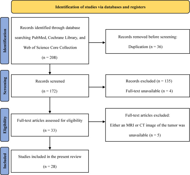
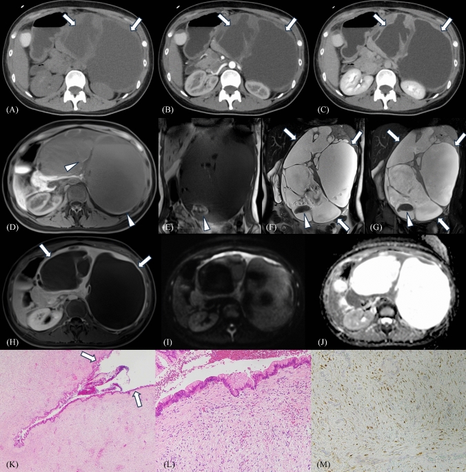
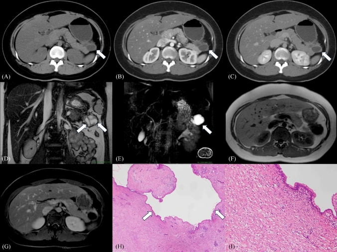
Purpose: This retrospective study aimed to investigate the radiological features of pancreatic desmoid-type fibromatosis (PDF) and systematically review the previous publications and two new cases. Methods: We searched PubMed, Cochrane Library, and Web of Science Core Collection and included 31 patients with pathologically proven PDFs with analyzable preoperative computed tomography (CT) and magnetic resonance imaging, including two patients from our institution and 29 patients from 28 publications. Two board-certified radiologists reviewed all images. Results: The median age of the patients was 39 years, with a male dominance observed (male, 54.8% vs. female, 45.2%). Abdominal pain was the most frequent symptom, occurring in 58.1% of cases. Surgical resection was performed in all cases of PDFs, resulting in a recurrence rate of 8.3% (2/24). The tumors were most commonly located in the pancreatic tail (23/31, 74.2%). In terms of morphology, a “solid” shape was most prevalent (14/31, 45.2%), followed by a “solid and cystic” shape (9/31, 29.0%) and a “cystic” shape (8/31, 25.8%). Characteristic radiological features included heterogeneous enhancement of the solid portion of the tumors on CT scans (13/20, 65%), moderate-to-weak enhancement in the late phase on CT (16/17, 94.1%), and a presence of cystic components in the tumors (17/31, 54.8%). In 16.1% (5/31) of PDFs, the cystic component was pathologically confirmed to be a dilated pancreatic duct. Conclusion: We summarized the clinical and imaging characteristics of PDF. Although the incidence may not be high, cystic components suggesting a dilated pancreatic duct within the tumor are unique imaging features in PDF.
Overview
The claims and statistics of this paper haven’t been checked yet.
Run the check — free, no account needed. It reads the open-access text and checks the sources it cites.
Integrity signals
Registry
Cites
Statistics
Figures
Verification
Claims the paper makes
The claims and statistics of this paper haven’t been checked yet.
Run the check — free, no account needed. It reads the open-access text and checks the sources it cites.
Self-check
Percentages, sums and reported differences re-computed from the paper’s own numbers.
The self-check needs the paper’s full text.
Citations
Cites
The PRISMA 2020 statement: An updated guideline for reporting systematic reviews
Page, M. J., McKenzie, J. E., Bossuyt, P. M., Boutron, I., Hoffmann, T. C., Mulrow, C. D., Shamseer, L., Tetzlaff, J. M., Akl, E. A., Brennan, S. E., et al. (2021) · BMJ · doi:10.1136/bmj.n71 · Cited by 108,283
Methodological quality and synthesis of case series and case reports
Murad, M. H., Sultan, S., Haffar, S., & Bazerbachi, F. (2018) · BMJ Evidence-Based Medicine · doi:10.1136/bmjebm-2017-110853 · Cited by 2,242
Declaración PRISMA 2020: una guía actualizada para la publicación de revisiones sistemáticas
Page, M. J., McKenzie, J. E., Bossuyt, P. M., Boutron, I., Hoffmann, T. C., Mulrow, C. D., Shamseer, L., Tetzlaff, J. M., Akl, E. A., Brennan, S. E., et al. (2021) · Revista Española de Cardiología · doi:10.1016/j.recesp.2021.06.016 · Cited by 2,095
Miettinen, M., & Lasota, J. P. (2006) · Archives of Pathology & Laboratory Medicine · doi:10.5858/2006-130-1466-gstrom · Cited by 1,499
Declaración PRISMA 2020: una guía actualizada para la publicación de revisiones sistemáticas
Page, M. J., McKenzie, J. E., Bossuyt, P. M., Boutron, I., Hoffmann, T. C., Mulrow, C. D., Shamseer, L., Tetzlaff, J. M., Akl, E. A., Brennan, S. E., et al. (2021) · Revista Española de Cardiología (English Edition) · doi:10.1016/j.rec.2021.07.010 · Cited by 1,313
The Desmoid Tumor. I.: Incidence, Sex-, Age- and Anatomical Distribution in the Finnish Population
Reitamo, J. J., Häyry, P., Nykyri, E., & Saxén, E. (1982) · American Journal of Clinical Pathology · doi:10.1093/ajcp/77.6.665 · Cited by 595
Salas, S., Dufresne, A., Bui, B., Blay, J., Terrier, P., Ranchère‐Vince, D., Bonvalot, S., Stoeckle, E., Guillou, L., Cesne, A. L., et al. (2011) · Journal of Clinical Oncology · doi:10.1200/jco.2010.33.5489 · Cited by 383
Bhattacharya, B., Dilworth, H. P., Iacobuzio–Donahue, C. A., Ricci, F., Weber, K. L., Furlong, M., Fisher, C., & Montgomery, E. A. (2005) · The American Journal of Surgical Pathology · doi:10.1097/01.pas.0000157938.95785.da · Cited by 322
A Prognostic Nomogram for Prediction of Recurrence in Desmoid Fibromatosis
Crago, A. M., Denton, B. T., Salas, S., Dufresne, A., Mezhir, J. J., Hameed, M., Gönen, M., Singer, S., & Brennan, M. F. (2013) · Annals of Surgery · doi:10.1097/sla.0b013e31828c8a30 · Cited by 279
Nieuwenhuis, M. H., Casparie, M. K., Mathus-Vliegen, L., Dekkers, O. M., Hogendoorn, P. C. W., & Vasen, H. F. A. (2010) · International Journal of Cancer · doi:10.1002/ijc.25664 · Cited by 225
Research works printed but not linked: 1
- Fritchie KJ, Crago AM, Van de Rijn M (2020) Soft Tissue and Bone Tumours, 5th. World Health Organization, pp 93-95
Cited by
Sorted by citations
A systematic review of cases of aortic penetration by inferior vena cava filters
Bai, Y., Wang, K., & Jia, Z. (2025) · Abdominal Radiology · doi:10.1007/s00261-025-05131-3 · Cited by 2
Clinical and morphological features of desmoid-type fibromatosis
Pilkevich, N. B., Khabibullin, R. R., Markovskaya, V. A., Yavorskaya, O. V., & Smirnova, A. P. (2025) · Yakut Medical Journal · doi:10.25789/ymj.2025.92.25
Metadata
- Authors
- Tanishima, T., Kurokawa, R., Sone, M., Nakai, Y., & Kusumoto, M.
- Affiliations
- National Cancer Centre Japan (JP)
- The University of Tokyo (JP)
- Venue
- Abdominal Radiology
- Published
- 2024-09-15
- Volume
- 50
- Issue
- 2
- Pages
- 839-850
- ISSN
- 2366-004X (ISSN-L)
- 2366-0058
- PMC
- PMC11794332
- OpenAlex
- W4402549875
- Cited by
- 2
- References
- 45
- Citations by year
- 2025: 2
- FWCI
- 0.15
- Type
- journal article
- Language
- en
- Open access
- hybrid
- doi.org
- Locations
- Abdominal Radiology (publishedVersion, cc-by)
- Research Square (acceptedVersion, cc-by)
- PubMed Central (publishedVersion, cc-by)
- PubMed (publishedVersion)
- Funders
- The University of Tokyo
- Topics
- Health Sciences › Medicine › Oncology › Soft tissue tumor case studies
- Health Sciences › Medicine › Oncology › Pancreatic and Hepatic Oncology Research
- Health Sciences › Medicine › Clinical Neurology › Neurofibromatosis and Schwannoma Cases
- Keywords
- aggressive fibromatosis, radiological features, computed tomography, magnetic resonance imaging, cystic tumor components, pancreatic duct dilatation, tumor morphology, tumor recurrence, surgical resection, pancreatic tail
- MeSH terms
- Adult, Female, Humans, Magnetic Resonance Imaging, Male, Middle Aged, Pancreatic Neoplasms, Retrospective Studies, Tomography, X-Ray Computed, Desmoid Tumors
About these checks
Registry, retraction, Cites and statistics results are computed, not judged, with one exception: a printed reference that no index lists is read once by a model before it counts as a citation error. Claims are extracted by a model and kept only when their quote appears verbatim in the text; each cited source is then checked, with a model’s help, for whether it says what the claim relies on. Those verdicts can be wrong — read the claim’s quote and the cited source before relying on one.
Statistics and arithmetic are recomputed, not judged.
Confidence bands are shown only for fields calibrated on our annotated test set. None is calibrated yet, so no band is shown.
What counts as a claim ›How we measure confidence ›How we check this ›
Are you an author, or do you see a problem with this page? Request a review
How to cite
Cite CoChat for the checks on this page, and the paper for its own findings.
- This page
- CoChat. (2026). Citation check: Radiological features of pancreatic desmoid-type fibromatosis: a case series and systematic review. https://app.cochat.ai/papers/10.1007/s00261-024-04570-8
- The paper
- Tanishima, T., Kurokawa, R., Sone, M., Nakai, Y., & Kusumoto, M. (2024). Radiological features of pancreatic desmoid-type fibromatosis: a case series and systematic review. Abdominal Radiology. https://doi.org/10.1007/s00261-024-04570-8
