

The DOI matches its Crossref record; 171 of the 171 references it cites were found in a registry or an index.
The cingulate cortex and limbic systems for emotion, action, and memory
Open accessAbstract
Evidence is provided for a new conceptualization of the connectivity and functions of the cingulate cortex in emotion, action, and memory. The anterior cingulate cortex receives information from the orbitofrontal cortex about reward and non-reward outcomes. The posterior cingulate cortex receives spatial and action-related information from parietal cortical areas. It is argued that these inputs allow the cingulate cortex to perform action-outcome learning, with outputs from the midcingulate motor area to premotor areas. In addition, because the anterior cingulate cortex connects rewards to actions, it is involved in emotion; and because the posterior cingulate cortex has outputs to the hippocampal system, it is involved in memory. These apparently multiple different functions of the cingulate cortex are related to the place of this proisocortical limbic region in brain connectivity.
Overview
The claims and statistics of this paper haven’t been checked yet.
Run the check — free, no account needed. It reads the open-access text and checks the sources it cites.
Integrity signals
Registry
Cites
Statistics
Figures
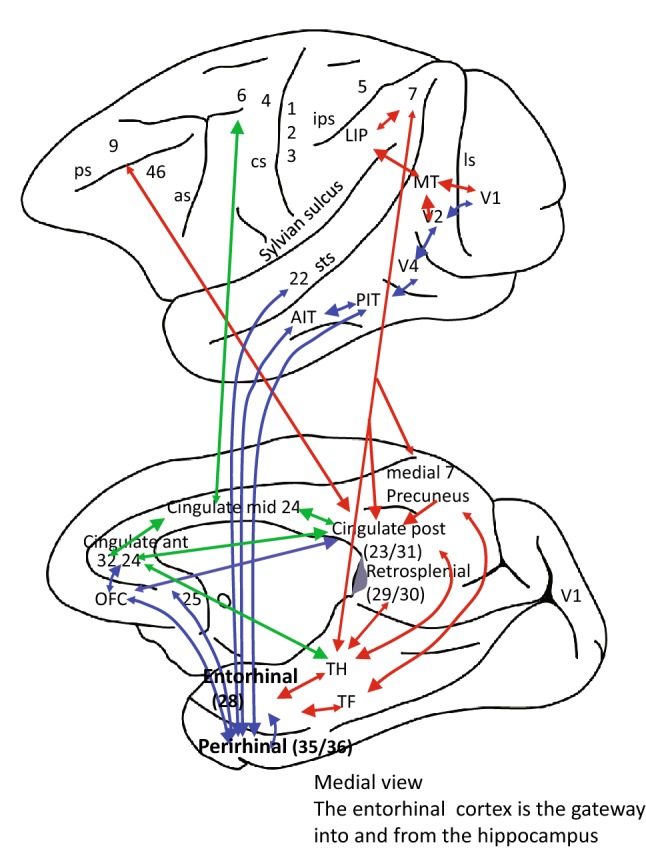
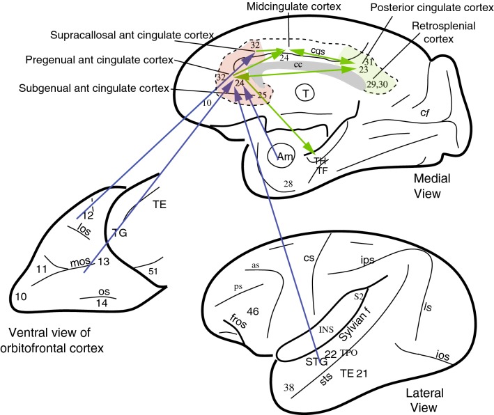
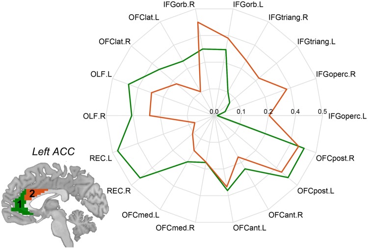
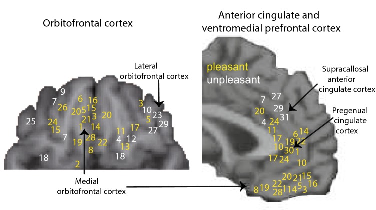
Verification
Claims the paper makes
The claims and statistics of this paper haven’t been checked yet.
Run the check — free, no account needed. It reads the open-access text and checks the sources it cites.
Claims other papers make about this one
Each is checked against this paper’s own text: does it say what the citing paper says it does?
The source backs part of the claim, not all of it — or backs it for a different population, condition or comparison.
In the citing paper
“Underactivated MCC in ADHD individuals may be construed as connected with the core symptom of inattention”
From Overlapping and differential neuropharmacological mechanisms of stimulants and nonstimulants for attention-deficit/hyperactivity disorder: A comparative neuroimaging analysis (2024) · cites this paper as Rolls, 2019
Experimental — accuracy not yet measuredThe source backs part of the claim, not all of it — or backs it for a different population, condition or comparison.
In the citing paper
“the MCC is considered a key area of emotion and cognition processing and subserves bottom-up attention diversion”
From Overlapping and differential neuropharmacological mechanisms of stimulants and nonstimulants for attention-deficit/hyperactivity disorder: A comparative neuroimaging analysis (2024) · cites this paper as Rolls, 2019
Experimental — accuracy not yet measured
Self-check
Percentages, sums and reported differences re-computed from the paper’s own numbers.
The self-check needs the paper’s full text.
Citations
Cites
Cognitive and emotional influences in anterior cingulate cortex
Bush, G., Luu, P., & Posner, M. I. (2000) · Trends in Cognitive Sciences · doi:10.1016/s1364-6613(00)01483-2 · Cited by 6,259
The precuneus: a review of its functional anatomy and behavioural correlates
Cavanna, A. E., & Trimble, M. R. (2006) · Brain · doi:10.1093/brain/awl004 · Cited by 5,128
Contributions of anterior cingulate cortex to behaviour
Devinsky, O., Morrell, M. J., & Vogt, B. A. (1995) · Brain · doi:10.1093/brain/118.1.279 · Cited by 3,377
Functional-anatomic fractionation of the brain’s default network
Andrews-Hanna, J. R., Reidler, J. S., Sepulcre, J., Poulin, R., & Buckner, R. L. (2010) · Neuron · doi:10.1016/j.neuron.2010.02.005 · Cited by 2,767
Ongur, D. (2000) · Cerebral Cortex · doi:10.1093/cercor/10.3.206 · Cited by 2,627
Gazzaniga, M. S., & Bizzi, E. (2000) · Cited by 2,547
(1995) · Choice Reviews Online · doi:10.5860/choice.32-5931 · Cited by 2,337
The role of the posterior cingulate cortex in cognition and disease
Leech, R., & Sharp, D. J. (2014) · Brain · doi:10.1093/brain/awt162 · Cited by 2,247
Emotion and motivation: the role of the amygdala, ventral striatum, and prefrontal cortex
Cardinal, R. N., Parkinson, J. A., Hall, J., & Everitt, B. J. (2002) · Neuroscience & Biobehavioral Reviews · doi:10.1016/s0149-7634(02)00007-6 · Cited by 2,129
A differential neural response in the human amygdala to fearful and happy facial expressions
Morris, J. S., Frith, C. D., Perrett, D. I., Rowland, D., Young, A. W., Calder, A. J., & Dolan, R. J. (1996) · Nature · doi:10.1038/383812a0 · Cited by 2,013
Research works printed but not linked: 2
- Rolls ET (2018a) The brain, emotion, and depression. Oxford University Press, Oxford
- Vogt BA (ed) (2019) Handbook of clinical neurology: cingulate cortex. handbook of clinical neurology, 3rd edn. Elsevier, New York
Cited by
Sorted by citations
Items 501–600 of 858
A cognitive model of economicsPreprint
Friesen, L. (2021) · doi:10.31234/osf.io/qtcrp · Cited by 3
Therapeutic potential of Garcinia kola against experimental toxoplasmosis in rats
Ahidjo, N., Yaya, F. M., Njamnshi, W. Y., Pambe, J. C. N., Ndianteng, E. W., Nwasike, C. N. C., Kemmo, C., Choupo, A. C., Zang, L. Y. M., Pieme, C. A., et al. (2024) · Brain Communications · doi:10.1093/braincomms/fcae255 · Cited by 2
Brain mechanisms discriminating enactive mental simulations of running and plogging
Philips, R. V., Baeken, C., Billieux, J., Harris, J. M., Maurage, P., Muela, I., Öz, İ. T., Pabst, A., Sescousse, G., Vögele, C., et al. (2024) · Human Brain Mapping · doi:10.1002/hbm.26807 · Cited by 3
Mindfulness in Cardiovascular Disease Patients: Improvements and Future Perspectives
Ricci, F., Bellomo, A., Berardis, D. D., & Ventriglio, A. (2024) · Heart and Mind · doi:10.4103/hm.hm-d-24-00084 · Cited by 2
Suárez‐Suárez, S., Cadaveira, F., Barrós‐Loscertales, A., García, J. M. P., Holguı́n, S. R., Blanco‐Ramos, J., & Doallo, S. (2025) · Addictive Behaviors Reports · doi:10.1016/j.abrep.2025.100585 · Cited by 3
Cortico-hippocampal networks underpin verbal memory encoding in temporal lobe epilepsy
Fiore, G., Giampiccolo, D., Xiao, F., Koepp, M. J., Iglesias, J. E., Vos, S. B., de Tisi, J., McEvoy, A. W., Bertani, G., Locatelli, M., et al. (2025) · Brain Communications · doi:10.1093/braincomms/fcaf067 · Cited by 2
Xiao, Y., Huang, C., Wang, J., Lin, Y., Quan, D., & Zheng, H. (2025) · Journal of Affective Disorders · doi:10.1016/j.jad.2025.03.088 · Cited by 3
Wi, J. H., Lee, H., Park, J. M., Heo, Y., Jo, S., Lee, J., Kim, Y., Jung, C., Kim, N., Song, G., et al. (2025) · Biomedicine & Pharmacotherapy · doi:10.1016/j.biopha.2025.117991 · Cited by 2
Neural effect of childhood maltreatment on neurovascular coupling in adolescent depression
Liu, S., Fan, D., He, C., Liu, X., Zhang, H., Zhang, H., Zhang, Z., Xie, C., & Pan, P. (2025) · European Child & Adolescent Psychiatry · doi:10.1007/s00787-025-02708-7 · Cited by 2
Teng, J.-F., Hu, X., Li, Y., Lu, X.-Y., Yao, J., Li, X., Yin, D.-N., Sui, J.-F., Liu, Y., Wang, P., et al. (2025) · Behavioural Brain Research · doi:10.1016/j.bbr.2025.115616 · Cited by 2
Koster, M., van der Pluijm, M., Fraikin, M., van Wingen, G. A., van de Giessen, E., de Haan, L., Vermeulen, J. M., & Ziermans, T. B. (2025) · Biological Psychiatry Global Open Science · doi:10.1016/j.bpsgos.2025.100539 · Cited by 2
Lin, J., Zeng, J., Huang, H., Lin, Y., & Chen, H. (2025) · Journal of Sleep Research · doi:10.1111/jsr.70121 · Cited by 2
Kochvar, A. P., Laviolette, S. R., Khan, A. S., Grin, B. M., & Dai, H. D. (2025) · Behavioural Brain Research · doi:10.1016/j.bbr.2025.115741 · Cited by 2
Evolutionary consideration of serial order processing by the normal rodent brain
Zlatkina, V., Martakis, K., Azizi, H., & Petrides, M. (2025) · Proceedings of the National Academy of Sciences · doi:10.1073/pnas.2509426122 · Cited by 2
Thomas, S. T., Palanikumar, L., Darby, D., & Brodtmann, A. G. (2025) · Cerebral Circulation - Cognition and Behavior · doi:10.1016/j.cccb.2025.100404 · Cited by 3
Ko, S.-L., Cheng, L., Hsu, J.-W., Chen, L., Bai, Y., Tu, P., & Chen, M. (2025) · Journal of the Formosan Medical Association · doi:10.1016/j.jfma.2025.10.039 · Cited by 3
Liu, Z., Wu, H., Xu, Y., Zhang, L., Tian, K., Long-fei, L., Zhang, Z., & Zhou, P. (2025) · BMC Psychiatry · doi:10.1186/s12888-025-07406-x · Cited by 2
Huang, Y., Lin, Z., Zhou, C., Wang, Y., & Luan, M. (2025) · Psychology of sport and exercise · doi:10.1016/j.psychsport.2025.103051 · Cited by 2
An EEG-based multi-scale hybrid attention and squeeze network for objective taste assessment
Yang, T., Xiao, Y., Li, Z., Chen, C., Ye, F., Cao, M., Liu, R., Zhu, Y., Qian, Z., & Rajapakse, J. C. (2025) · Future Foods · doi:10.1016/j.fufo.2025.100873 · Cited by 2
Caenegem, E. E. V., Verdú, M. M., Waltzing, B. M., Hamoline, G., Lennart, F., & Hardwick, R. M. (2025) · Imaging Neuroscience · doi:10.1162/imag.a.1095 · Cited by 4
Danstrom, I. A., Adkinson, J. A., Liu, Z., Robinson, M. E., Oswalt, D. N., Banks, G. P., Maheshwari, A., Lin, L., Shofty, B., Hasen, M. A., et al. (2026) · Brain stimulation · doi:10.1016/j.brs.2026.103049 · Cited by 1
Lammers‐Lietz, F., Borchers, F., Feinkohl, I., Kanar, Ç., Krampe, H., Lichtner, G., Sreekanth, J., Wiebach, J., Weygandt, M., Spies, C., et al. (2026) · European Journal of Anaesthesiology · doi:10.1097/eja.0000000000002365 · Cited by 2
Liu, Y., Rao, B., & Zhou, F. (2026) · Journal of Psychiatric Research · doi:10.1016/j.jpsychires.2026.02.043 · Cited by 2
Memories that touch deeply: Toward a neurobiological model of affective tactile memory
Bischoff, H., Meconi, F., & Crucianelli, L. (2026) · Neuroscience & Biobehavioral Reviews · doi:10.1016/j.neubiorev.2026.106685 · Cited by 2
Peng, Y., Liang, M., Fan, C., Han, D., Xia, Z., Zhou, Y., & Liang, X. (2026) · Building and Environment · doi:10.1016/j.buildenv.2026.114601 · Cited by 2
Structural and Functional Brain Alterations Induced by Noise Exposure: A Comprehensive Review
Venegas-Mora, H. V., Ispanixtlahuatl-Meráz, O., Martínez-Fernández, D. E., Aguilar-García, I. G., & Fernández-Quezada, D. (2026) · NeuroSci · doi:10.3390/neurosci7040075 · Cited by 3
A new model for freedom of movement using connectomic analysis
Rodríguez-Méndez, D. A., San‐Juan, D., Hallett, M., Antonopoulos, C. G., López-Reynoso, E., & Lara‐Ramírez, R. (2022) · PeerJ · doi:10.7717/peerj.13602 · Cited by 2
A study on brain neuronal activation based on the load in upper limb exercise (STROBE)
Choi, J., & Choi, M. (2022) · Medicine · doi:10.1097/md.0000000000030761 · Cited by 2
Cao, A., Hong, D., Che, C., Yu, X., Cai, Z., Yang, X., Zhang, D., & Yu, P. (2023) · Frontiers in Behavioral Neuroscience · doi:10.3389/fnbeh.2022.1039288 · Cited by 2
Wang, K., Zhao, X., Yuan, L., Chen, Q., Wang, Q., & Ai, L. (2023) · Frontiers in Neurology · doi:10.3389/fneur.2023.1100760 · Cited by 2
COVID-19 vaccine-hesitancy is associated with lower cortical volume in elderly individuals
Nabizadeh, F. (2023) · Neurology Letters · doi:10.52547/nl.2.1.35 · Cited by 2
Neuronal signatures of anger and fear in patients with psychosis
Shen, J., Kim, W., Tsogt, U., Odkhuu, S., Liu, C., Kang, N., Lee, K., Sui, J., Kim, S., & Chung, Y. (2023) · Psychiatry Research Neuroimaging · doi:10.1016/j.pscychresns.2023.111658 · Cited by 2
Yu, Z., Guinto, M. C., Lim, B. G., Tan, R. R. P., Yoshimoto, J., Ikeda, K., Ohta, Y., & Ohta, J. (2023) · Artificial Life and Robotics · doi:10.1007/s10015-023-00875-x · Cited by 2
Harel, Y., Pinsard, B., Boyle, J. A., Borghesani, V., Clei, M. L., Mignot, P.-H., St‐Laurent, M., Jerbi, K., & Bellec, P. (2026) · Imaging Neuroscience · doi:10.1162/imag.a.1256 · Cited by 2
Will you read how I will read? Naturalistic fMRI predictors of emergent reading
Wat, E. K., Jangraw, D. C., Finn, E. S., Bandettini, P. A., Preston, J. L., Landi, N., Hoeft, F., Frost, S. J., Lau, A. N.-L., Chen, G., et al. (2023) · Neuropsychologia · doi:10.1016/j.neuropsychologia.2023.108763 · Cited by 2
Lei, Z., Jin, Z., Xie, K., Li, S., Feng, L., Zhang, J., & Li, L. (2024) · NeuroImage · doi:10.1016/j.neuroimage.2024.120552 · Cited by 2
Correlations between Gustatory, Olfactory, Cognitive Function, and Age in Healthy Women
Sanna, F., Castelli, M. P., Mostallino, R., Loy, F., & Masala, C. (2024) · Nutrients · doi:10.3390/nu16111731 · Cited by 2
Lee, A. J., Initiative, A. D. N., Stark, J. H., & Hayes, S. M. (2024) · Journal of Alzheimer s Disease · doi:10.3233/jad-231468 · Cited by 4
Cortical Neurodynamics, Schizophrenia, Depression, and Obsessive-Compulsive Disorder
Rolls, E. T. (2024) · Springer eBooks · doi:10.1007/978-3-031-38391-5_11 · Cited by 2
Self-citationKoster, M., van der Pluijm, M., van de Giessen, E., Schrantee, A., van Hooijdonk, C. F. M., Selten, J., Booij, J., de Haan, L., Ziermans, T. B., & Vermeulen, J. M. (2024) · Schizophrenia Research · doi:10.1016/j.schres.2024.07.022 · Cited by 2
The impact of metabolic syndrome on the cerebral cortex: a Mendelian randomization study
Zhang, P., Zhang, X., Gao, B., Gao, Y., & Pan, Y. (2024) · Cerebral Cortex · doi:10.1093/cercor/bhae342 · Cited by 2
Buccilli, B., Paternò, G., Cortés-Contreras, A. P., Pineda, M. F. T., Monroy-Sosa, A., & Ascenzi, B. M. (2024) · Elsevier eBooks · doi:10.1016/b978-0-12-822404-5.00019-x · Cited by 2
Jiang, Y. B., Chen, Y., Wei, Y., Li, S., Wang, K., & Cheng, J. (2024) · Journal of Affective Disorders · doi:10.1016/j.jad.2024.08.207 · Cited by 2
Seyedmirzaei, H., Rasoulian, P., Parsaei, M., Hamidi, M., Ghanbari, A., Khaboushan, A. S., Fatehi, F., Kamali, A., & Sotoudeh, H. (2024) · Brain Imaging and Behavior · doi:10.1007/s11682-024-00934-2 · Cited by 2
Chen, Y., Yang, C., Gao, B., Chen, K., Keehn, R. J. J., Müller, R.-A., Yuan, L., & You, Y. (2024) · Biological Psychiatry Cognitive Neuroscience and Neuroimaging · doi:10.1016/j.bpsc.2024.10.014 · Cited by 3
Contrastive machine learning reveals species -shared and -specific brain functional architecture
Li, Y., Cao, G., Zhang, S., Zhang, W., Sun, Y., Zhou, J., Zhong, T., Yuan, Y., Liu, T., Liu, T., et al. (2024) · Medical Image Analysis · doi:10.1016/j.media.2024.103431 · Cited by 2
Targeted Brain Delivery of Therapeutics
Malaiya, A., Kewat, R., Paliwal, S. R., & Paliwal, R. (2024) · Springer eBooks · doi:10.1007/978-981-97-4970-6_13 · Cited by 2
Álvarez, J. D., García‐Gutiérrez, F., Bueso-Inchausti, P., Cabrera‐Martín, M. N., Delgado‐Alonso, C., Delgado‐Álvarez, A., Díez‐Cirarda, M., Carbó, A. V., Fernández‐Romero, L., Valles‐Salgado, M., et al. (2024) · Cortex · doi:10.1016/j.cortex.2024.11.022 · Cited by 3
Pereira-Rufino, L. D. S., Gobbo, D. R., Conte, R., de Sino, R. M., de Oliveira, N. N., Fidalgo, T. M., Sato, J. R., Carrete, H., Formigoni, M. L. O. D. S., Shi, Z., et al. (2025) · Psychiatry Research Neuroimaging · doi:10.1016/j.pscychresns.2025.111946 · Cited by 2
Cognitive impairment and associated neurobehavioral dysfunction in post-COVID syndrome
Schmidt, L., Klingner, C. M., Petersen, I., Volkmer, A., Schreiber, M., Schmidt, A., Reuken, P. A., Besteher, B., Geis, C., Ullsperger, M., et al. (2025) · Psychiatry Research · doi:10.1016/j.psychres.2025.116522 · Cited by 2
Tomoda, A., Nishitani, S., 藤澤, 隆., Takiguchi, S., Yao, A., kazuhiro, M., Hiraoka, D., Mizuno, Y., Ochiai, K., Kawata, N. Y. S., et al. (2025) · Biological Psychiatry · doi:10.1016/j.biopsych.2026.03.470 · Cited by 2
Flavor Processing in the Brain
Rolls, E. T. (2020) · Elsevier eBooks · doi:10.1016/b978-0-12-809324-5.23884-4 · Cited by 2
Self-citationDiabetes Mellitus Affects Working Memory
Braganza, D. C., Flores, E., Crew, L. A., Wirt, R. A., Ortiz, A. A., McNeela, A. M., Kinney, J. W., & Hyman, J. M. (2021) · Spectra Undergraduate Research Journal · doi:10.9741/2766-7227.1008 · Cited by 2
Colombo, B., & Iannello, P. (2022) · Journal of Cognitive Psychology · doi:10.1080/20445911.2022.2084546 · Cited by 4
Varanasi, S., Tuli, R., Han, F., Chen, R., & Choa, F. (2023) · Sensors · doi:10.3390/s23031603 · Cited by 2
Connectomic Networks and Their Impact on Clinical Outcomes in Glioma Treatment: A Review
Rawanduzy, C. A., Earl, E. R., Brooks, J. B., Khan, M., Dadario, N. B., Sughrue, M. E., & Karsy, M. J. (2023) · Indian Journal of Neurosurgery · doi:10.1055/s-0043-1771214 · Cited by 2
The Badly Behaving Brain: How Ultra-Processed Food Addiction Thwarts Sustained Weight Loss
Thompson, S. P., & Thaw, A. K. (2024) · IntechOpen eBooks · doi:10.5772/intechopen.1004428 · Cited by 6
The Synthesis and Decoding of Meaning
Schulze, H. G. (2021) · Journal of Artificial General Intelligence · doi:10.2478/jagi-2021-0002 · Cited by 2
Non-linear functional brain co-activations in short-term memory distortion tasksPreprint
Ceglarek, A., Ochab, J. K., Cifré, I., Fąfrowicz, M., Sikora-Wachowicz, B., Lewandowska, K., Bohaterewicz, B., Marek, T., & Chialvo, D. R. (2021) · bioRxiv (Cold Spring Harbor Laboratory) · doi:10.1101/2021.09.12.459960 · Cited by 2
Sensory Loss and DementiaPreprint
Lad, M., Sedley, W., & Griffiths, T. D. (2022) · doi:10.31234/osf.io/mt93g · Cited by 2
Sulaiman, S. K., Tsiga-Ahmed, F. I., Arora, T., Faris, M. E., Musa, M. S., Kareem, Y. A., Dayyab, F. M., Hussein, A., Sale, S., & Khan, M. A. (2022) · Research Square · doi:10.21203/rs.3.rs-1486892/v1 · Cited by 2
Long, T., Li, H., Shu, Y., Li, K., Xie, W., Zeng, Y., Huang, L., Zeng, L., Liu, X., & Peng, D. (2022) · Research Square · doi:10.21203/rs.3.rs-2220078/v1 · Cited by 2
Razza, L. B., Smet, S. D., Hoornweder, S. V., Witte, S. D., Luethi, M. S., Baeken, C., Brunoni, A. R., & Vanderhasselt, M. (2023) · bioRxiv (Cold Spring Harbor Laboratory) · doi:10.1101/2023.06.13.544810 · Cited by 2
Does dysthyroid optic neuropathy affect beyond the windows to the soul?
Liu, S. (2023) · European Radiology · doi:10.1007/s00330-023-10239-5 · Cited by 2
Meta-Analysis of Brain Volumetric Abnormalities in Patients with Remitted Major Depressive DisorderPreprint
Xu, X., Zhou, Q., Wen, F., & Yang, M. (2023) · SSRN Electronic Journal · doi:10.2139/ssrn.4613668 · Cited by 2
Kang, M. C., Gao, Y., Zhang, L.-J., Liang, R., Li, Q., Shu, H., Pan, Y., Ying, P., Xu, S., & Shao, Y. (2022) · Frontiers in Neurology · doi:10.3389/fneur.2022.874158 · Cited by 1
Мельников, М. Е., Bezmaternykh, D. D., Savelov, A. A., Petrovskiy, E. D., & Kochetova, A. V. (2022) · Bulletin of Experimental Biology and Medicine · doi:10.1007/s10517-022-05637-6 · Cited by 1
Reduced Cortical Complexity in Children with Developmental Delay in Saudi Arabia
Abujamea, A. H., Almosa, M., Uzair, M., Alabdullatif, N., & Bashir, S. (2023) · Cureus · doi:10.7759/cureus.48291 · Cited by 1
Yang, G., Haley, R. W., Guo, Y., & Gopinath, K. (2024) · Neuroimage Reports · doi:10.1016/j.ynirp.2024.100209 · Cited by 1
Ma, L., Zhang, M., Niu, X., Tao, Q., Dang, J., Sun, J., Mei, B., Wang, W., Han, S., Cheng, J., et al. (2024) · Journal of Affective Disorders · doi:10.1016/j.jad.2024.10.055 · Cited by 1
Pain modulates brain potentials and behavioral responses to unfairness
Hu, C., Wu, R., & Wang, C. (2024) · Social Cognitive and Affective Neuroscience · doi:10.1093/scan/nsae081 · Cited by 1
King, F. K., Song, Y., Fabbri, L., Perry, M. S., Papadelis, C. L., & Cooper, C. M. (2024) · Neuropsychologia · doi:10.1016/j.neuropsychologia.2024.109035 · Cited by 1
Yang, X., Wu, Z., Lai, H., Chen, L., Cao, D., & Liu, F. (2024) · Brain and Behavior · doi:10.1002/brb3.70196 · Cited by 1
Kim, Y., Jun, S. Y., Oh, J., Koo, J., & Lee, E. (2025) · Neurorehabilitation and neural repair · doi:10.1177/15459683241309580 · Cited by 1
Genetic markers of early response to lurasidone in acute schizophrenia
Yoshikawa, A., Li, J., Alliey‐Rodriguez, N., & Meltzer, H. Y. (2025) · The Pharmacogenomics Journal · doi:10.1038/s41397-024-00360-z · Cited by 2
Kim, G., Park, H. Y., & Kim, K. (2025) · Frontiers in Behavioral Neuroscience · doi:10.3389/fnbeh.2025.1514579 · Cited by 1
The Neuroanatomical Correlates of Dyspnea: An Activation Likelihood Estimation Meta-Analysis
Müller, C. F., Kerl, J., & Dellweg, D. (2025) · NeuroSci · doi:10.3390/neurosci6020036 · Cited by 1
Gerlach, A., Karim, H. T., Kahru, K., Tudorascu, D., Gross, J. J., Butters, M. A., & Andreescu, C. (2025) · Biological Psychiatry Cognitive Neuroscience and Neuroimaging · doi:10.1016/j.bpsc.2025.04.010 · Cited by 1
Xu, R., Yang, B., Li, J., Wei, X., Zhang, L., Tian, J., & Zhang, W. (2025) · Frontiers in Neurology · doi:10.3389/fneur.2025.1565046 · Cited by 1
Wang, J., Huang, J., Kang, X., Dong, H., & Lu, J. (2025) · Brain Imaging and Behavior · doi:10.1007/s11682-025-01012-x · Cited by 1
Auricular neuromodulation may act through interoception modulation
Canini, F., Claverie, D., Weill, F., Pailler, C., Paquier, B., Michel-Cherqui, M., Sonia Pellissier, A., Trousselard, M., & Sagui, E. (2025) · Brain Research · doi:10.1016/j.brainres.2025.149736 · Cited by 1
A depression–like phenotype is associated with discrete defects in the primary hippocampal circuit
Gunn, B. G., Yang, C. C., Lauterborn, J. C., Pruess, B. S., Quintanilla, J., Ge, K., Gall, C. M., & LYNCH, G. S. (2026) · Translational Psychiatry · doi:10.1038/s41398-026-04094-3 · Cited by 1
Shi, X., Zhu, Y., Zhang, Z., Ma, N., He, D., Wu, Y., Dai, Z., Qin, X., Chen, Y., Zhao, Y., et al. (2025) · Biology of Sex Differences · doi:10.1186/s13293-025-00723-6 · Cited by 1
Neurobiological Mechanisms and Therapeutic Interventions in Behavioral Addictions
Laadraoui, J., Aitlarradia, M., Oubella, K., Agouram, F., Oufquir, S., Aitbaba, A., Kabdy, H., & El‐Mansoury, B. (2025) · IGI Global eBooks · doi:10.4018/979-8-3373-0518-9.ch001 · Cited by 2
Comparative Anatomical Network Analysis of Chimpanzee and Human Craniocerebral Topology
Schuurman, T., & Bruner, E. (2025) · American Journal of Biological Anthropology · doi:10.1002/ajpa.70083 · Cited by 1
The aftermath of trauma on resting state source-level MEG microstate dynamics
Bertoli, M., Croce, P., Zappasodi, F., Matteo, R. D., Vellante, F., Pettorruso, M., Martinotti, G., & Brunetti, M. (2025) · European Journal of Psychotraumatology · doi:10.1080/20008066.2025.2520638 · Cited by 1
Tang, R., Bell, T. R., Elman, J. A., Reynolds, C. A., Gustavson, D. E., Puckett, O. K., Panizzon, M. S., Toomey, R., McKenzie, R. E., Lyons, M. J., et al. (2025) · GeroScience · doi:10.1007/s11357-025-01778-4 · Cited by 2
Dopamine D2/D3 receptor availability and working memory in stimulant use disorder
Dean, A. C., Allen, D. C., McClintick, M. N., Pochon, J.-B. F., Ghahremani, D. G., Mandelkern, M. A., & London, E. D. (2025) · The American Journal of Drug and Alcohol Abuse · doi:10.1080/00952990.2025.2534809 · Cited by 1
Identifying bipolar disorder and predicting its therapeutic response of cognitive impairment
Yuan, H., Xu, B., Wang, Y., Ou, Y., Qiu, Y., Teng, Z., Xu, X., Wu, G., Wang, B., Mao, W., et al. (2025) · Journal of Affective Disorders · doi:10.1016/j.jad.2025.119991 · Cited by 1
Song, Y. Y., Zeng, J., Cao, H., Cheng, B., & Yang, X. (2025) · Psychological Medicine · doi:10.1017/s0033291725101323 · Cited by 2
Daviddi, S., & Santangelo, V. (2025) · European Journal of Neuroscience · doi:10.1111/ejn.70244 · Cited by 3
Zhang, J. Y., Han, F., Wang, X., Wu, F., Song, X., Liu, Q., Wang, J., Grecucci, A., Zhang, Y., Yi, X., et al. (2025) · European Journal of Nuclear Medicine and Molecular Imaging · doi:10.1007/s00259-025-07585-5 · Cited by 1
Yan, H., Han, Y., Shan, X., Li, H., Liu, F., Li, P., Yuan, Y., Zhao, J., & Guo, W. (2025) · Journal of Affective Disorders · doi:10.1016/j.jad.2025.120449 · Cited by 1
Rodríguez‐Fernández, L., Zorzo, C., Gutiérrez‐Menéndez, A., Lobo, J. C., Valle, D. F. D., Castillo‐Escalona, E. G., Martínez, J. A., & Árias, J. L. (2025) · Physiology & Behavior · doi:10.1016/j.physbeh.2025.115135 · Cited by 1
Wang, H., Zhao, X., Pan, X., Gui, H., Xu, B., Tao, L., Chen, H., Wang, H., Lv, F., Luo, T., et al. (2025) · Parkinsonism & Related Disorders · doi:10.1016/j.parkreldis.2025.108104 · Cited by 1
Dynamics of ACC excitatory neurons during reversal learning: Implications for cognitive flexibility
Zhang, M., Shen, F., Liao, C., Liu, S., Wang, H., & Li, C. (2025) · Behavioural Brain Research · doi:10.1016/j.bbr.2025.115902 · Cited by 1
Ciarolla, A., Bobilev, A. M., Dybowski, F., Fang, Y., Sacco, C., Palka, J. M., Aslan, S., Liu, P., Lu, H., Tamminga, C. A., et al. (2025) · Schizophrenia Research · doi:10.1016/j.schres.2025.11.003 · Cited by 1
Large-scale brain mediation network based on resting-state functional MRI
Wang, B., Zhang, X., Pan, T., Li, T., Liu, T., & Yan, T. (2025) · Communications Biology · doi:10.1038/s42003-025-08948-2 · Cited by 1
Du, H., Gao, F., Han, Y., Xiao, C., Li, Q., Zhang, D., Wang, Z., Yu, Q., Cai, S., Fan, J., et al. (2025) · Depression and Anxiety · doi:10.1155/da/1087782 · Cited by 1
Hu, M., Song, K. S., Choi, J.-Y., Pyeon, A., Cho, H., Choi, J.-S., Choi, I., Chun, J.-W., & Kim, D.-J. (2025) · Life · doi:10.3390/life15121899 · Cited by 2
Metadata
- Authors
- Rolls, E. T.
- Affiliations
- University of Warwick (GB)
- Corresponding author
- Rolls, Edmund T.
- ORCID
- Rolls, Edmund T.
- Venue
- Brain Structure and Function
- Published
- 2019-08-26
- Volume
- 224
- Issue
- 9
- Pages
- 3001-3018
- ISSN
- 1863-2653 (ISSN-L)
- 1863-2661
- PMC
- PMC6875144
- OpenAlex
- W2970957161
- Cited by
- 859
- References
- 171
- Citations by year
- 2026: 110 · 2025: 175 · 2024: 173 · 2023: 114 · 2022: 148 · 2021: 94 · 2020: 39 · 2019: 6
- FWCI
- 9.11
- Type
- journal article
- Language
- en
- Open access
- hybrid
- doi.org
- Locations
- Brain Structure and Function (publishedVersion, cc-by)
- PubMed Central (publishedVersion, cc-by)
- PubMed (publishedVersion)
- Warwick Research Archive Portal (University of Warwick) (submittedVersion, cc-by)
- Topics
- Life Sciences › Neuroscience › Cognitive Neuroscience › Neural and Behavioral Psychology Studies
- Life Sciences › Neuroscience › Cognitive Neuroscience › Memory and Neural Mechanisms
- Life Sciences › Neuroscience › Cellular and Molecular Neuroscience › Neurotransmitter Receptor Influence on Behavior
- Keywords
- cingulate cortex, anterior cingulate cortex, posterior cingulate cortex, orbitofrontal cortex, reward processing, action-outcome learning, premotor cortex, parietal cortex, hippocampal system, emotions, memory, limbic system, brain connectivity
- MeSH terms
- Animals, Emotions, Gyrus Cinguli, Humans, Limbic System, Memory, Motor Activity, Neural Pathways, Reward
About these checks
Registry, retraction, Cites and statistics results are computed, not judged, with one exception: a printed reference that no index lists is read once by a model before it counts as a citation error. Claims are extracted by a model and kept only when their quote appears verbatim in the text; each cited source is then checked, with a model’s help, for whether it says what the claim relies on. Those verdicts can be wrong — read the claim’s quote and the cited source before relying on one.
Statistics and arithmetic are recomputed, not judged.
Confidence bands are shown only for fields calibrated on our annotated test set. None is calibrated yet, so no band is shown.
What counts as a claim ›How we measure confidence ›How we check this ›
Are you an author, or do you see a problem with this page? Request a review
