

The DOI matches its Crossref record; 1 of the 1 references it cites were found in a registry or an index.
Distribution of Matrix Metalloproteinases in Human Atherosclerotic Carotid Plaques and Their Production by Smooth Muscle Cells and Macrophage Subsets
Open accessAbstract
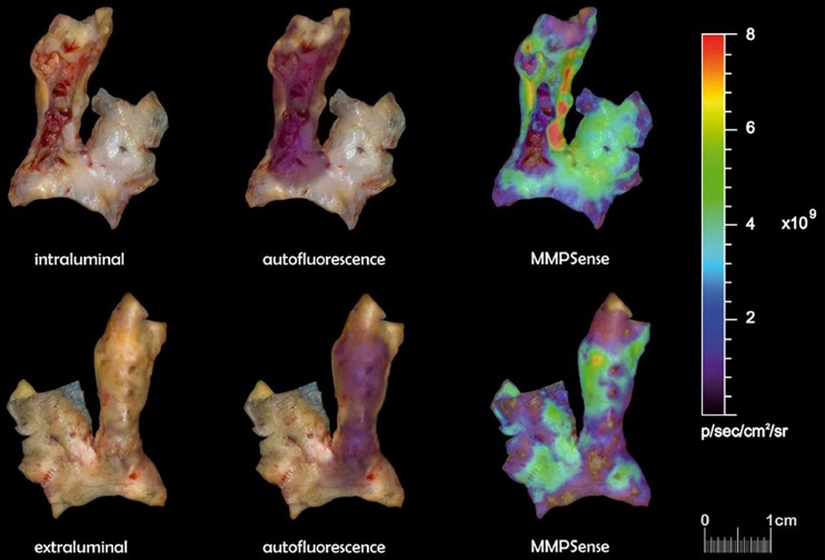
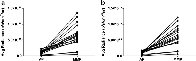
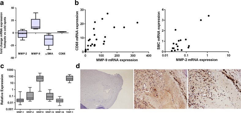
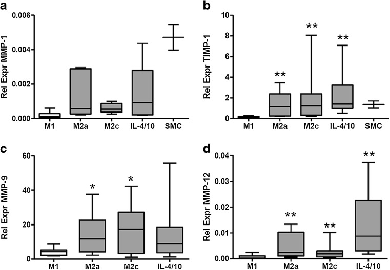
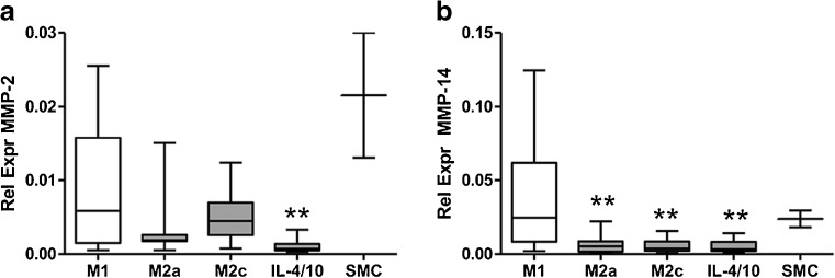
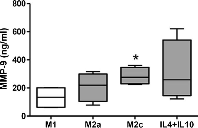
PURPOSE: In this study, the potential of matrix metalloproteinase (MMP) sense for detection of atherosclerotic plaque instability was explored. Secondly, expression of MMPs by macrophage subtypes and smooth muscle cells (SMCs) was investigated. PROCEDURES: Twenty-three consecutive plaques removed during carotid endarterectomy were incubated in MMPSense™ 680 and imaged with IVIS® Spectrum. mRNA levels of MMPs, macrophage markers, and SMCs were determined in plaque specimens, and in in vitro differentiated M1 and M2 macrophages. RESULTS: There was a significant difference between autofluorescence signals and MMPSense signals, both on the intraluminal and extraluminal sides of plaques. MMP-9 and CD68 messenger RNA (mRNA) expression was higher in hot spots, whereas MMP-2 and αSMA expression was higher in cold spots. In vitro M2 macrophages had higher mRNA expression of MMP-1, MMP-9, MMP-12, and TIMP-1 compared to M1 macrophages. CONCLUSION: MMP-9 is most dominantly MMP present in atherosclerotic plaques and is produced by M2 rather than M1 macrophages.
Overview
The claims and statistics of this paper haven’t been checked yet.
Run the check — free, no account needed. It reads the open-access text and checks the sources it cites.
Integrity signals
Registry
Cites
Statistics
Figures
Verification
Claims the paper makes
The claims and statistics of this paper haven’t been checked yet.
Run the check — free, no account needed. It reads the open-access text and checks the sources it cites.
Claims other papers make about this one
Each is checked against this paper’s own text: does it say what the citing paper says it does?
The source backs part of the claim, not all of it — or backs it for a different population, condition or comparison.
In the citing paper
“Among several types of M2 macrophages, M2c macrophages are known to orchestrate matrix remodeling by releasing MMPs and TIMPs (tissue inhibitors of metalloproteinase)15,39.”
From Bioengineered liver crosslinked with nano-graphene oxide enables efficient liver regeneration via MMP suppression and immunomodulation (2023) · cites this paper as [39]
Experimental — accuracy not yet measuredThe source backs part of the claim, not all of it — or backs it for a different population, condition or comparison.
In the citing paper
“TIMP-1 is predominantly expressed by M2c macrophages, although some MMPs are also expressed in M2c macrophages39.”
From Bioengineered liver crosslinked with nano-graphene oxide enables efficient liver regeneration via MMP suppression and immunomodulation (2023) · cites this paper as [39]
Experimental — accuracy not yet measuredThe source backs part of the claim, not all of it — or backs it for a different population, condition or comparison.
In the citing paper
“the proteolytic balance, a ratio of MMP-9 to TIMP-1, is known to be higher in M1 macrophages39,40.”
From Bioengineered liver crosslinked with nano-graphene oxide enables efficient liver regeneration via MMP suppression and immunomodulation (2023) · cites this paper as [39]
Experimental — accuracy not yet measured
Self-check
Percentages, sums and reported differences re-computed from the paper’s own numbers.
The self-check needs the paper’s full text.
Citations
Cites
Imaging of atherosclerotic cardiovascular disease
Sanz, J., & Fayad, Z. A. (2008) · Nature · doi:10.1038/nature06803 · Cited by 558
Research works printed but not linked: 32
- Libby P (2012) Inflammation in atherosclerosis. Arterioscler Thromb Vasc Biol 32:2045–2051 · DOI
- Shah PK (2009) Inflammation and plaque vulnerability. Cardiovasc Drugs Ther 23:31–40 · DOI
- Ross R (1999) Atherosclerosis--an inflammatory disease. N Engl J Med 340:115–126 · DOI · Paper page
- Newby AC, George SJ, Ismail Y et al (2009) Vulnerable atherosclerotic plaque metalloproteinases and foam cell phenotypes. Thromb Haemost 101:1006–1011 · DOI
- Newby AC (2005) Dual role of matrix metalloproteinases (matrixins) in intimal thickening and atherosclerotic plaque rupture. Physiol Rev 85:1–31 · DOI · Paper page
- Medbury HJ, James V, Ngo J et al (2013) Differing association of macrophage subsets with atherosclerotic plaque stability. Int Angiol 32:74–84
- Jager NA, Teteloshvili N, Zeebregts CJ et al (2012) Macrophage folate receptor-beta (FR-beta) expression in auto-immune inflammatory rheumatic diseases: a forthcoming marker for cardiovascular risk? Autoimmun Rev 11:621–626 · DOI
- Mantovani A, Sica A, Sozzani S et al (2004) The chemokine system in diverse forms of macrophage activation and polarization. Trends Immunol 25:677–686 · DOI · Paper page
- Wolfs IM, Donners MM, de Winther MP (2011) Differentiation factors and cytokines in the atherosclerotic plaque micro-environment as a trigger for macrophage polarisation. Thromb Haemost 106:763–771 · DOI · Paper page
- Raffetto JD, Khalil RA (2008) Matrix metalloproteinases and their inhibitors in vascular remodeling and vascular disease. Biochem Pharmacol 75:346–359 · DOI
- Loftus IM, Naylor AR, Goodall S et al (2000) Increased matrix metalloproteinase-9 activity in unstable carotid plaques. A potential role in acute plaque disruption. Stroke 31:40–47 · DOI
- Alvarez B, Ruiz C, Chacon P et al (2004) Serum values of metalloproteinase-2 and metalloproteinase-9 as related to unstable plaque and inflammatory cells in patients with greater than 70% carotid artery stenosis. J Vasc Surg 40:469–475 · DOI
- Taurino M, Raffa S, Mastroddi M et al (2007) Metalloproteinase expression in carotid plaque and its correlation with plasma levels before and after carotid endarterectomy. Vasc Endovascular Surg 41:516–521 · DOI
- Sapienza P, di Marzo L, Borrelli V et al (2004) Basic fibroblast growth factor mediates carotid plaque instability through metalloproteinase-2 and −9 expression. Eur J Vasc Endovasc Surg 28:89–97 · DOI
- Kunte H, Amberger N, Busch MA et al (2008) Markers of instability in high-risk carotid plaques are reduced by statins. J Vasc Surg 47:513–522 · DOI
- Eldrup N, Gronholdt ML, Sillesen H, Nordestgaard BG (2006) Elevated matrix metalloproteinase-9 associated with stroke or cardiovascular death in patients with carotid stenosis. Circulation 114:1847–1854 · DOI
- Turu MM, Krupinski J, Catena E et al (2006) Intraplaque MMP-8 levels are increased in asymptomatic patients with carotid plaque progression on ultrasound. Atherosclerosis 187:161–169 · DOI
- Johnson JL, George SJ, Newby AC, Jackson CL (2005) Divergent effects of matrix metalloproteinases 3, 7, 9, and 12 on atherosclerotic plaque stability in mouse brachiocephalic arteries. Proc Natl Acad Sci U S A 102:15575–15580 · DOI
- Wallis de Vries BM, van Dam GM et al (2008) Current imaging modalities to visualize vulnerability within the atherosclerotic carotid plaque. J Vasc Surg 48:1620–1629 · DOI
- Deguchi JO, Aikawa M, Tung CH et al (2006) Inflammation in atherosclerosis: visualizing matrix metalloproteinase action in macrophages in vivo. Circulation 114:55–62 · DOI
- Jaffer FA, Kim DE, Quinti L et al (2007) Optical visualization of cathepsin K activity in atherosclerosis with a novel, protease-activatable fluorescence sensor. Circulation 115:2292–2298 · DOI
- Wallis de Vries BM, Hillebrands JL, van Dam GM et al (2009) Images in cardiovascular medicine. Multispectral near-infrared fluorescence molecular imaging of matrix metalloproteinases in a human carotid plaque using a matrix-degrading metalloproteinase-sensitive activatable fluorescent probe. Circulation 119:e534–e536 · DOI
- Meerwaldt R, Hermus L, Reijnen MM, Zeebregts CJ (2010) Carotid endarterectomy: current consensus and controversies. Surg Technol Int 20:283–291
- Jager NA, Westra J, van Dam GM et al (2012) Targeted folate receptor beta fluorescence imaging as a measure of inflammation to estimate vulnerability within human atherosclerotic carotid plaque. J Nucl Med 53:1222–1229 · DOI
- Jager NA, Westra J, Golestani R et al (2014) Folate receptor-beta imaging using 99mTc-folate to explore distribution of polarized macrophage populations in human atherosclerotic plaque. J Nucl Med 55:1945–1951 · DOI
- Jaffer FA, Vinegoni C, John MC et al (2008) Real-time catheter molecular sensing of inflammation in proteolytically active atherosclerosis. Circulation 118:1802–1809 · DOI
- Choudhary S, Higgins CL, Chen IY et al (2006) Quantitation and localization of matrix metalloproteinases and their inhibitors in human carotid endarterectomy tissues. Arterioscler Thromb Vasc Biol 26:2351–2358 · DOI
- Sluijter JP, Pulskens WP, Schoneveld AH et al (2006) Matrix metalloproteinase 2 is associated with stable and matrix metalloproteinases 8 and 9 with vulnerable carotid atherosclerotic lesions: a study in human endarterectomy specimen pointing to a role for different extracellular matrix metalloproteinase inducer glycosylation forms. Stroke 37:235–239 · DOI
- Verhoeven B, Hellings WE, Moll FL et al (2005) Carotid atherosclerotic plaques in patients with transient ischemic attacks and stroke have unstable characteristics compared with plaques in asymptomatic and amaurosis fugax patients. J Vasc Surg 42:1075–1081 · DOI
- Heo SH, Cho CH, Kim HO et al (2011) Plaque rupture is a determinant of vascular events in carotid artery atherosclerotic disease: involvement of matrix metalloproteinases 2 and 9. J Clin Neurol 7:69–76 · DOI
- Tziakas DN, Lazarides MK, Tentes IK et al (2005) Gelatinases [matrix metalloproteinase-2 (MMP-2) and MMP-9] induce carotid plaque instability but their systemic levels are not predictive of local events. Ann Vasc Surg 19:529–533 · DOI
- Zajac E, Schweighofer B, Kupriyanova TA et al (2013) Angiogenic capacity of M1- and M2-polarized macrophages is determined by the levels of TIMP-1 complexed with their secreted proMMP-9. Blood 122:4054–4067 · DOI
Cited by
Sorted by citations
Kim, D.-H., Kim, M.-J., Kwak, S.-Y., Jeong, J., Choi, D., Choi, S. W., Ryu, J., & Kang, K.-S. (2023) · Nature Communications · doi:10.1038/s41467-023-35941-2 · Cited by 53
Metadata
- Authors
- Jager, N. A., Wallis de Vries, B. M., Hillebrands, J.-L., Harlaar, N. J., Tio, R. A., Slart, R. H. J. A., van Dam, G. M., Boersma, H. H., Zeebregts, C. J., & Westra, J.
- Affiliations
- University Medical Center Groningen (NL)
- Corresponding author
- Zeebregts, Clark J.
- ORCID
- Venue
- Molecular Imaging and Biology
- Published
- 2015-09-16
- Volume
- 18
- Issue
- 2
- Pages
- 283-291
- ISSN
- 1536-1632 (ISSN-L)
- 1860-2002
- PMC
- PMC4783451
- OpenAlex
- W2173658293
- Cited by
- 55
- References
- 1
- Citations by year
- 2026: 6 · 2025: 3 · 2024: 3 · 2023: 12 · 2022: 3 · 2021: 7 · 2020: 6 · 2019: 4 · 2018: 5 · 2017: 2 · 2016: 4
- FWCI
- 2.90
- Type
- journal article
- Language
- en
- Open access
- hybrid
- doi.org
- Locations
- Molecular Imaging and Biology (publishedVersion, cc-by)
- Europe PMC (PubMed Central) (publishedVersion, cc-by)
- PubMed Central (publishedVersion, cc-by)
- PubMed (publishedVersion)
- University of Groningen research database (University of Groningen / Centre for Information Technology) (publishedVersion, cc-by)
- University of Groningen research database (University of Groningen / Centre for Information Technology) (submittedVersion, cc-by)
- Topics
- Health Sciences › Medicine › Cardiology and Cardiovascular Medicine › Cerebrovascular and Carotid Artery Diseases
- Life Sciences › Biochemistry, Genetics and Molecular Biology › Cancer Research › Protease and Inhibitor Mechanisms
- Life Sciences › Immunology and Microbiology › Immunology › Atherosclerosis and Cardiovascular Diseases
- Keywords
- matrix metalloproteinases, atherosclerotic carotid plaque, plaque vulnerability, MMP-9, macrophage polarization, M1 macrophages, M2 macrophages, smooth muscle cells, autofluorescence, CD68, alpha-smooth muscle actin, MMP-2, MMP-12, TIMP-1, carotid endarterectomy
- MeSH terms
- Aged, Aged, 80 and over, Demography, Enzyme-Linked Immunosorbent Assay, Female, Fluorescence, Humans, Macrophages, Male, Middle Aged, Risk Factors, RNA, Messenger, Gene Expression Regulation, Enzymologic, Carotid Stenosis, Matrix Metalloproteinase 2, Matrix Metalloproteinase 9, Myocytes, Smooth Muscle, Plaque, Atherosclerotic
About these checks
Registry, retraction, Cites and statistics results are computed, not judged, with one exception: a printed reference that no index lists is read once by a model before it counts as a citation error. Claims are extracted by a model and kept only when their quote appears verbatim in the text; each cited source is then checked, with a model’s help, for whether it says what the claim relies on. Those verdicts can be wrong — read the claim’s quote and the cited source before relying on one.
Statistics and arithmetic are recomputed, not judged.
Confidence bands are shown only for fields calibrated on our annotated test set. None is calibrated yet, so no band is shown.
What counts as a claim ›How we measure confidence ›How we check this ›
Are you an author, or do you see a problem with this page? Request a review
