

The DOI matches its Crossref record; 56 of the 56 references it cites were found in a registry or an index.
The role of magnetic resonance imaging in assessing residual disease and pathologic complete response in breast cancer patients receiving neoadjuvant chemotherapy: a systematic review
Open accessAbstract
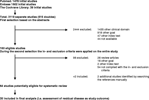
OBJECTIVES: This systematic review aimed to assess the role of magnetic resonance imaging (MRI) in evaluating residual disease extent and the ability to detect pathologic complete response (pCR) after neoadjuvant chemotherapy for invasive breast cancer. METHODS: PubMed, the Cochrane Library, MEDLINE, and Embase databases were searched for relevant studies published until 1 July 2012. After primary selection, two reviewers independently assessed the content of each eligible study using a standardised extraction form and pre-defined inclusion and exclusion criteria. RESULTS: A total of 35 eligible studies were selected. Correlation coefficients of residual tumour size assessed by MRI and pathology were good, with a median value of 0.698. Reported sensitivity, specificity, positive predictive value and negative predictive value for predicting pCR with MRI ranged from 25 to 100 %, 50-97 %, 47-73 % and 71-100 %, respectively. Both overestimation and underestimation were observed. MRI proved more accurate in determining residual disease than physical examination, mammography and ultrasound. Diagnostic accuracy of MRI after neoadjuvant chemotherapy could be influenced by treatment regimen and breast cancer subtype. CONCLUSIONS: Breast MRI accuracy for assessing residual disease after neoadjuvant chemotherapy is good and surpasses other diagnostic means. However, both overestimation and underestimation of residual disease extent could be observed. MAIN MESSAGES: • Breast MRI accuracy for assessing residual disease is good and surpasses other diagnostic means. • Correlation coefficients of residual tumour size assessed by MRI and pathology were considered good. • However, both overestimation and underestimation of residual disease were observed. • Diagnostic accuracy of MRI seems to be affected by treatment regimen and breast cancer subtype.
Overview
The claims and statistics of this paper haven’t been checked yet.
Run the check — free, no account needed. It reads the open-access text and checks the sources it cites.
Integrity signals
Registry
Cites
Statistics
Figures
Verification
Claims the paper makes
The claims and statistics of this paper haven’t been checked yet.
Run the check — free, no account needed. It reads the open-access text and checks the sources it cites.
Claims other papers make about this one
Each is checked against this paper’s own text: does it say what the citing paper says it does?
The source backs part of the claim, not all of it — or backs it for a different population, condition or comparison.
In the citing paper
“MRI is superior to mammography and US in determining tumor extent and morphology before and after NAC because it provides comprehensive imaging information about tumors (6).”
From A multimodal and fully automated system for prediction of pathological complete response to neoadjuvant chemotherapy in breast cancer (2025) · cites this paper as [6]
Experimental — accuracy not yet measured
Self-check
Percentages, sums and reported differences re-computed from the paper’s own numbers.
The self-check needs the paper’s full text.
Citations
Cites
STATISTICAL METHODS FOR ASSESSING AGREEMENT BETWEEN TWO METHODS OF CLINICAL MEASUREMENT
Martin Bland, J., & Altman, D. (1986) · The Lancet · doi:10.1016/s0140-6736(86)90837-8 · Cited by 47,965
Berg, W. A., Gutierrez, L., NessAiver, M. S., Carter, W. B., Bhargavan, M., Lewis, R. S., & Ioffe, O. B. (2004) · Radiology · doi:10.1148/radiol.2333031484 · Cited by 1,441
Neoadjuvant Versus Adjuvant Systemic Treatment in Breast Cancer: A Meta-Analysis
Mauri, D., Pavlidis, N., & Ioannidis, J. P. A. (2005) · JNCI Journal of the National Cancer Institute · doi:10.1093/jnci/dji021 · Cited by 1,214
Carney, P. A., Miglioretti, D. L., Yankaskas, B. C., Kerlikowske, K. M., Rosenberg, R., Rutter, C. M., Geller, B. M., Abraham, L., Taplin, S. H., Dignan, M., et al. (2003) · Annals of Internal Medicine · doi:10.7326/0003-4819-138-3-200302040-00008 · Cited by 1,144
Overview of resistance to systemic therapy in patients with breast cancer
Gonzalez-Angulo, A. M., Morales-Vasquez, F., & Hortobagyi, G. N. (2007) · Advances in Experimental Medicine and Biology · doi:10.1007/978-0-387-74039-3_1 · Cited by 904
Hylton, N. M., Blume, J. D., Bernreuter, W. K., Pisano, E. D., Rosen, M. A., Morris, E. A., Weatherall, P. T., Lehman, C. D., Newstead, G. M., Polin, S. M., et al. (2012) · Radiology · doi:10.1148/radiol.12110748 · Cited by 499
Yeh, E. D., Slanetz, P. J., Kopans, D. B., Rafferty, E. A., Georgian-Smith, D., Moy, L. P., Halpern, E. F., Moore, R. H., Kuter, I., & Taghian, A. G. (2005) · American Journal of Roentgenology · doi:10.2214/ajr.184.3.01840868 · Cited by 400
Partridge, S. C., Gibbs, J., Lü, Y., Esserman, L. J., Sudilovsky, D., & Hylton, N. M. (2002) · American Journal of Roentgenology · doi:10.2214/ajr.179.5.1791193 · Cited by 337
Loo, C. E., Straver, M. E., Rodenhuis, S., Muller, S. H., Wesseling, J., Peeters, M.-J. T. F. D. V., & Gilhuijs, K. G. A. (2011) · Journal of Clinical Oncology · doi:10.1200/jco.2010.31.1258 · Cited by 303
Park, S. H., Moon, W. K., Cho, N., Song, I. C., Chang, J. M., Park, I.-A., Han, W., & Noh, D. (2010) · Radiology · doi:10.1148/radiol.10092021 · Cited by 280
Cited by
Sorted by citations
Items 101–200 of 237
Chan, S., Chang, Y.-C., Huang, P., Ouyang, Y., Chang, Y., Chang, R., Chai, J., Chen, C. C., Chen, H., Chang, C.-I., et al. (2019) · BioMed Research International · doi:10.1155/2019/3843295 · Cited by 13
Pasquero, G., Surace, A., Ponti, A., Bortolini, M., Tota, D., Mano, M. P., Arisio, R., Benedetto, C., & Baù, M. G. (2020) · In Vivo · doi:10.21873/invivo.11857 · Cited by 13
Antolín, S., Nebril, B. A., Albaina, L., Concha, Á., Santiago, P., García‐Caballero, T., Mosquera, J., Varela, J. R., Soler, R., & Calvo, L. (2018) · Breast Cancer Targets and Therapy · doi:10.2147/bctt.s179750 · Cited by 12
Kim, S., Lee, J., Cho, N., & Kim, Y.-G. (2024) · Scientific Reports · doi:10.1038/s41598-024-74276-w · Cited by 11
Negrão, E. M. S., Bitencourt, A. G. V., de Souza, J. A., & Marques, E. F. (2019) · Radiologia Brasileira · doi:10.1590/0100-3984.2018.0149 · Cited by 11
Murphy, C., Mukaro, V. R., Tobler, R., Asher, R., Gibbs, E., West, L., Giuffrè, B., Baron-Hay, S. E., & Khasraw, M. (2017) · Internal Medicine Journal · doi:10.1111/imj.13617 · Cited by 11
Svecic, A., Mansour, R., Tang, A., & Kadoury, S. (2021) · PLoS ONE · doi:10.1371/journal.pone.0259692 · Cited by 11
Savaridas, S., Vinnicombe, S., Warwick, V., & Evans, A. J. (2023) · British Journal of Radiology · doi:10.1259/bjr.20220921 · Cited by 10
Onishi, N., Bareng, T. J., Gibbs, J., Li, W., Price, E. R., Joe, B. N., Kornak, J., Esserman, L. J., Newitt, D. C., Hylton, N. M., et al. (2023) · Radiology Imaging Cancer · doi:10.1148/rycan.220126 · Cited by 10
Breast Magnetic Resonance Imaging Performance
Melsaether, A. N., & Gudi, A. (2014) · Topics in Magnetic Resonance Imaging · doi:10.1097/rmr.0000000000000035 · Cited by 10
Moustafa, A. F. I., Kamal, R. M., Gomaa, M. M. M., Mostafa, S., Mubarak, R. I., & Eladawy, M. (2019) · The Egyptian Journal of Radiology and Nuclear Medicine · doi:10.1186/s43055-019-0041-8 · Cited by 10
Cho, I.-H., & Kong, E. (2016) · Nuclear Medicine and Molecular Imaging · doi:10.1007/s13139-016-0446-5 · Cited by 10
Liedtke, C., Thill, M., Hanf, V., & Schütz, F. (2014) · Breast Care · doi:10.1159/000363591 · Cited by 9
Breast Magnetic Resonance Imaging for Patients With Newly Diagnosed Breast Cancer: A Review
Kim, S., & Cho, N. (2022) · Journal of Breast Cancer · doi:10.4048/jbc.2022.25.e35 · Cited by 9
Verma, M., Abdelrahman, L., Collado‐Mesa, F., & Abdel-Mottaleb, M. S. (2023) · Diagnostics · doi:10.3390/diagnostics13132251 · Cited by 9
Myers, K. S., Stern, E., Ambinder, E. B., & Oluyemi, E. T. (2020) · British Journal of Radiology · doi:10.1259/bjr.20201202 · Cited by 9
Yaghoobpoor, S., Fathi, M., Ghorani, H., Valizadeh, P., Jannatdoust, P., Tavasol, A., Zarei, M., & Arian, A. (2024) · European Journal of Radiology Open · doi:10.1016/j.ejro.2024.100561 · Cited by 8
Magnetic Resonance Imaging of the Breast
Holbrook, A. I., & Newell, M. S. (2016) · Clinical Obstetrics & Gynecology · doi:10.1097/grf.0000000000000189 · Cited by 8
Hu, X., Li, Y., Jin, G., Lai, S., Huang, X., & Su, D. (2017) · Oncotarget · doi:10.18632/oncotarget.18999 · Cited by 8
Soliman, A. H., & Osman, A. (2018) · The Egyptian Journal of Radiology and Nuclear Medicine · doi:10.1016/j.ejrnm.2018.06.010 · Cited by 8
Kamal, R. M., Saad, S. M., Moustafa, A. F. I., Gomaa, M. M. M., Mokhtar, O., Gouda, I., Hassan, A., Hilal, A., & ElZayat, A. (2020) · The Egyptian Journal of Radiology and Nuclear Medicine · doi:10.1186/s43055-020-00275-2 · Cited by 8
Omitting Surgery in Complete Responders After Neoadjuvant Chemotherapy: The Quest Continues
Mamounas, E. P. (2018) · Annals of Surgical Oncology · doi:10.1245/s10434-018-6678-0 · Cited by 8
Zhu, C., Chen, M., Liu, Y., Li, P., Ye, W., Ye, H., Ye, Y., Liu, Z., Liang, C., & Liu, C. (2023) · Quantitative Imaging in Medicine and Surgery · doi:10.21037/qims-22-1170 · Cited by 7
Vidali, S., Irmici, G., Depretto, C., Bellini, C., Pugliese, F., Incardona, L. A., Naro, F. D., Benedetto, D. D., Filippo, G. D., Ferraro, F., et al. (2024) · Cancers · doi:10.3390/cancers16152694 · Cited by 7
Yin, L., Wei, X. C., Zhang, Q., Xiang, L., Zhang, Y., Wang, D., Chen, P., Cao, X. D., Shaibu, Z., & Qin, R. (2024) · The Breast · doi:10.1016/j.breast.2024.103797 · Cited by 7
Apte, A., Marsh, S., Chandrasekharan, S., & Chakravorty, A. (2021) · Annals of Medicine and Surgery · doi:10.1016/j.amsu.2021.102380 · Cited by 7
Savaridas, S., Whelehan, P., Warwick, V., Vinnicombe, S., & Evans, A. J. (2022) · British Journal of Radiology · doi:10.1259/bjr.20210779 · Cited by 7
Kwon, M.-R., Chu, J., Kook, S. H., & Kim, E. Y. (2022) · Clinical Imaging · doi:10.1016/j.clinimag.2022.05.002 · Cited by 7
Bear, H. D. (2014) · Breast Diseases A Year Book Quarterly · doi:10.1016/j.breastdis.2014.01.047 · Cited by 7
Weiss, A., Agnese, D. M., Al‐Hilli, Z., Cabıoğlu, N., Farr, D., Kantor, O., Obeng‐Gyasi, S., & Wilke, L. G. (2025) · Annals of Surgical Oncology · doi:10.1245/s10434-025-17405-7 · Cited by 6
Reis, J. S., Thomas, O. M. T., Lahooti, M., Lyngra, M., Schandiz, H., Boavida, J., Gjesdal, K., Sauer, T., Geisler, J., & Geitung, J. T. (2021) · Breast Cancer Research and Treatment · doi:10.1007/s10549-021-06343-z · Cited by 6
Shawky-Abdelfatah, N. O., Abdallah, R. H., Ibrahim, S. F., & Ahmed, A. I. (2022) · The Egyptian Journal of Radiology and Nuclear Medicine · doi:10.1186/s43055-022-00740-0 · Cited by 6
Browne, R. C., McAnena, P. F., O’Halloran, N., Moloney, B., Crilly, E., Kerin, M. J., & Lowery, A. (2022) · Breast Cancer Basic and Clinical Research · doi:10.1177/11782234221103504 · Cited by 6
Perfusion Imaging by Magnetic Resonance
Sánchez‐Gonzalez, J., Luna, A., & da Cruz, L. C. H. (2013) · Springer eBooks · doi:10.1007/978-3-642-40412-2_16 · Cited by 6
Bufi, E., Belli, P., Matteo, M. D., Giuliani, M., Tumino, M., Rinaldi, P. M., Nardone, L., Franceschini, G., Mulè, A., & Bonomo, L. (2016) · Clinical Breast Cancer · doi:10.1016/j.clbc.2016.06.007 · Cited by 6
Bayoumi, D., Zaky, M. M., Ibrahim, D. A., Abdallah, A., & Abouelkhair, K. M. (2019) · Polish Journal of Radiology · doi:10.5114/pjr.2019.92282 · Cited by 6
Zhang, J., Wu, Q., Peng, L., Zhu, X., & Li, B. (2025) · Journal of Applied Clinical Medical Physics · doi:10.1002/acm2.70296 · Cited by 6
Arian, A., Hashemi, M. G., Talebi, V., Ahmadinejad, N., Eslami, B., Sedighi, N., & Omranipour, R. (2023) · European Journal of Radiology Open · doi:10.1016/j.ejro.2023.100517 · Cited by 5
Wang, P. N., Velikina, J., Bancroft, L. C. H., Samsonov, A. A., Kelcz, F., Strigel, R. M., & Holmes, J. H. (2022) · Tomography · doi:10.3390/tomography8030128 · Cited by 5
Eom, H. J., Kim, H. H., Kim, H. J., Choi, W. J., Chae, E. Y., Shin, H. J., & Cha, J. H. (2025) · European Radiology · doi:10.1007/s00330-025-11640-y · Cited by 4
Qadir, A., Singh, N., Dean, J., Brown, K., TACEY, M. A., Mann, B. A., Kron, T., Cahoon, G. D., Lau, E. W. F., Scott, A. M., et al. (2024) · Pilot and Feasibility Studies · doi:10.1186/s40814-024-01557-6 · Cited by 4
Lin, Y., Cheng, M., Wu, C., Huang, Y., Zhu, T., Li, J., Gao, H., & Wang, K. (2024) · The Lancet Regional Health - Western Pacific · doi:10.1016/j.lanwpc.2024.101254 · Cited by 4
Han, Y., Jung, J. G., Kim, J., Lim, C., Kim, H. K., Lee, H., Moon, H., & Han, W. (2023) · International Journal of Surgery · doi:10.1097/js9.0000000000000754 · Cited by 4
Qadir, A., Singh, N., Moe, A. A. K., Cahoon, G. D., Lye, J., Chao, M., Foroudi, F., & Uribe, S. A. (2024) · Clinical Breast Cancer · doi:10.1016/j.clbc.2024.05.010 · Cited by 4
Yeh, E. D., Rives, A. F., Nakhlis, F., Bay, C. P., Harrison, B. T., Bellon, J. R., Remolano, M. C., Jacene, H. A., Giess, C. S., & Overmoyer, B. A. (2021) · Academic Radiology · doi:10.1016/j.acra.2021.08.003 · Cited by 4
Peng, Y., Yuan, F., Xie, F., Yang, H., Wang, S.-H., Wang, C., Yang, Y., Du, W., Liu, M., & Wang, S.-H. (2023) · Clinical Radiology · doi:10.1016/j.crad.2022.12.002 · Cited by 3
Theunissen, J. E. M., van Haaren, E. R. M., Frotscher, C., Körver‐Steeman, R., Janssen, A., Vissers, Y. L. J., van Bastelaar, J., Valentijn-Morsing, A., Bouwman, L. H., & Lobbes, M. B. I. (2024) · Journal of Cancer · doi:10.7150/jca.98597 · Cited by 3
Self-citationTumour volume analysis applied to imaging and histological examinations in breast cancer
Gordon, A. B., Sheeka, A., Cleator, S., Leff, D. R., & Lim, A. K. P. (2025) · European Journal of Surgical Oncology · doi:10.1016/j.ejso.2025.109578 · Cited by 3
CEM immediately after contrast-enhanced CT: a one-step staging of breast cancer
Ancona, A., Telegrafo, M., Fella, R. R., Iamele, D., Cantore, S., & Moschetta, M. (2024) · European Radiology Experimental · doi:10.1186/s41747-024-00440-6 · Cited by 3
A Practical Guide to Wireless Breast Localization Devices
Sabato, J. J. D., Kim, J., Sehareen, S., Vincoff, N. S., & Kalantarova, S. (2024) · Applied Radiology · doi:10.37549/ar-d-24-0001 · Cited by 3
Cuadriello, E. F., Fernández‐Guinea, O., Eiró, N., González, L. O., Junquera, S., & Vizoso, F. J. (2016) · Magnetic Resonance Imaging · doi:10.1016/j.mri.2016.04.005 · Cited by 3
Breast Imaging and Image-Guided Biopsy Techniques
Ganott, M. A., Griffith, B. M., & Rudzinski, S. M. (2019) · Springer eBooks · doi:10.1007/978-3-030-04606-4_5 · Cited by 3
Chotai, N. C., Loke, S. C., & Yu, T. E. (2021) · Advances in Breast Cancer Research · doi:10.4236/abcr.2021.104016 · Cited by 2
Kwon, M.-R., Ko, E. Y., Lee, J. E., Han, B., Ko, E. S., Choi, J. S., Kim, H., Kim, M. K., Yu, J. H., Lee, H., et al. (2024) · Breast Cancer · doi:10.1007/s12282-024-01638-7 · Cited by 2
Park, G. E., Mun, H. S., Kim, S. H., & Kang, B. J. (2025) · Academic Radiology · doi:10.1016/j.acra.2025.04.010 · Cited by 2
Park, G. E., Shin, K., Mun, H. S., & Kang, B. J. (2025) · Tomography · doi:10.3390/tomography11070072 · Cited by 2
Steyerová, P. (2017) · Springer eBooks · doi:10.1007/978-3-319-56673-3_12 · Cited by 2
Quantitative DCE-MRI of the Breast
Huang, W. (2022) · Advances in magnetic resonance technology and applications · doi:10.1016/b978-0-12-822729-9.00007-2 · Cited by 2
Physicians’ Hierarchy of Tumor Biomarkers for Optimizing Chemotherapy in Breast Cancer Care
Thannickal, H. H., Eltoum, N., Henderson, N. L., Wallner, L. P., Wagner, L. I., Wolff, A. C., & Rocque, G. B. (2023) · The Oncologist · doi:10.1093/oncolo/oyad198 · Cited by 2
Hernández-Cortes, G., Fuertes, S., Martínez, V., Rubio, M., Vera, U., Murillo, R., González‐Cortijo, L., del Cerro, E., Mohedano, R., & de la Cuesta, R. S. (2020) · Revista Española de Medicina Nuclear e Imagen Molecular · doi:10.1016/j.remn.2020.09.001 · Cited by 2
Mirza, S. M., O'Brien, J. P., & Aitken, J. (2016) · Breast Cancer Management · doi:10.2217/bmt-2016-0011 · Cited by 2
Hilal, T., & Patel, B. (2018) · Annals of Surgical Oncology · doi:10.1245/s10434-018-6899-2 · Cited by 2
Dodhia, P., Meepagala, S., Moallem, G., Rubin, D. L., Bean, G. R., & Rusu, M. (2024) · SPIE Medical Imaging: Imaging Informatics for Healthcare, Research, and Applications · doi:10.1117/12.3006495 · Cited by 1
Martínez, A. M., Bravo, E. I., Alvà, H. P., Martínez, P. R., Tomas, M., Miguel, I. P., & Puyalto, P. (2024) · Radiología (English Edition) · doi:10.1016/j.rxeng.2024.04.001 · Cited by 1
Bansal, A., Vyas, S., Hari, S., Thulkar, S. P., Gogia, A., Deo, S. V. S., Srivastava, A. K., & Mathur, S. R. (2025) · Indian Journal of Breast Imaging · doi:10.25259/ijbi_25_2024 · Cited by 1
Iamsamang, A., Thiravit, S., Suvannarerg, V., Rushatamukayanunt, P., Korpraphong, P., Chuthapisith, S., & Imruetaicharoenchoke, W. (2025) · Siriraj Medical Journal/San Sirirat · doi:10.33192/smj.v77i9.276157 · Cited by 1
Verma, M., Collado‐Mesa, F., & Abdel-Mottaleb, M. S. (2026) · ACM Transactions on Computing for Healthcare · doi:10.1145/3793536 · Cited by 1
Ali, M., Imran, M. J., Hussain, J., Zakria, M., Abbasi, A. E., & Sultan, A. (2026) · Biomarkers · doi:10.1080/1354750x.2026.2633567 · Cited by 1
Hong, L. I., Ruofan, H., Huang, W. Q., Huang, S., Xu, X., Shao, B., & Chen, H. (2026) · Academic Radiology · doi:10.1016/j.acra.2026.04.042 · Cited by 1
Therapy Response Imaging in Breast Cancer
Kataoka, M. (2020) · Medical radiology · doi:10.1007/978-3-030-31171-1_5 · Cited by 1
Contrast-Enhanced Mammography for Practicing Radiologists
Ngo, M., Kim, G., Phillips, J., Fishman, M. D. C., & Slanetz, P. J. (2022) · Advances in Clinical Radiology · doi:10.1016/j.yacr.2022.04.012 · Cited by 1
Bae, S. J., Chun, J. W., Lee, S. B., Ryu, J. M., Nam, S. J., Jeong, J., Park, H. S., & Ahn, S. G. (2024) · Breast Cancer Research · doi:10.1186/s13058-024-01807-8 · Cited by 1
Martínez, A. M., Bravo, E. I., Alvà, H. P., Martínez, P. R., Tomas, M., Miguel, I. P., & de Pablo, P. P. (2024) · Radiología · doi:10.1016/j.rx.2024.04.003 · Cited by 1
Breast cancer response post neoadjuvant chemotherapy using MRI measurements
Sivaranjini, S., & Nirmala, K. (2017) · International Conference on Signal Processing, Communication and Networking (ICSCN) · doi:10.1109/icscn.2017.8085419 · Cited by 1
Contrast-Enhanced Mammography in Neoadjuvant Therapy Response Monitoring
Iotti, V., & Rossi, P. G. (2019) · Springer eBooks · doi:10.1007/978-3-030-11063-5_8 · Cited by 1
Kanani, A. N., Sharma, N., & Buckley, D. L. (2019) · Current Breast Cancer Reports · doi:10.1007/s12609-019-0311-5 · Cited by 1
Douma, E.-S. A. M., Bendary, S. H., El-Agamawi, A. Y., Zayed, A. M., & El-Hameid, N. M. A. (2020) · Al-Azhar Medical Journal · doi:10.21608/amj.2020.120555 · Cited by 1
Contrast-Enhanced In-Phase Dixon Sequence: Impact on Biopsy Clip Detection on Breast MRI
Taylor-Cho, M. W., Robertson, S. H., Knight, J. R., Miller, M. M., & Baker, J. A. (2022) · American Journal of Roentgenology · doi:10.2214/ajr.22.28024 · Cited by 1
Aslan, A. A., Erdemli, S., Erdoğdu, İ. H., Gültekin, S., Uras, C., Tokat, F., Arıcan, A., Özer, L., Behzatoğlu, K., Meydan, N., et al. (2023) · Acta Oncologica Turcica · doi:10.5505/aot.2023.60973 · Cited by 1
Mri-Based Radiomics in Breast Cancer
Granzier, R. W. Y. (2022) · doi:10.26481/dis.20220922rg · Cited by 2
Papel de la resonancia magnética de mama en la valoración de la respuesta a la neoadyuvancia
Mur, C. G., Ciria, S. C., & Senar, A. C. (2015) · Revista de Senología y Patología Mamaria · doi:10.1016/j.senol.2015.06.002
Comprehensive breast MRI: an update
Vinnicombe, S., & Petralia, G. (2014) · Cancer Imaging · doi:10.1186/1470-7330-14-s1-o39
Breast Imaging and Image-Guided Biopsy Techniques
Ganott, M. A., Griffith, B. M., & Rudzinski, S. M. (2015) · Springer eBooks · doi:10.1007/978-3-319-22843-3_5
Hylton, N. M., Gatsonis, C. A., Rosen, M. A., Lehman, C. D., Newitt, D. C., Partridge, S. C., Bernreuter, W. K., Pisano, E. D., Morris, E. A., Weatherall, P. T., et al. (2016)
Braga, S. A. (2016) · Methods in molecular biology · doi:10.1007/978-1-4939-3347-1_15
Forme avancée de carcinome infiltrant
Mery, E., Charitansky, H., Dalenc, F., Boulet, B., & Massabeau, C. (2016) · Elsevier eBooks · doi:10.1016/b978-2-294-74449-5.00019-8
Magnetic Resonance Imaging and Neoadjuvant Chemotherapy
Le‐Petross, H. T., Lim, B., & Hylton, N. M. (2017) · Springer eBooks · doi:10.1007/978-3-319-42563-4_5
Personalized Medicine, Biomarkers of Risk and Breast MRI
Sutton, E. J., Purvis, N., Pinker, K., & Morris, E. A. (2017) · Springer eBooks · doi:10.1007/978-3-319-42563-4_17
Dratwa, C., Sebbag-Sfez, D., & Thibault, F. E. (2017) · Imagerie de la Femme · doi:10.1016/j.femme.2017.09.001
Molière, S., Roedlich, M. N., Bodin, F., Robert, É., & Mathelin, C. (2019) · Imagerie de la Femme · doi:10.1016/j.femme.2019.01.002
Gooch, J. C., Timothy, B., Chun, J., Elianna, K., Guth, A. A., Deborah, A., Richard, S., Linda, M. S., & Freya, S. (2019) · International Journal of Radiology and Imaging Technology · doi:10.23937/2572-3235.1510056
Fernandez, J. J. M., García, M. L., Rodrigo, S. P., Agustín, M. C. D., & Muñoz, L. F. (2018) · Seram
Neoadjuvant Chemotherapy and Biomarkers
Chotai, N. C., & Kulkarni, S. R. (2020) · Springer eBooks · doi:10.1007/978-981-15-1412-8_15
Černeka, E., Miletić, D., Avirović, M., Grebić, D., & Zujić, P. V. (2020) · Medicina Fluminensis · doi:10.21860/medflum2020_237302
Response Assessment and Follow-Up by Imaging in Breast Tumors
Goethem, M. V., Castello, A., Lobbes, M. B. I., Rose, F. D., Scorsetti, M., & Lopci, E. (2020) · Medical radiology · doi:10.1007/978-3-030-38261-2_24
Self-citationSyed, A., Premi, V., Varghese, R. P., Arora, M., Kapoor, A., Satish Chaitanya, K., Sharda, P., Gupta, M., & Ravi, B. (2020) · The Breast Journal · doi:10.1111/tbj.13996
Magnetic resonance-guided ROLL/SNOLL in breast cancer patients treated with neoadjuvant chemotherapy
Hernández-Cortes, G., Fuertes, S., Martinez, V., Rubio, M., Vera, U., Murillo, R., González‐Cortijo, L., del Cerro, E., Mohedano, R., & de la Cuesta, R. S. (2020) · Revista Española de Medicina Nuclear e Imagen Molecular (English Edition) · doi:10.1016/j.remnie.2020.12.003
Bi, Z., Zhang, Z.-P., Xu, C.-J., Chen, P., Qiu, P., Zhao, T., & Wang, Y. (2021) · Research Square · doi:10.21203/rs.3.rs-960396/v1
Metadata
- Authors
- Lobbes, M. B. I., Prevos, R., Smidt, M., Tjan-Heijnen, V. C. G., van Goethem, M., Schipper, R., Beets-Tan, R. G., & Wildberger, J. E.
- Affiliations
- Maastricht University Medical Centre (NL)
- Corresponding author
- Lobbes, M. B. I.
- Venue
- Insights into Imaging
- Published
- 2013-01-29
- Volume
- 4
- Issue
- 2
- Pages
- 163-175
- ISSN
- 1869-4101 (ISSN-L)
- PMC
- PMC3609956
- OpenAlex
- W2057016707
- Cited by
- 237
- References
- 56
- Citations by year
- 2026: 10 · 2025: 15 · 2024: 27 · 2023: 21 · 2022: 22 · 2021: 25 · 2020: 15 · 2019: 23 · 2018: 16 · 2017: 24 · 2016: 16 · 2015: 11 · 2014: 8 · 2013: 4
- FWCI
- 1.84
- Type
- journal article
- Language
- en
- Open access
- gold
- doi.org
- Locations
- Insights into Imaging (publishedVersion, cc-by)
- Europe PMC (PubMed Central) (publishedVersion, cc-by-nc)
- PubMed Central (publishedVersion, cc-by-nc)
- PubMed (publishedVersion)
- Research Publications (Maastricht University) (publishedVersion, cc-by)
- Research Publications (Maastricht University) (submittedVersion, cc-by)
- Topics
- Health Sciences › Medicine › Radiology, Nuclear Medicine and Imaging › MRI in cancer diagnosis
- Health Sciences › Medicine › Pathology and Forensic Medicine › Breast Lesions and Carcinomas
- Life Sciences › Biochemistry, Genetics and Molecular Biology › Cancer Research › Breast Cancer Treatment Studies
- Keywords
- breast MRI, residual disease, pathological complete response, neoadjuvant chemotherapy, invasive breast cancer, diagnostic accuracy, breast cancer subtypes
About these checks
Registry, retraction, Cites and statistics results are computed, not judged, with one exception: a printed reference that no index lists is read once by a model before it counts as a citation error. Claims are extracted by a model and kept only when their quote appears verbatim in the text; each cited source is then checked, with a model’s help, for whether it says what the claim relies on. Those verdicts can be wrong — read the claim’s quote and the cited source before relying on one.
Statistics and arithmetic are recomputed, not judged.
Confidence bands are shown only for fields calibrated on our annotated test set. None is calibrated yet, so no band is shown.
What counts as a claim ›How we measure confidence ›How we check this ›
Are you an author, or do you see a problem with this page? Request a review
