

The DOI matches its Crossref record; 56 of the 56 references it cites were found in a registry or an index.
The role of magnetic resonance imaging in assessing residual disease and pathologic complete response in breast cancer patients receiving neoadjuvant chemotherapy: a systematic review
Open accessAbstract
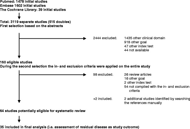
OBJECTIVES: This systematic review aimed to assess the role of magnetic resonance imaging (MRI) in evaluating residual disease extent and the ability to detect pathologic complete response (pCR) after neoadjuvant chemotherapy for invasive breast cancer. METHODS: PubMed, the Cochrane Library, MEDLINE, and Embase databases were searched for relevant studies published until 1 July 2012. After primary selection, two reviewers independently assessed the content of each eligible study using a standardised extraction form and pre-defined inclusion and exclusion criteria. RESULTS: A total of 35 eligible studies were selected. Correlation coefficients of residual tumour size assessed by MRI and pathology were good, with a median value of 0.698. Reported sensitivity, specificity, positive predictive value and negative predictive value for predicting pCR with MRI ranged from 25 to 100 %, 50-97 %, 47-73 % and 71-100 %, respectively. Both overestimation and underestimation were observed. MRI proved more accurate in determining residual disease than physical examination, mammography and ultrasound. Diagnostic accuracy of MRI after neoadjuvant chemotherapy could be influenced by treatment regimen and breast cancer subtype. CONCLUSIONS: Breast MRI accuracy for assessing residual disease after neoadjuvant chemotherapy is good and surpasses other diagnostic means. However, both overestimation and underestimation of residual disease extent could be observed. MAIN MESSAGES: • Breast MRI accuracy for assessing residual disease is good and surpasses other diagnostic means. • Correlation coefficients of residual tumour size assessed by MRI and pathology were considered good. • However, both overestimation and underestimation of residual disease were observed. • Diagnostic accuracy of MRI seems to be affected by treatment regimen and breast cancer subtype.
Overview
The claims and statistics of this paper haven’t been checked yet.
Run the check — free, no account needed. It reads the open-access text and checks the sources it cites.
Integrity signals
Registry
Cites
Statistics
Figures
Verification
Claims the paper makes
The claims and statistics of this paper haven’t been checked yet.
Run the check — free, no account needed. It reads the open-access text and checks the sources it cites.
Claims other papers make about this one
Each is checked against this paper’s own text: does it say what the citing paper says it does?
The source backs part of the claim, not all of it — or backs it for a different population, condition or comparison.
In the citing paper
“MRI is superior to mammography and US in determining tumor extent and morphology before and after NAC because it provides comprehensive imaging information about tumors (6).”
From A multimodal and fully automated system for prediction of pathological complete response to neoadjuvant chemotherapy in breast cancer (2025) · cites this paper as [6]
Experimental — accuracy not yet measured
Self-check
Percentages, sums and reported differences re-computed from the paper’s own numbers.
The self-check needs the paper’s full text.
Citations
Cites
STATISTICAL METHODS FOR ASSESSING AGREEMENT BETWEEN TWO METHODS OF CLINICAL MEASUREMENT
Martin Bland, J., & Altman, D. (1986) · The Lancet · doi:10.1016/s0140-6736(86)90837-8 · Cited by 47,965
Berg, W. A., Gutierrez, L., NessAiver, M. S., Carter, W. B., Bhargavan, M., Lewis, R. S., & Ioffe, O. B. (2004) · Radiology · doi:10.1148/radiol.2333031484 · Cited by 1,441
Neoadjuvant Versus Adjuvant Systemic Treatment in Breast Cancer: A Meta-Analysis
Mauri, D., Pavlidis, N., & Ioannidis, J. P. A. (2005) · JNCI Journal of the National Cancer Institute · doi:10.1093/jnci/dji021 · Cited by 1,214
Carney, P. A., Miglioretti, D. L., Yankaskas, B. C., Kerlikowske, K. M., Rosenberg, R., Rutter, C. M., Geller, B. M., Abraham, L., Taplin, S. H., Dignan, M., et al. (2003) · Annals of Internal Medicine · doi:10.7326/0003-4819-138-3-200302040-00008 · Cited by 1,144
Overview of resistance to systemic therapy in patients with breast cancer
Gonzalez-Angulo, A. M., Morales-Vasquez, F., & Hortobagyi, G. N. (2007) · Advances in Experimental Medicine and Biology · doi:10.1007/978-0-387-74039-3_1 · Cited by 904
Hylton, N. M., Blume, J. D., Bernreuter, W. K., Pisano, E. D., Rosen, M. A., Morris, E. A., Weatherall, P. T., Lehman, C. D., Newstead, G. M., Polin, S. M., et al. (2012) · Radiology · doi:10.1148/radiol.12110748 · Cited by 499
Yeh, E. D., Slanetz, P. J., Kopans, D. B., Rafferty, E. A., Georgian-Smith, D., Moy, L. P., Halpern, E. F., Moore, R. H., Kuter, I., & Taghian, A. G. (2005) · American Journal of Roentgenology · doi:10.2214/ajr.184.3.01840868 · Cited by 400
Partridge, S. C., Gibbs, J., Lü, Y., Esserman, L. J., Sudilovsky, D., & Hylton, N. M. (2002) · American Journal of Roentgenology · doi:10.2214/ajr.179.5.1791193 · Cited by 337
Loo, C. E., Straver, M. E., Rodenhuis, S., Muller, S. H., Wesseling, J., Peeters, M.-J. T. F. D. V., & Gilhuijs, K. G. A. (2011) · Journal of Clinical Oncology · doi:10.1200/jco.2010.31.1258 · Cited by 303
Park, S. H., Moon, W. K., Cho, N., Song, I. C., Chang, J. M., Park, I.-A., Han, W., & Noh, D. (2010) · Radiology · doi:10.1148/radiol.10092021 · Cited by 280
Cited by
Sorted by citations
Items 201–237 of 237
Breast Cancer Imaging Pre- and Post-Neoadjuvant Systemic Therapy
Chung, S. H., & Raza, S. (2021) · Springer eBooks · doi:10.1007/978-3-030-88020-0_9
Intervencionismo ecográfico en imagen mamaria
Goldaracena, J. M. O. (2022) · Radiología · doi:10.1016/j.rx.2021.11.003
Ultrasound-guided interventional procedures in breast imaging
Goldaracena, J. M. O. (2022) · Radiología (English Edition) · doi:10.1016/j.rxeng.2021.09.006
Savaridas, S., Vinnicombe, S., Warwick, V., & Evans, A. J. (2022) · medRxiv · doi:10.1101/2022.08.26.22279254
Disease and Treatment Monitoring
Li, W., Newitt, D. C., Partridge, S. C., & Hylton, N. M. (2022) · Elsevier eBooks · doi:10.1016/b978-0-323-79702-3.00005-8
Neoadjuvant therapy response assessment with breast MRI
Arjmandi, F. K., & Doğan, B. E. (2022) · Advances in magnetic resonance technology and applications · doi:10.1016/b978-0-12-822729-9.00009-6
Iquebal, A. S., Zhou, S., Pfeiffer, N., Ranjbar, S., Banerjee, I., Swanson, K. R., Batalini, F., Anderson, K. S., Murtaza, M., Pockaj, B. A., et al. (2022) · Research Square · doi:10.21203/rs.3.rs-2232518/v1
Kim, E. Y., Yun, J.-S., Park, Y. L., Park, C. H., Kwon, M.-R., Moon, J. H., Youn, I., Choi, Y. J., & Kook, S. H. (2022) · Journal of Breast Disease · doi:10.14449/jbd.2022.10.2.69
Cortés, G. H., de Vega, V. M., Cabero, S. F., Alonso, M. R., Cortijo, L. G., García, R. M., & Abbad, R. S. D. L. C. (2023) · Revista de Senología y Patología Mamaria · doi:10.1016/j.senol.2022.100438
Bae, S. J., Chun, J. W., Lee, S. B., Ryu, J. M., Nam, S. J., Jeong, J., Park, H. S., & Ahn, S. G. (2023) · Research Square · doi:10.21203/rs.3.rs-3036122/v1
ERKAN, S., Yabanoğlu, H., GÜNDOĞDU, R., Avcı, T., & Çakmak, E. (2023) · Cukurova Anestezi ve Cerrahi Bilimler Dergisi · doi:10.36516/jocass.1294672
(2023) · International Journal on Oncology and Radiotherapy · doi:10.51626/ijor.2023.04.00025
Ko, T., Lai, A., Leung, B., Law, M., Wong, A., Chin, K., & Wong, W. (2024) · Hong Kong Journal of Radiology · doi:10.12809/hkjr2217606
THE METAL CLIPS AS BREAST CANCER MARKERS IN ASSESSMENT OF RESPONSE AFTER NEOADJUVANT CHEMOTHERAPY
Gambarova, G. H., Asgarova, H. E., Aghamaliyeva, A. J., PANAKHOVA, M. S., AGABEYLY, L. Z., & Qorçiyeva, G. S. (2024) · World of Medicine and Biology · doi:10.26724/2079-8334-2024-2-88-28-32
Yashaswinii, P., Christina, E. P., Ramaswami, S., Sl, H., & Natarajan, P. (2024) · Cureus · doi:10.7759/cureus.65355
Narasimhamurthy, M., O’Brien, S. R., Andrejeva, L., & Nayak, A. (2024) · Springer eBooks · doi:10.1007/978-3-031-65711-5_9
Park, G. E., Kang, B. J., Kim, S. H., & Mun, H. S. (2024) · Life · doi:10.3390/life14111449
Guo, S., Wang, D., Zhao, Q., Bi, Z., Li, W., & Zhu, J. (2024) · Quantitative Imaging in Medicine and Surgery · doi:10.21037/qims-24-511
Optimization of radiotherapy in low-risk breast cancer patients
Civil, Y. A. (2025) · doi:10.5463/thesis.1151
Musah, T. (2025) · Lecture notes in computer science · doi:10.1007/978-3-032-05559-0_8
Claassens, E. L., van Amstel, F. J. G., de Boer, M., Lalji, U. C., Simons, J. M., Kooreman, L. F. S., Smidt, M. L., & van Nijnatten, T. J. A. (2025) · European Journal of Surgical Oncology · doi:10.1016/j.ejso.2025.111205
Self-citationWright, G. C., Glover, L., & Hubbard, T. J. E. (2025) · The Breast · doi:10.1016/j.breast.2025.104663
Contrast-Enhanced Mammography: A Comprehensive Overview of Current Status and Future Directions
Renganathan, R., Palanisamy, S., Kini, D. V., Subramaniam, P., Krishnappa, L., & Chang, J. M. (2025) · Journal of the Korean Society of Radiology · doi:10.3348/jksr.2025.0053
Graeser, M., Schrading, S., Gluz, O., Strobel, K., Herzog, C., Umutlu, L., Frydrychowicz, A., Rjosk-Dendorfer, D., Würstlein, R., Culemann, R., et al. (2021) · Figshare · doi:10.6084/m9.figshare.14247656
Graeser, M., Schrading, S., Gluz, O., Strobel, K., Herzog, C., Umutlu, L., Frydrychowicz, A., Rjosk-Dendorfer, D., Würstlein, R., Culemann, R., et al. (2021) · Figshare · doi:10.6084/m9.figshare.14247659
Graeser, M., Schrading, S., Gluz, O., Strobel, K., Herzog, C., Umutlu, L., Frydrychowicz, A., Rjosk-Dendorfer, D., Würstlein, R., Culemann, R., et al. (2021) · Figshare · doi:10.6084/m9.figshare.14247653
Bae, S. J., Chun, J. W., Lee, S. B., Ryu, J. M., Nam, S. J., Jeong, J., Park, H. S., & Ahn, S. G. (2024) · Figshare · doi:10.6084/m9.figshare.26708189
Graeser, M., Schrading, S., Gluz, O., Strobel, K., Herzog, C., Umutlu, L., Frydrychowicz, A., Rjosk-Dendorfer, D., Würstlein, R., Culemann, R., et al. (2021) · Figshare · doi:10.6084/m9.figshare.14247662
van Amstel, F. J. G., van Mierlo, R. G. M., Nelemans, P. J., Engelen, S. M. E., Houwers, J. B., Kooreman, L. F. S., Tjan‐Heijnen, V. C. G., Siesling, S., Smidt, M. L., & van Nijnatten, T. J. A. (2025) · European Radiology · doi:10.1007/s00330-025-12249-x
Self-citationMRI response to neoadjuvant chemotherapy and prognostic implications in breast cancer patients
Damião, S. Q., da Cunha, E. F. R., Tavares, M. C., Sanches, S. M., Osorio, C. A. B. D. T., Brot, M. D., Makdissi, F. B. A., & Bitencourt, A. G. V. (2026) · British Journal of Radiology · doi:10.1093/bjr/tqag023
Yılmaz, E., Mısır, M. E., Şeker, M. E., Güldoğan, N., Turk, E. B., Gorucu, H., & Arıbal, E. (2026) · Clinical Radiology · doi:10.1016/j.crad.2026.107349
Xu, M., Wang, A., Shan, D., Chen, X., Zhang, R., Xu, C., Yuan, J., Li, J., An, G., Shen, Z., et al. (2026) · BMC Medical Imaging · doi:10.1186/s12880-026-02311-5
Bellini, C., Bicchierai, G., Maiello, C., Amato, F., Benedetto, D. D., Naro, F. D., Vidali, S., Tonelli, P., Vanzi, E., Boeri, C., et al. (2026) · European Radiology · doi:10.1007/s00330-026-12633-1
di Micco, R., Saraçoğlu, S., Pfob, A., Canevari, C., Gallivanone, F., Antunovic, L., Gelardi, F., Scifo, P., Neri, I., Losio, C., et al. (2026) · Frontiers in Oncology · doi:10.3389/fonc.2026.1845216
Sun, X., Wang, D., Zhou, Q., Liu, W., Gao, X., Shen, X., Yang, X., Ma, T., Ma, T., Liu, C., et al. (2026) · European Journal of Radiology · doi:10.1016/j.ejrad.2026.113214
D’Archi, S., Carnassale, B., Scardina, L., Accetta, C., Lauretis, F. D., Guglielmo, E. D., Franco, A., Gambaro, E., Magno, S., Moschella, F., et al. (2026) · European Journal of Surgical Oncology · doi:10.1016/j.ejso.2026.112159
Metadata
- Authors
- Lobbes, M. B. I., Prevos, R., Smidt, M., Tjan-Heijnen, V. C. G., van Goethem, M., Schipper, R., Beets-Tan, R. G., & Wildberger, J. E.
- Affiliations
- Maastricht University Medical Centre (NL)
- Corresponding author
- Lobbes, M. B. I.
- Venue
- Insights into Imaging
- Published
- 2013-01-29
- Volume
- 4
- Issue
- 2
- Pages
- 163-175
- ISSN
- 1869-4101 (ISSN-L)
- PMC
- PMC3609956
- OpenAlex
- W2057016707
- Cited by
- 237
- References
- 56
- Citations by year
- 2026: 10 · 2025: 15 · 2024: 27 · 2023: 21 · 2022: 22 · 2021: 25 · 2020: 15 · 2019: 23 · 2018: 16 · 2017: 24 · 2016: 16 · 2015: 11 · 2014: 8 · 2013: 4
- FWCI
- 1.84
- Type
- journal article
- Language
- en
- Open access
- gold
- doi.org
- Locations
- Insights into Imaging (publishedVersion, cc-by)
- Europe PMC (PubMed Central) (publishedVersion, cc-by-nc)
- PubMed Central (publishedVersion, cc-by-nc)
- PubMed (publishedVersion)
- Research Publications (Maastricht University) (publishedVersion, cc-by)
- Research Publications (Maastricht University) (submittedVersion, cc-by)
- Topics
- Health Sciences › Medicine › Radiology, Nuclear Medicine and Imaging › MRI in cancer diagnosis
- Health Sciences › Medicine › Pathology and Forensic Medicine › Breast Lesions and Carcinomas
- Life Sciences › Biochemistry, Genetics and Molecular Biology › Cancer Research › Breast Cancer Treatment Studies
- Keywords
- breast MRI, residual disease, pathological complete response, neoadjuvant chemotherapy, invasive breast cancer, diagnostic accuracy, breast cancer subtypes
About these checks
Registry, retraction, Cites and statistics results are computed, not judged, with one exception: a printed reference that no index lists is read once by a model before it counts as a citation error. Claims are extracted by a model and kept only when their quote appears verbatim in the text; each cited source is then checked, with a model’s help, for whether it says what the claim relies on. Those verdicts can be wrong — read the claim’s quote and the cited source before relying on one.
Statistics and arithmetic are recomputed, not judged.
Confidence bands are shown only for fields calibrated on our annotated test set. None is calibrated yet, so no band is shown.
What counts as a claim ›How we measure confidence ›How we check this ›
Are you an author, or do you see a problem with this page? Request a review
