

The DOI matches its Crossref record; 40 of the 40 references it cites were found in a registry or an index.
Periodontal Ehlers-Danlos Syndrome Is Caused by Mutations in C1R and C1S , which Encode Subcomponents C1r and C1s of Complement
Open accessAbstract
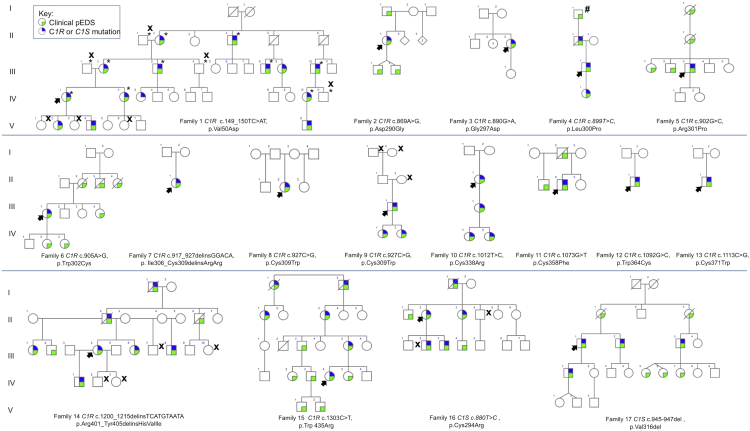
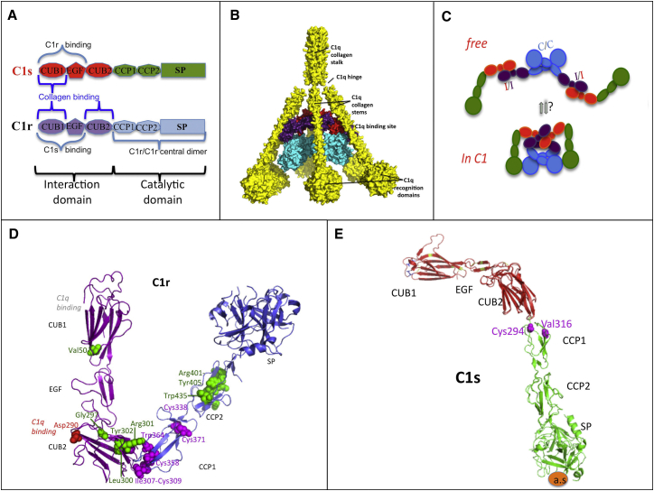
Periodontal Ehlers-Danlos syndrome (pEDS) is an autosomal-dominant disorder characterized by early-onset periodontitis leading to premature loss of teeth, joint hypermobility, and mild skin findings. A locus was mapped to an approximately 5.8 Mb region at 12p13.1 but no candidate gene was identified. In an international consortium we recruited 19 independent families comprising 107 individuals with pEDS to identify the locus, characterize the clinical details in those with defined genetic causes, and try to understand the physiological basis of the condition. In 17 of these families, we identified heterozygous missense or in-frame insertion/deletion mutations in C1R (15 families) or C1S (2 families), contiguous genes in the mapped locus that encode subunits C1r and C1s of the first component of the classical complement pathway. These two proteins form a heterotetramer that then combines with six C1q subunits. Pathogenic variants involve the subunit interfaces or inter-domain hinges of C1r and C1s and are associated with intracellular retention and mild endoplasmic reticulum enlargement. Clinical features of affected individuals in these families include rapidly progressing periodontitis with onset in the teens or childhood, a previously unrecognized lack of attached gingiva, pretibial hyperpigmentation, skin and vascular fragility, easy bruising, and variable musculoskeletal symptoms. Our findings open a connection between the inflammatory classical complement pathway and connective tissue homeostasis.
Overview
The claims and statistics of this paper haven’t been checked yet.
Run the check — free, no account needed. It reads the open-access text and checks the sources it cites.
Integrity signals
Registry
Cites
Statistics
Figures
Verification
Claims the paper makes
The claims and statistics of this paper haven’t been checked yet.
Run the check — free, no account needed. It reads the open-access text and checks the sources it cites.
Claims other papers make about this one
Each is checked against this paper’s own text: does it say what the citing paper says it does?
The cited source doesn’t support the claim as written.
In the citing paper
“In 93 individuals with C1R or C1S mutations reported by Kapferer-Seebacher et al. [2016] joint hypermobility was reported in 56% of patients, mostly affecting the fingers (30%), the elbows (19%), knees (11%), hips, wrist, and ankle (3%).”
From The Ehlers–Danlos syndromes, rare types (2017) · cites this paper as Kapferer-Seebacher et al., 2016
Checked by a reasoning model — may be wrong.
Experimental — accuracy not yet measuredThe source backs part of the claim, not all of it — or backs it for a different population, condition or comparison.
In the citing paper
“Marfanoid habitus, scoliosis, osteoarthritis, flat feet, and hernia were not consistent features [Kapferer-Seebacher et al., 2016].”
From The Ehlers–Danlos syndromes, rare types (2017) · cites this paper as Kapferer-Seebacher et al., 2016
Experimental — accuracy not yet measuredThe source backs part of the claim, not all of it — or backs it for a different population, condition or comparison.
In the citing paper
“Atrophic scars were present in 50% of cases [Kapferer-Seebacher et al., 2016].”
From The Ehlers–Danlos syndromes, rare types (2017) · cites this paper as Kapferer-Seebacher et al., 2016
Experimental — accuracy not yet measuredThe source backs part of the claim, not all of it — or backs it for a different population, condition or comparison.
In the citing paper
“Kapferer-Seebacher et al. [2016] reported a prevalence of 16% for vascular complications (cerebral aneurisms or aortic dissection), and one individual with three events of organ rupture (once duodenum, twice lung).”
From The Ehlers–Danlos syndromes, rare types (2017) · cites this paper as Kapferer-Seebacher et al., 2016
Experimental — accuracy not yet measuredThe source backs part of the claim, not all of it — or backs it for a different population, condition or comparison.
In the citing paper
“Heterozygous missense or in-frame insertion/deletion variants in C1R were detected in 15 families (comprising 76 affected individuals), and heterozygous missense or in-frame insertion/ deletion variants in C1S were detected in two families (comprising 16 affected individuals).”
From The Ehlers–Danlos syndromes, rare types (2017) · cites this paper as Kapferer-Seebacher et al., 2016
Experimental — accuracy not yet measured
The source backs part of the claim, not all of it — or backs it for a different population, condition or comparison.
In the citing paper
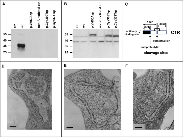
“Additionally, patients' fibroblasts showed an increased proportion of dilated rough ER (RER) cisternae [Reinstein et al., 2012;Kapferer-Seebacher et al., 2016].”
From The Ehlers–Danlos syndromes, rare types (2017) · cites this paper as Kapferer-Seebacher et al., 2016
Experimental — accuracy not yet measuredThe source backs part of the claim, not all of it — or backs it for a different population, condition or comparison.
In the citing paper
“There were single patients with autoimmunologic disorders like Sj€ ogren syndrome, rosacea, and Crohn's disease [Kapferer-Seebacher et al., 2016].”
From The Ehlers–Danlos syndromes, rare types (2017) · cites this paper as Kapferer-Seebacher et al., 2016
Experimental — accuracy not yet measuredThe cited source says what the claim attributes to it.
In the citing paper
“In 17 of these families, heterozygous missense or in-frame insertion/deletion mutations in C1R (15 families) or C1S (two families) were identified.”
From The Ehlers–Danlos syndromes, rare types (2017) · cites this paper as Kapferer-Seebacher et al., 2016
The cited source says what the claim attributes to it.
In the citing paper
“Variants never involved the catalytic domain of C1r or C1s [Kapferer-Seebacher et al., 2016].”
From The Ehlers–Danlos syndromes, rare types (2017) · cites this paper as Kapferer-Seebacher et al., 2016
The cited source says what the claim attributes to it.
In the citing paper
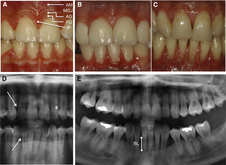
“A characteristic recently described feature is a striking lack of attached gingiva causing oral tissue fragility (Fig. 1) [Kapferer-Seebacher et al., 2016].”
From The Ehlers–Danlos syndromes, rare types (2017) · cites this paper as Kapferer-Seebacher et al., 2016
The cited source says what the claim attributes to it.
In the citing paper
“In 2016, an international consortium published 19 independent families comprising 107 individuals with pEDS to identify the genetic locus [Kapferer-Seebacher et al., 2016].”
From The Ehlers–Danlos syndromes, rare types (2017) · cites this paper as Kapferer-Seebacher et al., 2016
The cited source says what the claim attributes to it.
In the citing paper
“Biochemical analysis of collagen in cultured skin fibroblasts did not show abnormalities in the production and secretion of type I, III, and V collagens in several case reports and also in eight individuals with confirmed mutations in C1S/C1R [Biesecker et al., 1991;Cunniff and Williamson-Kruse, 1995;Kapferer-Seebacher et al., 2016].”
From The Ehlers–Danlos syndromes, rare types (2017) · cites this paper as Kapferer-Seebacher et al., 2016
The cited source says what the claim attributes to it.
In the citing paper
“In 93 individuals with C1R or C1S mutations reported by Kapferer-Seebacher et al. [2016], consistent cutaneous findings were pretibial discolorations (83%), easy bruising (96%), skin fragility (83%), and (mild) hyperelasticity of the skin (73%).”
From The Ehlers–Danlos syndromes, rare types (2017) · cites this paper as Kapferer-Seebacher et al., 2016
The cited source says what the claim attributes to it.
In the citing paper
“Electron microscopy examination of skin reported in seven individuals with C1R/C1S mutations showed decreased collagen content, abnormal variation in collagen fibril diameter and some abnormally shaped fibrils [Rahman et al., 2003;Reinstein et al., 2012Reinstein et al., , 2013;;Kapferer-Seebacher et al., 2016], which is in line with further reports [Kobayasi, 2004;Mataix et al., 2008].”
From The Ehlers–Danlos syndromes, rare types (2017) · cites this paper as Kapferer-Seebacher et al., 2016
The cited source says what the claim attributes to it.
In the citing paper
“Kapferer-Seebacher et al. [2016] reported a prevalence of 40% for recurrent infections like otitis media, herpes zoster, bladder infections, empyema, kidney infections, or pneumonia.”
From The Ehlers–Danlos syndromes, rare types (2017) · cites this paper as Kapferer-Seebacher et al., 2016
Self-check
Percentages, sums and reported differences re-computed from the paper’s own numbers.
The self-check needs the paper’s full text.
Citations
Cites
The PyMOL Molecular Graphics System
DeLano, W. L. (2002) · Medical Entomology and Zoology · Cited by 18,147
Peer Bork, B., & Beckmann, G. (1993) · Journal of Molecular Biology · doi:10.1006/jmbi.1993.1305 · Cited by 582
Bonadio, J. F., & Byers, P. H. (1985) · Nature · doi:10.1038/316363a0 · Cited by 194
Self-citationAggressive periodontitis: case definition and diagnostic criteria
Albandar, J. M. (2014) · Periodontology 2000 · doi:10.1111/prd.12014 · Cited by 183
The bradykinin-forming cascade and its role in hereditary angioedema
Kaplan, A. P., & Joseph, K. (2010) · Annals of Allergy Asthma & Immunology · doi:10.1016/j.anai.2010.01.007 · Cited by 165
Steiglitz, B. M., Keene, D. R., & Greenspan, D. S. (2002) · Journal of Biological Chemistry · doi:10.1074/jbc.m209891200 · Cited by 165
Baumann, M., Giunta, C., Krabichler, B., Rüschendorf, F., Zoppi, N., Colombi, M., Bittner, R. E., Quijano-Roy, S., Muntoni, F., Cirak, S., et al. (2012) · The American Journal of Human Genetics · doi:10.1016/j.ajhg.2011.12.004 · Cited by 162
Self-citationFrank, I., Piatak, M., Stoessel, H., Romani, N., Bonnyay, D., Lifson, J. D., & Pope, M. (2002) · Journal of Virology · doi:10.1128/jvi.76.6.2936-2951.2002 · Cited by 137
Self-citationMaekawa, T., Abe, T., Hajishengallis, E., Hosur, K. B., DeAngelis, R. A., Ricklin, D., Lambris, J. D., & Hajishengallis, G. (2014) · The Journal of Immunology · doi:10.4049/jimmunol.1400569 · Cited by 115
Budayova‐Spano, M., Lacroix, M., Thielens, N. M., Arlaud, G. J., Fontecilla‐Camps, J. C., & Gaboriaud, C. (2002) · The EMBO Journal · doi:10.1093/emboj/21.3.231 · Cited by 111
Self-citation
Cited by
Sorted by citations
Tangye, S. G., Al–Herz, W., Bousfiha, A. A., Chatila, T. A., Cunningham‐Rundles, C., Etzioni, A., Franco, J. L., Holland, S. M., Klein, C., Morio, T., et al. (2020) · Journal of Clinical Immunology · doi:10.1007/s10875-019-00737-x · Cited by 1,221
Tinkle, B., Castori, M., Berglund, B., Cohen, H., Grahame, R., Kazkaz, H., & Levy, H. (2017) · American Journal of Medical Genetics Part C: Seminars in Medical Genetics · doi:10.1002/ajmg.c.31538 · Cited by 499
Diagnosis, natural history, and management in vascular Ehlers-Danlos syndrome
Byers, P. H., Belmont, J., Black, J., De Backer, J., Frank, M., Jeunemaitre, X., Johnson, D., Pepin, M., Robert, L., Sanders, L., et al. (2017) · American Journal of Medical Genetics Part C: Seminars in Medical Genetics · doi:10.1002/ajmg.c.31553 · Cited by 381
Self-citationMalfait, F., Castori, M., Francomano, C. A., Giunta, C., Kosho, T., & Byers, P. H. (2020) · Nature Reviews Disease Primers · doi:10.1038/s41572-020-0194-9 · Cited by 346
Self-citationAlbandar, J. M., Susin, C., & Hughes, F. J. (2018) · Journal of Periodontology · doi:10.1002/jper.16-0480 · Cited by 253
The Ehlers–Danlos syndromes, rare types
Brady, A. F., Demirdas, S., Fournel‐Gigleux, S., Ghali, N., Giunta, C., Kapferer‐Seebacher, I., Kosho, T., Mendoza‐Londono, R., Pope, M. F., Rohrbach, M., et al. (2017) · American Journal of Medical Genetics Part C Seminars in Medical Genetics · doi:10.1002/ajmg.c.31550 · Cited by 258
Self-citationAlbandar, J. M., Susin, C., & Hughes, F. J. (2018) · Journal of Clinical Periodontology · doi:10.1111/jcpe.12947 · Cited by 215
Gensemer, C., Burks, R., Kautz, S. A., Judge, D. P., Lavallee, M. E., & Norris, R. A. (2020) · Developmental Dynamics · doi:10.1002/dvdy.220 · Cited by 163
Vascular aspects of the Ehlers-Danlos Syndromes
Malfait, F. (2018) · Matrix Biology · doi:10.1016/j.matbio.2018.04.013 · Cited by 113
Genetic Susceptibility Contributing to Periodontal and Cardiovascular Disease
Aarabi, G., Zeller, T., Seedorf, H., Reissmann, D. R., Heydecke, G., Schäefer, A. S., & Seedorf, U. (2017) · Journal of Dental Research · doi:10.1177/0022034517699786 · Cited by 101
Vascular phenotypes in nonvascular subtypes of the Ehlers-Danlos syndrome: a systematic review
D’hondt, S., Damme, T. V., & Malfait, F. (2017) · Genetics in Medicine · doi:10.1038/gim.2017.138 · Cited by 92
Ghali, N., Sobey, G. J., & Burrows, N. (2019) · BMJ · doi:10.1136/bmj.l4966 · Cited by 62
Role of intracellular complement activation in kidney fibrosis
Portilla, D., & Xavier, S. (2021) · British Journal of Pharmacology · doi:10.1111/bph.15408 · Cited by 59
Periodontal manifestations of Ehlers–Danlos syndromes: A systematic review
Kapferer‐Seebacher, I., Lundberg, P., Malfait, F., & Zschocke, J. (2017) · Journal Of Clinical Periodontology · doi:10.1111/jcpe.12807 · Cited by 51
Self-citationViiklepp, K., Nissinen, L. M., Ojalill, M., Riihilä, P., Kallajoki, M. A., Meri, S., Heino, J., & Kähäri, V. (2021) · Journal of Investigative Dermatology · doi:10.1016/j.jid.2021.10.008 · Cited by 51
Olsen, I., & Singhrao, S. K. (2019) · Journal of Oral Microbiology · doi:10.1080/20002297.2019.1676486 · Cited by 48
Complement C1s as a diagnostic marker and therapeutic target: Progress and propective
Ye, J., Yang, P., Yang, Y., & Xia, S. (2022) · Frontiers in Immunology · doi:10.3389/fimmu.2022.1015128 · Cited by 40
Kaur, G., Grover, V., Bhaskar, N., Kaur, R. K., & Jain, A. K. (2018) · Inflammation and Regeneration · doi:10.1186/s41232-018-0065-x · Cited by 37
C1R mutations trigger constitutive complement 1 activation in periodontal Ehlers-Danlos syndrome
Gröbner, R., Kapferer-Seebacher, I., Amberger, A., Redolfi, R., Dalonneau, F., Björck, E., Milnes, D., Bally, I., Rossi, V., Thielens, N., et al. (2019) · Frontiers in Immunology · doi:10.3389/fimmu.2019.02537 · Cited by 35
Self-citationKapferer‐Seebacher, I., Oakley-Hannibal, E., Lepperdinger, U., Johnson, D. S., Ghali, N., Brady, A. F., Sobey, G. J., Zschocke, J., & van Dijk, F. S. (2020) · Genetics in Medicine · doi:10.1038/s41436-020-00985-y · Cited by 34
Self-citationExome and genome sequencing in adults with undiagnosed disease: a prospective cohort study
Shickh, S., Salazar, M., Zakoor, K.-R., Lázaro, C., Gu, J. Y. J., Goltz, J., Kleinman, D., Noor, A., Khalouei, S., Mighton, C. E., et al. (2020) · Journal of Medical Genetics · doi:10.1136/jmedgenet-2020-106936 · Cited by 32
Self-citationEhlers-Danlos syndromes and their manifestations in the visual system
Asanad, S., Bayomi, M., Brown, D., Buzzard, J., Lai, E. W., Ling, C. J. M., Miglani, T., Mohammed, T. K., Tsai, J., Uddin, O., et al. (2022) · Frontiers in Medicine · doi:10.3389/fmed.2022.996458 · Cited by 31
Angwin, C., Ghali, N., Baker, D., Brady, A. F., Pope, F. M., Vandersteen, A., Wagner, B. E., Ferguson, D. J. P., & van Dijk, F. S. (2019) · British Journal of Dermatology · doi:10.1111/bjd.18165 · Cited by 29
Self-citationPeriodontal Ehlers–Danlos syndrome is associated with leukoencephalopathy
Kapferer‐Seebacher, I., Waisfisz, Q., Boesch, S., Bronk, M., van Tintelen, J. P., Gizewski, E. R., Gröbner, R., Zschocke, J., & van der Knaap, M. S. (2018) · Neurogenetics · doi:10.1007/s10048-018-0560-x · Cited by 27
Self-citationMonogenic systemic lupus erythematosus: insights in pathophysiology
Batu, E. D. (2018) · Rheumatology International · doi:10.1007/s00296-018-4048-7 · Cited by 27
New primary immunodeficiencies 2021 context and future
Demirdağ, Y. Y., Fuleihan, R., Orange, J. S., & Yu, J. E. (2021) · Current Opinion in Pediatrics · doi:10.1097/mop.0000000000001075 · Cited by 27
Common and rare genetic variants of complement components in human disease
de Jorge, E. G., López‐Lera, A., Bayarri‐Olmos, R., Yébenes, H., López‐Trascasa, M., & de Córdoba, S. R. (2018) · Molecular Immunology · doi:10.1016/j.molimm.2018.06.011 · Cited by 25
Complement C1r serine protease contributes to kidney fibrosis
Xavier, S., Sahu, R. K., Bontha, S. V., Mas, V., Taylor, R. P., Megyesi, J. K., Thielens, N. M., & Portilla, D. (2019) · American Journal of Physiology-Renal Physiology · doi:10.1152/ajprenal.00357.2019 · Cited by 24
Self-citationCurtis, D., Bakaya, K., Sharma, L., & Bandyopadhyay, S. (2020) · Annals of Human Genetics · doi:10.1111/ahg.12375 · Cited by 24
Complement System and Alarmin HMGB1 Crosstalk: For Better or Worse
Gaboriaud, C., Lorvellec, M., Rossi, V., Dumestre‐Pérard, C., & Thielens, N. M. (2022) · Frontiers in Immunology · doi:10.3389/fimmu.2022.869720 · Cited by 24
Self-citationOral manifestations of Ehlers‐Danlos syndromes
Lepperdinger, U., Zschocke, J., & Kapferer‐Seebacher, I. (2021) · American Journal of Medical Genetics Part C Seminars in Medical Genetics · doi:10.1002/ajmg.c.31941 · Cited by 21
Self-citationBehrmann, A., Zhong, D., Li, L., Cheng, S., Mead, M., Ramachandran, B., Sabaeifard, P., Goodarzi, M., Lemoff, A. S., Kronenberg, H. M., et al. (2020) · Circulation Research · doi:10.1161/circresaha.119.316141 · Cited by 21
Henneton, P., Albuisson, J., Adham, S., Legrand, A., Mazzella, J. M., Jeunemaı̂tre, X., & Frank, M. (2019) · Circulation Genomic and Precision Medicine · doi:10.1161/circgen.117.001996 · Cited by 20
Bally, I., Dalonneau, F., Chouquet, A., Gröbner, R., Amberger, A., Kapferer‐Seebacher, I., Stoiber, H., Zschocke, J., Thielens, N. M., Rossi, V., et al. (2019) · Frontiers in Immunology · doi:10.3389/fimmu.2019.02962 · Cited by 19
Self-citationRadanova, M. A., Vasilev, V. V., Mihaylova, G., Kosturkova, M., Kishore, U., & Roumenina, L. T. (2022) · International Journal of Molecular Sciences · doi:10.3390/ijms23169281 · Cited by 19
Mechanobiology in the Comorbidities of Ehlers Danlos Syndrome
Royer, S. P., & Han, S. J. (2022) · Frontiers in Cell and Developmental Biology · doi:10.3389/fcell.2022.874840 · Cited by 19
Kovács, A. L., Kárteszi, J., Prohászka, Z., Kalmár, T., Késmárky, G., Koltai, K., Nagy, Z., Sebők, J., Vas, T., Molnár, K., et al. (2022) · Frontiers in Immunology · doi:10.3389/fimmu.2022.919411 · Cited by 18
C3-targeted host-modulation approaches to oral inflammatory conditions
Kajikawa, T., Mastellos, D. C., Hastürk, H., Kotsakis, G. A., Yancopoulou, D., Lambris, J. D., & Hajishengallis, G. (2022) · Seminars in Immunology · doi:10.1016/j.smim.2022.101608 · Cited by 18
The yin and the yang of early classical pathway complement disorders
Sullivan, K. E. (2022) · Clinical & Experimental Immunology · doi:10.1093/cei/uxac056 · Cited by 17
Schäefer, A. S., Nibali, L., Zoheir, N., Moutsopoulos, N. M., & Loos, B. G. (2025) · Periodontology 2000 · doi:10.1111/prd.12622 · Cited by 19
Wu, B., Li, J., Wang, Y., Cheng, Y., Wang, C., & Shu, X. (2020) · Heart and Vessels · doi:10.1007/s00380-020-01710-0 · Cited by 16
Ehlers-Danlos Syndromes, Joint Hypermobility and Hypermobility Spectrum Disorders
Micale, L., Fusco, C., & Castori, M. (2021) · Advances in experimental medicine and biology · doi:10.1007/978-3-030-80614-9_9 · Cited by 17
High risk of peri‐implant disease in periodontal Ehlers–Danlos Syndrome. A case series
Rinner, A., Zschocke, J., Schossig, A. S., Gröbner, R., Strobl, H., & Kapferer‐Seebacher, I. (2018) · Clinical Oral Implants Research · doi:10.1111/clr.13373 · Cited by 15
Self-citationUnderstanding the basis of Ehlers–Danlos syndrome in the era of the next-generation sequencing
Cortini, F., Villa, C., Marinelli, B., Combi, R., Pesatori, A. C., & Bassotti, A. (2019) · Archives of Dermatological Research · doi:10.1007/s00403-019-01894-0 · Cited by 15
Vagdouti, T., & Tsilingaridis, G. (2018) · International Journal of Oral and Dental Health · doi:10.23937/2469-5734/1510055 · Cited by 15
Oelerich, O., Kleinheinz, J., Reissmann, D. R., Köppe, J., & Hanisch, M. R. (2020) · International Journal of Environmental Research and Public Health · doi:10.3390/ijerph17218243 · Cited by 15
Pathogenic mechanisms in genetically defined Ehlers–Danlos syndromes
Syx, D., & Malfait, F. (2024) · Trends in Molecular Medicine · doi:10.1016/j.molmed.2024.06.001 · Cited by 15
Exome Sequencing of 5 Families with Severe Early-Onset Periodontitis
Richter, G. M., Wagner, G., Reichenmiller, K., Staufenbiel, I., Martins, O., Löscher, B.-S., Holtgrewe, M., Jepsen, S. V., Dommisch, H., & Schäefer, A. S. (2021) · Journal of Dental Research · doi:10.1177/00220345211029266 · Cited by 14
Wang, T. J., Stecco, A., Hakim, A. J., & Schleip, R. (2025) · International Journal of Molecular Sciences · doi:10.3390/ijms26125587 · Cited by 14
Lacroix, M., Tessier, A., Dumestre‐Pérard, C., Goff, S. V., Gout, E., Bruckner‐Tuderman, L., Kiritsi, D., Nyström, A., Ricard‐Blum, S., Moali, C., et al. (2017) · Scientific Reports · doi:10.1038/s41598-017-17318-w · Cited by 14
Self-citationThe Ehlers–Danlos Syndromes against the Backdrop of Inborn Errors of Metabolism
Damme, T. V., Colman, M., Syx, D., & Malfait, F. (2022) · Genes · doi:10.3390/genes13020265 · Cited by 15
Oral characteristics in adult individuals with periodontal Ehlers‐Danlos syndrome
Lepperdinger, U., Angwin, C., Milnes, D., Sobey, G. J., Ghali, N., Johnson, D. S., Brady, A. F., Kammin, T., Bowen, J. M., Gröbner, R., et al. (2022) · Journal Of Clinical Periodontology · doi:10.1111/jcpe.13698 · Cited by 13
Self-citationTseng, C., Lai, Y., Kuo, T., Su, J., Sung, P., Feng, C., Lin, Y., Chen, P.-C., Tai, M., Cheng, S.-Y., et al. (2019) · Marine Drugs · doi:10.3390/md17030138 · Cited by 13
Doolan, B. J., Lavallee, M. E., Haußer, I., Pope, F. M., Seneviratne, S. L., Winship, I., & Burrows, N. (2023) · Journal of the American Academy of Dermatology · doi:10.1016/j.jaad.2023.01.034 · Cited by 12
Self-citationGenetic complexity of diagnostically unresolved Ehlers-Danlos syndrome
Vandersteen, A. M., Weerakkody, R. A., Parry, D. A., Kanonidou, C., Toddie-Moore, D., Vandrovcová, J., Darlay, R., Santoyo‐López, J., Meynert, A. M., BioResource, N., et al. (2023) · Journal of Medical Genetics · doi:10.1136/jmg-2023-109329 · Cited by 14
Self-citationExosome-mediated regulation of inflammatory pathway during respiratory viral disease
Gheitasi, H., Sabbaghian, M., Shekarchi, A. A., Mirmazhary, A., & Poortahmasebi, V. (2024) · Virology Journal · doi:10.1186/s12985-024-02297-y · Cited by 11
Degradation of collagen I by activated C1s in periodontal Ehlers-Danlos Syndrome
Amberger, A., Pertoll, J., Traunfellner, P., Kapferer‐Seebacher, I., Stoiber, H., Klimaschewski, L. P., Thielens, N. M., Gaboriaud, C., & Zschocke, J. (2023) · Frontiers in Immunology · doi:10.3389/fimmu.2023.1157421 · Cited by 11
Self-citationStock, F., Hanisch, M. R., Lechner, S., Biskup, S., Bohring, A., Zschocke, J., & Kapferer‐Seebacher, I. (2021) · Biomolecules · doi:10.3390/biom11020149 · Cited by 11
Self-citationChehadeh, S. E., Legrand, A., Stoetzel, C., Geoffroy, V., Billon, C., Adham, S., Jeunemaı̂tre, X., Jaussaud, R., Muller, J., Schaefer, É., et al. (2021) · Clinical Genetics · doi:10.1111/cge.13972 · Cited by 11
Curtis, D., Bakaya, K., Sharma, L., & Bandyopadhyay, S. (2019) · bioRxiv (Cold Spring Harbor Laboratory) · doi:10.1101/596007 · Cited by 11
Burrows, N. (2019) · Wiley eBooks · doi:10.1002/9781119142812.ch93 · Cited by 11
Whole-exome sequencing of Finnish patients with vascular cognitive impairment
Mönkäre, S., Kuuluvainen, L., Kun‐Rodrigues, C., Carmona, S., Schleutker, J., Brás, J., Pöyhönen, M., Guerreiro, R. J., & Myllykangas, L. (2020) · European Journal of Human Genetics · doi:10.1038/s41431-020-00775-9 · Cited by 10
Wu, J., Yang, J., Zhao, J., Wu, J.-R., Zhang, X., Leung, W. K., & Sun, W. (2018) · Journal Of Clinical Periodontology · doi:10.1111/jcpe.12988 · Cited by 10
Rare Genetic Disorders Affecting the Periodontal Supporting Tissues in Adolescence
Kapferer‐Seebacher, I., Foradori, L., Zschocke, J., & Schilke, R. (2021) · Frontiers in Dental Medicine · doi:10.3389/fdmed.2021.687510 · Cited by 9
Self-citationAngwin, C., Zschocke, J., Kammin, T., Björck, E., Bowen, J., Brady, A. F., Burns, H., Cummings, C. A., Gardner, R., Ghali, N., et al. (2023) · Frontiers in Genetics · doi:10.3389/fgene.2023.1136339 · Cited by 8
Self-citationGermain, D. P. (2017) · Annales de Dermatologie et de Vénéréologie · doi:10.1016/j.annder.2017.06.017 · Cited by 8
HMGB1 cleavage by complement C1s and its potent anti-inflammatory product
Lorvellec, M., Chouquet, A., Koch, J., Bally, I., Signor, L., Vigne, J., Dalonneau, F., Thielens, N. M., Rabilloud, T., Dalzon, B., et al. (2023) · Frontiers in Immunology · doi:10.3389/fimmu.2023.1151731 · Cited by 7
Self-citationOrthodontics in patients with significant medical co-morbidities
Alawsi, F., Sawbridge, D., & Fitzgerald, R. C. (2020) · Journal of Orthodontics · doi:10.1177/1465312520949881 · Cited by 7
Genetic determinants of complement activation in the general population
Noce, D., Foco, L., Orth‐Höller, D., König, E., Barbieri, G., Pietzner, M., Ghasemi-Semeskandeh, D., Coassin, S., Fuchsberger, C., Gögele, M., et al. (2023) · Cell Reports · doi:10.1016/j.celrep.2023.113611 · Cited by 6
The Role of Cutaneous Manifestations in the Diagnosis of the Ehlers-Danlos Syndromes
Stembridge, N. S., Doolan, B. J., Lavallee, M. E., Haußer, I., Pope, F. M., Seneviratne, S. L., Winship, I., & Burrows, N. (2022) · Skin Health and Disease · doi:10.1002/ski2.140 · Cited by 7
Self-citationPeriodontal Ehlers–Danlos syndrome in early childhood: A case report of loss of deciduous teeth
Martins, R. S., Muniz, F. W. M. G., Gondim, J. O., Maurique, L. D. S., Nolasco-Lopes, C. M., de Oliveira, B. M., & Carvalho, R. D. S. (2023) · Journal of Indian Society of Periodontology · doi:10.4103/jisp.jisp_266_22 · Cited by 6
Heggie, C., Al‐Diwani, H., Arundel, P., & Balmer, R. C. (2024) · International Journal of Paediatric Dentistry · doi:10.1111/ipd.13188 · Cited by 5
Nikolaeva, E. A., & Semyachkina, A. N. (2021) · Rossiyskiy Vestnik Perinatologii i Pediatrii (Russian Bulletin of Perinatology and Pediatrics) · doi:10.21508/1027-4065-2021-66-1-22-30 · Cited by 5
Almatrafi, A., Hashmi, J. A., Fadhli, F., Alharbi, A., Afzal, S., Ramzan, K., & Basit, S. (2020) · Global Medical Genetics · doi:10.1055/s-0041-1722873 · Cited by 4
Collagens in the Physiopathology of the Ehlers–Danlos Syndromes
Malfait, F., Vroman, R., Colman, M., & Syx, D. (2021) · Biology of extracellular matrix · doi:10.1007/978-3-030-67592-9_3 · Cited by 4
Geenen, R., & Lumley (2018) · Utrecht University Repository (Utrecht University) · doi:10.3233/978-1-61499-878-5-i
Oral manifestations of primary immunodeficiencies
Долгих, М. А., Bolkov, M. A., Тузанкина, И. А., Саркисян, Н. Г., & Hovsepyan, N. A. (2019) · Bulletin of Siberian Medicine · doi:10.20538/1682-0363-2019-3-144-154 · Cited by 3
Ehlers-Danlos syndrome: a review
Nayani, S., Dick, J., & Curl, C. (2019) · Dental Update · doi:10.12968/denu.2019.46.7.634 · Cited by 3
Classification, nosology and diagnostics of Ehlers-Danlos syndrome
Hamel, B. C. J. (2019) · Journal of Biomedicine and Translational Research · doi:10.14710/jbtr.v5i2.4531 · Cited by 4
Complement Gene Mutation and Ehlers-Danlos Syndrome
Wilson, G. N., Tonk, S. S., Tonk, V. S., & Lampe, R. M. (2020) · Journal of Biosciences and Medicines · doi:10.4236/jbm.2020.86003 · Cited by 3
Nakajima, K., Suzuki, H., Yamamoto, M., Yamamoto, T., Kawai, T., Nakabayashi, K., Hata, K., Kosaki, K., Nakajima, H., Sano, S., et al. (2022) · The Journal of Dermatology · doi:10.1111/1346-8138.16372 · Cited by 3
Frank, M. M., & Sullivan, K. E. (2020) · Elsevier eBooks · doi:10.1016/b978-0-12-816768-7.00042-9 · Cited by 3
Liao, Z., Zhao, T., Wang, N., Chen, J., Sun, W., & Wu, J. (2022) · Frontiers in Genetics · doi:10.3389/fgene.2022.834928 · Cited by 2
No prominent role for complement C1-esterase inhibitor in Marfan syndrome mice
Hibender, S., Li, S., Postma, A. V., Hoogeland, M. E., Klaver, D., Pouw, R. B., Niessen, H. W. M., Driessen, A. H. G., Koolbergen, D. R., de Vries, C. J. M., et al. (2022) · Vascular Biology · doi:10.1530/vb-22-0016 · Cited by 2
Thielens, N. M., Gaboriaud, C., & Rossi, V. (2017) · Elsevier eBooks · doi:10.1016/b978-0-12-810420-0.00011-0 · Cited by 2
Self-citationEhlers-Danlos syndrome VIII with novel C1R variant accompanying white matter changes
Seo, G. H., Kim, Y.-M., Ghang, B., Kim, G.-H., & Lee, B. H. (2019) · Journal of genetic medicine · doi:10.5734/jgm.2019.16.1.43 · Cited by 2
Clinical and genetic parallels of the classification and diagnosis of Ehlers-Danlos syndrome
Nadyrshina, D. D., Тюрин, А. В., Хуснутдинова, Э. К., & Хусаинова, Р. И. (2021) · Nauchno-prakticheskii zhurnal «Medicinskaia genetika» · doi:10.25557/2073-7998.2021.06.14-26 · Cited by 2
Hyphae and HealthspanPreprint
Chambers, P. W. (2024) · Qeios · doi:10.32388/yzhinh · Cited by 2
Poulsen, S. E., Staudinger, E. M., Steffensen, M. A., Jensen, P. Ø., Vilhardt, F., & Nissen, M. H. (2026) · Scientific Reports · doi:10.1038/s41598-026-51289-1 · Cited by 1
Oral and maxillofacial clinical features of Ehlers–Danlos syndrome: a systematic review
Antunes, D., Nicot, R., Bovis, M., & Ferri, J. (2025) · Oral Surgery Oral Medicine Oral Pathology and Oral Radiology · doi:10.1016/j.oooo.2025.05.010 · Cited by 1
Atrophic violaceus pretibial plaques: when the clue is the oral mucosa
Martínez-Mera, C., Moreno‐Torres, V., González‐Vioque, E., Mellor, S., & Alfageme, F. (2020) · Clinical and Experimental Dermatology · doi:10.1111/ced.14440 · Cited by 1
Nilius, M., Nilius, M. H., Müller, C., Lauer, G., Berit, K., & Kohlhaas, M. (2022) · Clinical Case Reports · doi:10.1002/ccr3.6490 · Cited by 1
Qu, C., & Holm, C. K. (2025) · Cells · doi:10.3390/cells14070479 · Cited by 1
Thielens, N. M., & Gaboriaud, C. (2017) · Elsevier eBooks · doi:10.1016/b978-0-12-810420-0.00010-9 · Cited by 1
Self-citationSobotková, M. (2020) · Vnitřní lékařství · doi:10.36290/vnl.2020.100 · Cited by 1
Inherited Complement Deficiencies
Sullivan, K. E. (2022) · Elsevier eBooks · doi:10.1016/b978-0-12-812534-2.00001-1 · Cited by 1
Integrated Clinical Genetics/Syndromology for the Orthodontist
Hartsfield, J. K., Morford, L. A., & Shafi, A. M. (2023) · Wiley eBooks · doi:10.1002/9781119870081.ch4 · Cited by 1
Self-citationHyphae and HealthspanPreprint
Chambers, P. W. (2024) · Qeios · doi:10.32388/yzhinh.2 · Cited by 1
Problemy periodontologiczno‑zębowe w zespole Ehlersa‑Danlosa — przegląd
Kicerman, M., Frankiewicz, M., & Matthews‐Brzozowska, T. (2020) · Dental Forum · doi:10.20883/df.2020.9
Structures of the MASP Proteases and Comparison with Complement C1r and C1s
Gaboriaud, C., Rossi, V., & Thielens, N. M. (2021) · Springer eBooks · doi:10.1007/978-3-030-67048-1_4
Self-citation
Metadata
- Authors
- Kapferer-Seebacher, I., Pepin, M., Werner, R., Aitman, T. J., Nordgren, A., Stoiber, H., Thielens, N., Gaboriaud, C., Amberger, A., Schossig, A., Gruber, R., Giunta, C., Bamshad, M., Björck, E., Chen, C., Chitayat, D., Dorschner, M., Schmitt-Egenolf, M., Hale, C. J., Hanna, D., Hennies, H. C., Heiss-Kisielewsky, I., Lindstrand, A., Lundberg, P., Mitchell, A. L., Nickerson, D. A., Reinstein, E., Rohrbach, M., Romani, N., Schmuth, M., Silver, R., Taylan, F., Vandersteen, A., Vandrovcova, J., Weerakkody, R., Yang, M., Pope, F. M., Byers, P. H., Zschocke, J., Aleck, K., Banki, Z., Dudas, J., Dumfahrt, H., Haririan, H., Hartsfield, J. K., Kagen, C. N., Lindert, U., Meitinger, T., Posch, W., Pritz, C., Ross, D., Schroer, R. J., Wick, G., Wildin, R., & Wilflingseder, D.
- Affiliations
- University of Washington (US)
- Innsbruck Medical University (AT)
- Institute of Genetics and Cancer (GB)
- Karolinska University Hospital (SE)
- Centre National de la Recherche Scientifique (FR)
- University Children's Hospital Zurich (CH)
- Mount Sinai Hospital (CA)
- University of Huddersfield (GB)
- Umeå University (SE)
- Case Western Reserve University (US)
- Meir Medical Center (IL)
- Karolinska Institutet (SE)
- Izaak Walton Killam Health Centre (CA)
- Imperial College London (GB)
- West Middlesex University Hospital (GB)
- Corresponding author
- Byers, Peter H.
- Zschocke, Johannes
- ORCID
- Aitman, Timothy J.
- Nordgren, Ann
- Stoiber, Heribert
- Thielens, Nicole
- Gaboriaud, Christine
- Gruber, Robert
- Giunta, Cecilia
- Bamshad, Michael
- Björck, Erik
- Chitayat, David
- Hennies, Hans Christian
- Lindstrand, Anna
- Lundberg, Pernilla
- Mitchell, Anna L.
- Nickerson, Deborah A.
- Reinstein, Eyal
- Rohrbach, Marianne
- Romani, Nikolaus
- Schmuth, Matthias
- Taylan, Fulya
- Weerakkody, Ruwan
- Yang, Margaret
- Byers, Peter H.
- Zschocke, Johannes
- Haririan, Hady
- Hartsfield, James K.
- Lindert, Uschi
- Meitinger, Thomas
- Posch, Wilfried
- Pritz, Christian
- Ross, David
- Wildin, Robert
- Wilflingseder, Doris
- Venue
- The American Journal of Human Genetics
- Publisher
- Elsevier BV
- Published
- 2016-11
- Volume
- 99
- Issue
- 5
- Pages
- 1005-1014
- ISSN
- 0002-9297 (ISSN-L)
- PMC
- PMC5097948
- OpenAlex
- W2531200591
- Cited by
- 123
- References
- 40
- Citations by year
- 2026: 8 · 2025: 4 · 2024: 10 · 2023: 11 · 2022: 20 · 2021: 14 · 2020: 18 · 2019: 15 · 2018: 13 · 2017: 10
- FWCI
- 7.68
- Type
- journal article
- Language
- en
- Open access
- hybrid
- doi.org
- Locations
- The American Journal of Human Genetics (publishedVersion, cc-by)
- Europe PMC (PubMed Central) (publishedVersion, cc-by)
- PubMed Central (publishedVersion, cc-by)
- UWA Profiles and Research Repository (UWA) (publishedVersion, cc-by)
- PubMed (publishedVersion)
- Kölner Universitäts PublikationsServer (Universität zu Köln) (publishedVersion)
- Zurich Open Repository and Archive (University of Zurich) (acceptedVersion, cc-by)
- HAL (submittedVersion, other-oa)
- University of Huddersfield Repository (University of Huddersfield) (submittedVersion)
- DiVA at Umeå University (Umeå University) (submittedVersion, other-oa)
- UCL Discovery (University College London) (submittedVersion)
- Institutional Research Information System University of Turin (University of Turin) (submittedVersion)
- Huddersfield Research Portal (University of Huddersfield) (submittedVersion, cc-by)
- Edinburgh Research Explorer (University of Edinburgh) (submittedVersion, other-oa)
- Zurich Open Repository and Archive (University of Zurich) (submittedVersion, cc-by)
- Zurich Open Repository and Archive (University of Zurich)
- Funders
- Oesterreichische Nationalbank (15408)
- Propter Homines Foundation
- FWF (W1253-B24)
- SNF (310030_138288)
- UK MRC Clinical Sciences Centre
- Wellcome Clinical Training Fellowship (100565/Z/12/Z)
- Freudmann Fund for Translational Research in Ehlers Danlos syndrome, the Ehlers Danlos Research Fund, and the Center for Precision Diagnostics at the University of Washington
- National Heart, Lung, and Blood Institute (U54HG006493)
- Topics
- Life Sciences › Immunology and Microbiology › Immunology › Complement system in diseases
- Health Sciences › Dentistry › Periodontics › Oral microbiology and periodontitis research
- Health Sciences › Dentistry › Periodontics › Oral and gingival health research
- Keywords
- C1r, C1s, classical complement pathway, C1q, missense mutations, insertion-deletion mutations, periodontitis, endoplasmic reticulum retention, autosomal dominant disorder, gene mapping, joint hypermobility, skin fragility
- MeSH terms
- Adolescent, Adult, Child, Child, Preschool, Chromosome Mapping, Chromosomes, Human, Pair 12, Complement C1s, Ehlers-Danlos Syndrome, Endoplasmic Reticulum, Female, Humans, Male, Pedigree, Periodontitis, Protein Conformation, Complement C1r, Gene Deletion, Mutation, Missense, Young Adult, Genetic Loci, Exome
About these checks
Registry, retraction, Cites and statistics results are computed, not judged, with one exception: a printed reference that no index lists is read once by a model before it counts as a citation error. Claims are extracted by a model and kept only when their quote appears verbatim in the text; each cited source is then checked, with a model’s help, for whether it says what the claim relies on. Those verdicts can be wrong — read the claim’s quote and the cited source before relying on one.
Statistics and arithmetic are recomputed, not judged.
Confidence bands are shown only for fields calibrated on our annotated test set. None is calibrated yet, so no band is shown.
What counts as a claim ›How we measure confidence ›How we check this ›
Are you an author, or do you see a problem with this page? Request a review
