

The DOI matches its Crossref record; 55 of the 55 references it cites were found in a registry or an index.
Longitudinal MRI-based fusion novel model predicts pathological complete response in breast cancer treated with neoadjuvant chemotherapy: a multicenter, retrospective study
Open accessAbstract
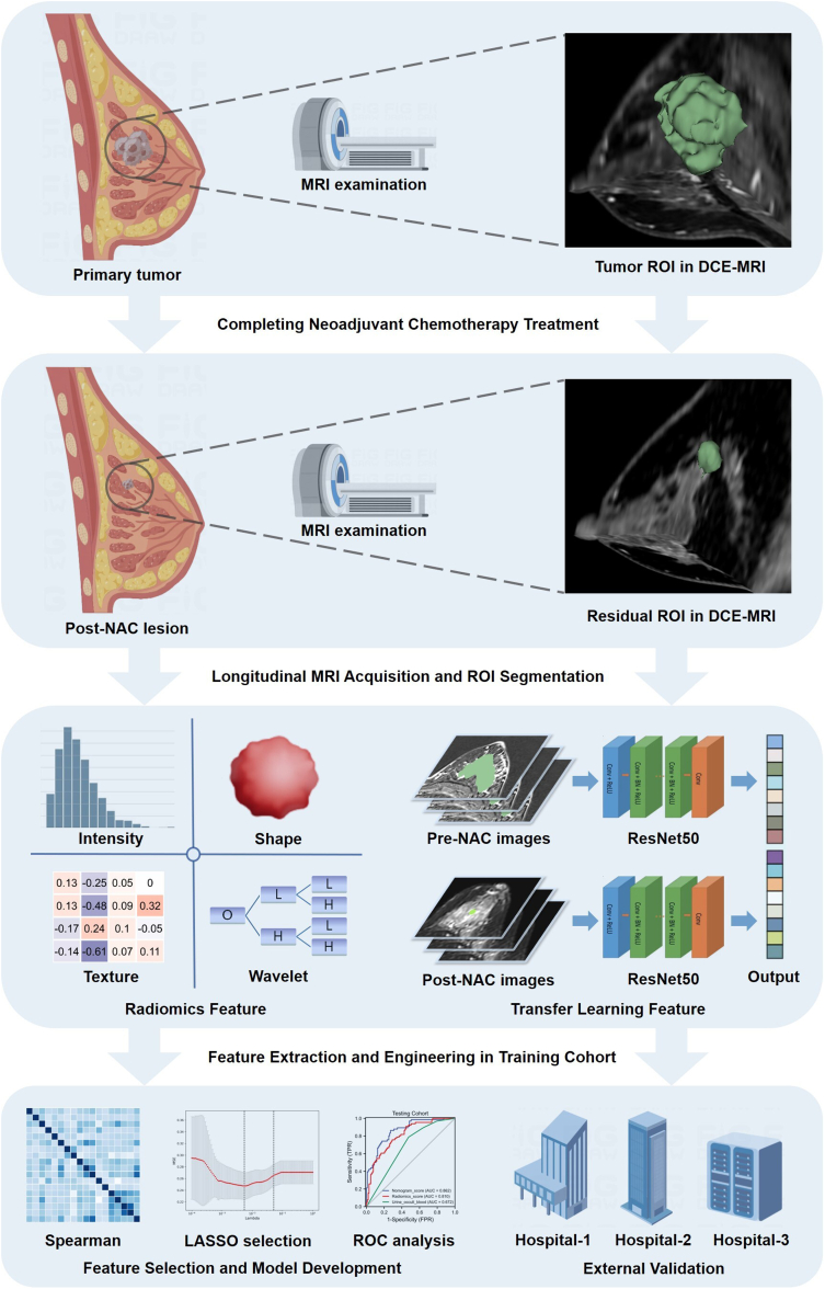
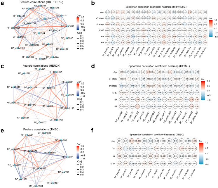
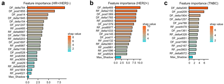
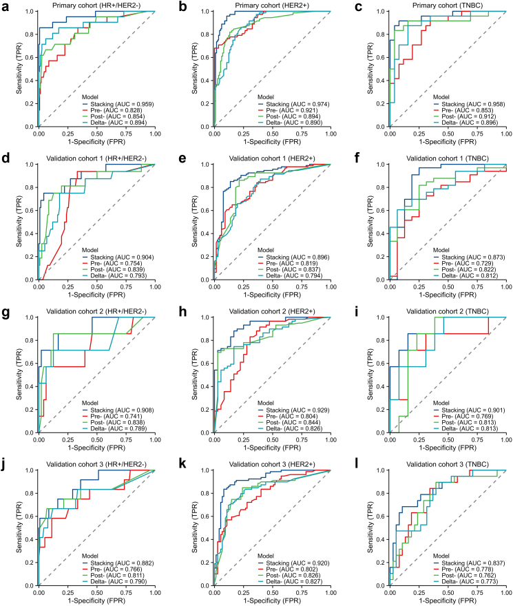
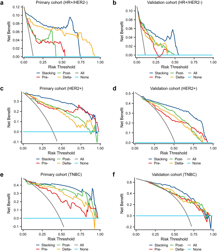
Background: Accurate identification of pCR to neoadjuvant chemotherapy (NAC) is essential for determining appropriate surgery strategy and guiding resection extent in breast cancer. However, a non-invasive tool to predict pCR accurately is lacking. Our study aims to develop ensemble learning models using longitudinal multiparametric MRI to predict pCR in breast cancer. Methods: From July 2015 to December 2021, we collected pre-NAC and post-NAC multiparametric MRI sequences per patient. We then extracted 14,676 radiomics and 4096 deep learning features and calculated additional delta-value features. In the primary cohort (n = 409), the inter-class correlation coefficient test, U-test, Boruta and the least absolute shrinkage and selection operator regression were used to select the most significant features for each subtype of breast cancer. Five machine learning classifiers were then developed to predict pCR accurately for each subtype. The ensemble learning strategy was used to integrate the single-modality models. The diagnostic performances of models were evaluated in the three external cohorts (n = 343, 170 and 340, respectively). Findings: A total of 1262 patients with breast cancer from four centers were enrolled in this study, and pCR rates were 10.6% (52/491), 54.3% (323/595) and 37.5% (66/176) in HR+/HER2-, HER2+ and TNBC subtype, respectively. Finally, 20, 15 and 13 features were selected to construct the machine learning models in HR+/HER2-, HER2+ and TNBC subtypes, respectively. The multi-Layer Perception (MLP) yields the best diagnostic performances in all subtypes. For the three subtypes, the stacking model integrating pre-, post- and delta-models yielded the highest AUCs of 0.959, 0.974 and 0.958 in the primary cohort, and AUCs of 0.882-0.908, 0.896-0.929 and 0.837-0.901 in the external validation cohorts, respectively. The stacking model had accuracies of 85.0%-88.9%, sensitivities of 80.0%-86.3%, and specificities of 87.4%-91.5% in the external validation cohorts. Interpretation: Our study established a novel tool to predict the responses of breast cancer to NAC and achieve excellent performance. The models could help to determine post-NAC surgery strategy for breast cancer. Funding: This study is supported by grants from the National Natural Science Foundation of China (82171898, 82103093), the Deng Feng project of high-level hospital construction (DFJHBF202109), the Guangdong Basic and Applied Basic Research Foundation (grant number, 2020A1515010346, 2022A1515012277), the Science and Technology Planning Project of Guangzhou City (202002030236), the Beijing Medical Award Foundation (YXJL-2020-0941-0758), and the Beijing Science and Technology Innovation Medical Development Foundation (KC2022-ZZ-0091-5). Funding sources were not involved in the study design, data collection, analysis and interpretation, writing of the report, or decision to submit the article for publication.
Overview
The claims and statistics of this paper haven’t been checked yet.
Run the check — free, no account needed. It reads the open-access text and checks the sources it cites.
Integrity signals
Registry
Cites
Statistics
Figures
Verification
Claims the paper makes
The claims and statistics of this paper haven’t been checked yet.
Run the check — free, no account needed. It reads the open-access text and checks the sources it cites.
Claims other papers make about this one
Each is checked against this paper’s own text: does it say what the citing paper says it does?
The source backs part of the claim, not all of it — or backs it for a different population, condition or comparison.
In the citing paper
“Researchers found that predicting NAC response in patients with locally advanced breast cancer based on a deep learning algorithm is feasible (13).”
From A multimodal and fully automated system for prediction of pathological complete response to neoadjuvant chemotherapy in breast cancer (2025) · cites this paper as [13]
Experimental — accuracy not yet measuredThe cited source says what the claim attributes to it.
In the citing paper
“Many researchers found that deep learning can improve the prediction ability of NAC response prediction in breast cancer (13, 22).”
From A multimodal and fully automated system for prediction of pathological complete response to neoadjuvant chemotherapy in breast cancer (2025) · cites this paper as [13]
The cited source says what the claim attributes to it.
In the citing paper
“the model was developed based on single-center data.”
From A multimodal and fully automated system for prediction of pathological complete response to neoadjuvant chemotherapy in breast cancer (2025) · cites this paper as [13]
Self-check
Percentages, sums and reported differences re-computed from the paper’s own numbers.
The self-check needs the paper’s full text.
Citations
Cites
DeLong, E. R., DeLong, D. M., & Clarke-Pearson, D. L. (1988) · Biometrics · doi:10.2307/2531595 · Cited by 22,649
3D Slicer as an image computing platform for the Quantitative Imaging Network
Fedorov, A., Beichel, R., Kalpathy–Cramer, J., Finet, J., Fillion‐Robin, J., Pujol, S., Bauer, C., Jennings, D. L., Fennessy, F., Milan Sonka, A., et al. (2012) · Magnetic Resonance Imaging · doi:10.1016/j.mri.2012.05.001 · Cited by 9,095
Computational Radiomics System to Decode the Radiographic Phenotype
(2017) · doi:10.1158/0008-5472.can-17-0339 · Cited by 6,593
Radiomics: extracting more information from medical images using advanced feature analysis
Lambin, P., Rios-Velazquez, E., Leijenaar, R., Carvalho, S., van Stiphout, R. G. P. M., Granton, P., Zegers, C. M. L., Gillies, R., Boellard, R., Dekker, A., et al. (2012) · European Journal of Cancer · doi:10.1016/j.ejca.2011.11.036 · Cited by 6,084
Cortazar, P., Zhang, L., Untch, M., Mehta, K. M., Costantino, J. P., Wolmark, N., Bonnefoi, H. R., Cameron, D., Gianni, L., Valagussa, P., et al. (2014) · The Lancet · doi:10.1016/s0140-6736(13)62422-8 · Cited by 4,777
Self-citationHammond, M. E., Hayes, D. F., Dowsett, M., Allred, D. C., Hagerty, K. L., Badve, S., Fitzgibbons, P. L., Francis, G. D., Goldstein, N. S., Hayes, M. M., et al. (2010) · Journal of Clinical Oncology · doi:10.1200/jco.2009.25.6529 · Cited by 4,217
Zwanenburg, A., Vallières, M., Abdalah, M. A., Aerts, H. J. W. L., Andrearczyk, V., Apte, A., Ashrafinia, S., Bakas, S., Beukinga, R. J., Boellaard, R., et al. (2020) · Radiology · doi:10.1148/radiol.2020191145 · Cited by 4,177
Wolff, A. C., Hammond, M. E. H., Hicks, D. G., Dowsett, M., McShane, L. M., Allison, K. H., Allred, D. C., Bartlett, J. M. S., Bilous, M., Fitzgibbons, P. L., et al. (2013) · Journal of Clinical Oncology · doi:10.1200/jco.2013.50.9984 · Cited by 4,080
von Minckwitz, G., Untch, M., Blohmer, J.-U., Costa, S. D., Eidtmann, H., Fasching, P. A., Gerber, B., Eiermann, W., Hilfrich, J., Huober, J., et al. (2012) · Journal of Clinical Oncology · doi:10.1200/jco.2011.38.8595 · Cited by 2,659
RECIST 1.1—Update and clarification: From the RECIST committee
Schwartz, L. H., Litière, S., de Vries, E., Ford, R., Gwyther, S., Mandrekar, S., Shankar, L., Bogaerts, J., Chen, A., Dancey, J., et al. (2016) · European Journal of Cancer · doi:10.1016/j.ejca.2016.03.081 · Cited by 1,906
Cited by
Sorted by citations
Items 101–175 of 175
Wang, X., Zhang, Y., Yang, M., Wu, N., Wang, S., Chen, H., Zhou, T., Zhang, Y., Wang, X., Jin, Z., et al. (2024) · Scientific Reports · doi:10.1038/s41598-024-80409-y · Cited by 3
Gong, J., Meng, F., Liu, C., Lu, J., Li, J., Yang, Z., Sun, H., Teng, X., Zhang, J., Cai, J., et al. (2025) · Insights into Imaging · doi:10.1186/s13244-025-02044-z · Cited by 3
Xu, J., Gao, S., Zhu, Q., Dai, F., Sun, C., Lee, W., Ye, Y., Deng, G., Huang, Z., Li, X., et al. (2025) · Abdominal Radiology · doi:10.1007/s00261-025-04956-2 · Cited by 2
Liang, M., Zhu, T., Huang, N., Zhang, L., Yang, C., Gao, H., Zhang, X., Li, P., Cheng, M., & Wang, K. (2025) · Clinical & Translational Oncology · doi:10.1007/s12094-025-03993-z · Cited by 2
Self-citationWang, J., Wu, S., Shi, A., Zhang, H., Niu, M., Tian, X., & Liu, J. (2025) · Annals of Nuclear Medicine · doi:10.1007/s12149-025-02121-9 · Cited by 2
Lin, R., Chen, Y., Wang, Y., Tang, L., & Huang, W. (2025) · BMC Medical Imaging · doi:10.1186/s12880-025-02017-0 · Cited by 2
Huang, Y., Gong, H., Hu, W., Ren, H., Cao, Y. J., Lan, X., Chen, H., Wang, L., Wang, X., & Zhang, J. (2025) · npj Precision Oncology · doi:10.1038/s41698-025-01220-0 · Cited by 2
Self-citationMa, Q., Ren, J., Ge, Y., Yuan, Y., & Tao, X. (2025) · Cancer Imaging · doi:10.1186/s40644-025-00982-x · Cited by 2
Zhu, P.-F., Zhang, X., Mao, Y., Zhou, P., Lin, J.-J., Shi, L., Cui, X., & He, Y. (2026) · BMC Cancer · doi:10.1186/s12885-025-15153-1 · Cited by 2
Self-citationHou, W.-T., Pu, Z.-F., Xu, Z.-Y., Wu, A.-H., Liu, Z., Zhao, K., Duan, C.-L., Guo, J., Chen, K., Qiu, S.-Q., et al. (2026) · Medical Image Analysis · doi:10.1016/j.media.2026.104069 · Cited by 2
3D multi-omics tumour atlases: from technology to biology and clinical translation
Liu, M., Villazon, J. I., Forjaz, A., Tian, X., Fan, R., Wang, S., Shi, L., Wirtz, D., & Kiemen, A. L. (2026) · Nature reviews. Cancer · doi:10.1038/s41568-026-00940-0 · Cited by 3
Ding, L., Wu, J. I. A. H. U. I., Liang, Y., Ding, J., Zhou, S., Li, L., Lin, L., Zeng, J., & Chen, C. (2026) · Cancer Medicine · doi:10.1002/cam4.72035 · Cited by 2
Yin, S., Ding, N., Ji, Y., Qiao, Z., Yuan, J., Chi, J., & Jin, L. H. (2024) · Acta Radiologica · doi:10.1177/02841851241245970 · Cited by 2
Yang, C., Li, P., Chen, Y., Zheng, J., Zhang, X., Gao, H., Zhang, L., & Wang, K. (2024) · Supportive Care in Cancer · doi:10.1007/s00520-024-08610-3 · Cited by 2
Self-citationMao, H., Chen, K.-G., Zhu, B., Guo, W., & Shi, S.-L. (2025) · BMC Medical Imaging · doi:10.1186/s12880-025-01579-3 · Cited by 2
Shang, J., Chen, J., Gao, X., Wan, Z., Yang, R., Lei, Z., Chen, S., Chen, M., Quan, Y., & Bai, J. (2025) · International Journal of Surgery · doi:10.1097/js9.0000000000003070 · Cited by 2
Zhang, X.-P., He, Y., Rao, T., Qiu, X.-T., Lai, Q., Zhang, Y.-C., & Zhang, G. (2025) · La radiologia medica · doi:10.1007/s11547-025-02077-w · Cited by 2
Self-citationWei, Y., Zhu, Y., Dong, Q., Wang, W., Yu, T., Zhang, J., & Dong, Y. (2025) · European Journal of Radiology · doi:10.1016/j.ejrad.2025.112256 · Cited by 1
Wang, H., Gu, Y., Wu, G., Yang, Y., Zhang, W., Mu, G., Xue, W., Fu, C., Yang, X., Chen, L., et al. (2025) · Computer Methods and Programs in Biomedicine · doi:10.1016/j.cmpb.2025.108986 · Cited by 1
Zou, Y., Zhao, B., Mao, Y., Lv, M., Wang, Y., Su, X., Zaixian, Z., Wu, J., & Wang, Q. (2026) · Frontiers in Medicine · doi:10.3389/fmed.2025.1639977 · Cited by 1
Jia, M., Li, H., Xiao, W.-L., Xue, J., Wang, Z., He, X., Wang, X. S., & Men, D. (2026) · Ultrasound in Medicine & Biology · doi:10.1016/j.ultrasmedbio.2025.12.009 · Cited by 1
Tan, Y., Zhang, W., Xie, L.-X., Lan, Q.-X., Li, L., & Dai, W. B. (2026) · BMC Public Health · doi:10.1186/s12889-026-27345-w · Cited by 1
Radiomics: Current Applications and Future Directions
Shao, J., Wei, M., Li, K., Lv, G., Liu, K., & Guo, Y. C. (2026) · MedComm · doi:10.1002/mco2.70773 · Cited by 2
Jiang, T., Li, X., Shen, J., Yan, Y. J., Sui, L., Zhou, L., Jin, Z., Ou, D., Wang, L., Chuan, Z., et al. (2026) · Academic Radiology · doi:10.1016/j.acra.2026.06.010 · Cited by 1
Carlini, A., Foca, F., Sirico, M., Farolfi, A., Casadei, C., Gianni, C., Palleschi, M., Gentili, N., Mariotti, M., Menna, G. D., et al. (2026) · The Breast · doi:10.1016/j.breast.2026.104861 · Cited by 1
Sun, S., Chen, Y., Liu, Y., Li, C., Miao, S., Yang, B., & Yu, F. (2024) · Academic Radiology · doi:10.1016/j.acra.2024.09.065 · Cited by 1
Wang, K., Qi, L., Zhang, T., Li, J., Zhang, M., & Du, H. (2024) · Research Square · doi:10.21203/rs.3.rs-4479368/v1 · Cited by 1
Self-citationCao, Z., Shi, Z., Huang, X., Han, C., Dong, X., Zhao, Z., Wang, D., Xu, P., Liu, Z., & Liu, W. (2023) · Lecture notes in computer science · doi:10.1007/978-981-99-4749-2_26
Qian, F., Mao, Y., Dong, J., Xie, F., Fang, X. L., Zhang, Q., Xia, P., Han, X., & Lu, N. (2024) · Translational Cancer Research · doi:10.21037/tcr-23-770
Application of Artificial Intelligence in Breast Imaging
Peng, W., Gong, J., & Wang, T. (2024) · Springer eBooks · doi:10.1007/978-981-99-8441-1_11
Huang, M., Jin, Y., Wang, M., Song, Q., Fan, Y., Zhang, Y., Tian, C., Zhang, C., & Liu, S. (2024) · Breast Cancer Targets and Therapy · doi:10.2147/bctt.s478110
Chen, K., & Ying, H. (2024) · Gastrointestinal Endoscopy · doi:10.1016/j.gie.2024.09.016
Liu, X., Wang, X., Wang, G., Zhu, J., Zhuo, S., Shi, Y., Wen, X., Han, H., Chen, J., Zhong, Q., et al. (2025) · SSRN Electronic Journal · doi:10.2139/ssrn.5219322
Liu, W., Wang, W., Zhang, H., & Guo, M. (2025) · Journal of Imaging Informatics in Medicine · doi:10.1007/s10278-025-01561-z
Liu, P., Liu, Y., Xie, Y., Hu, S., Zhou, H., & Yang, L. M. (2025) · Journal of Surgical Oncology · doi:10.1002/jso.70058
Chen, Y., Sun, Y., Chen, Y., Zhang, J., Zhang, H., Liu, K., Dong, L., Zhang, X., Zhou, R., Wang, J., et al. (2025) · European Journal of Nuclear Medicine and Molecular Imaging · doi:10.1007/s00259-025-07536-0
Self-citationRadiomics-based intelligent diagnosis and treatment
Fang, M., Wang, Z., Fu, J., Wang, Y., Zhao, Y., Qin, Z., Zhang, C., Xie, X., Tang, L., & Dong, D. (2025) · Elsevier eBooks · doi:10.1016/b978-0-443-30142-1.00006-7
Review of Breast Cancer Datasets and Repositories: Emphasis on Longitudinal Component
Chitaouy, I., Haddouchi, M., & Berrado, A. (2025) · International Conference on Intelligent Systems: Theories and Applications · doi:10.1109/sita67914.2025.11273590
Zheng, X., Huang, Y., Lin, Y., Zhu, T., Zou, J., Wang, S., & Wang, K. (2024) · Figshare · doi:10.6084/m9.figshare.26650089
Self-citationFan, M., Wang, K., Pan, D., Cao, X., Li, Z., He, S., Xie, S., You, C., Gu, Y., & Li, L. (2024) · Figshare · doi:10.6084/m9.figshare.26746175
Self-citationRavi, R., Li, R., Abdelfatah, T., Chan, S. Y. T., & Chen, X. (2026) · Informatics in Medicine Unlocked · doi:10.1016/j.imu.2026.101812
Lai, X., He, H., Liang, B., Xu, Z., Yang, L., Wu, T., Han, K., Li, W., Liu, Q., Zhu, C., et al. (2026) · Academic Radiology · doi:10.1016/j.acra.2026.01.018
Self-citation王, 秋. (2026) · Advances in Clinical Medicine · doi:10.12677/acm.2026.162533
Gong, J., Yang, Z., Li, J., Wang, Q., Meng, F., Liu, M., Sun, H., & Zhao, L. (2026) · Radiotherapy and Oncology · doi:10.1016/j.radonc.2026.111471
Zhai, M., Bi, J., Ke, Z., Hu, M., Pi, G., Li, Y., He, H., Li, Y., Zheng, H., Wu, X., et al. (2026) · Breast Cancer Targets and Therapy · doi:10.2147/bctt.s577469
Fan, M., He, S., Wan, C., Zhang, J., Tan, T., Zhou, S., Chen, S., You, C., Gu, Y., & Li, L. (2026) · BMC Cancer · doi:10.1186/s12885-026-15889-4
Shi, Y., Wang, P., Ye, M., Wu, A., & Zhou, Y. (2026) · International Journal of Intelligent Systems · doi:10.1155/int/1564250
Zhou, M., Zhu, L., Zhang, K., Wei, S., Wang, H., He, S., Liu, X., & Huang, X. (2026) · Frontiers in Oncology · doi:10.3389/fonc.2026.1764094
Schnitzler, T. (2026) · Radiology Artificial Intelligence · doi:10.1148/ryai.260266
Liu, Y., Li, J., Ning, Y., Wu, Y., He, P., & Dong, X. (2026) · Frontiers in Oncology · doi:10.3389/fonc.2026.1718061
An, R., Sun, Y., Yang, R., Wang, M., Chen, X., Pang, X., Zhao, J. W., Chen, X., Shi, F., Fu, Q., et al. (2026) · European Journal of Nuclear Medicine and Molecular Imaging · doi:10.1007/s00259-026-07920-4
Li, H., Huang, J., Hou, J., Chen, X., Zhi, C., & Li, Z. (2026) · PLoS ONE · doi:10.1371/journal.pone.0350085
Gao, Y., Wu, C., Zhu, X., Yin, L., Chen, J., Liu, H., Chen, Y., Ye, J., Xu, L., Duan, X., et al. (2026) · Research Square · doi:10.21203/rs.3.rs-9636896/v1
王, 一. (2026) · Advances in Clinical Medicine · doi:10.12677/acm.2026.1662199
Li, Z.-H., Weng, J., Zeng, Y.-H., Lin, S.-Y., Li, S., Bai, K., & Xu, G.-L. (2026) · World Journal of Gastroenterology · doi:10.3748/wjg.120899
Yi, C., Xu, T., You, R., Zhang, Q., Liu, R., & Yang, X. (2026) · BMC Cancer · doi:10.1186/s12885-026-16330-6
Predicting breast cancer pathological complete response with clinical and imaging data
Tao, X., Li, Y., Ye, Y., Liang, X., Qiu, X., & Zhao, J. (2026) · Future Oncology · doi:10.1080/14796694.2026.2706356
Wu, H., Yin, X., Cheng, J., Zhang, J., Zheng, L., Zhang, L., Wu, F., Zhao, Q., Yang, J., & Wang, H. (2026) · Insights into Imaging · doi:10.1186/s13244-026-02359-5
Self-citationZhao, X., Bai, J., Dong, L.-A., Li, Z.-H., He, J.-Z., Chen, Y., Liu, Y., Hou, W.-T., Xu, Z.-Y., Du, Z.-C., et al. (2026) · Breast Cancer Research · doi:10.1186/s13058-026-02354-0
Kessler, D. A., Bobowicz, M., Kosno, M., Joshi, S., Kostas Marias, A., Díaz, O., & Lekadir, K. (2026) · European Journal of Radiology · doi:10.1016/j.ejrad.2026.113101 · Cited by 1
Li, Q., Chen, J., Yu, W., Li, X., & Deng, Y. (2026) · Frontiers in Oncology · doi:10.3389/fonc.2026.1829672
Ding, S., Meng, H., Cui, X., Yang, Y., Xing, L., Zuo, Z., Liu, C., Xue, L., & Yin, X. (2026) · Cancer Medicine · doi:10.1002/cam4.72141
Li, Y., Wang, P., Zou, X., Zhang, Q., & Wang, W. (2026) · Magnetic Resonance Imaging · doi:10.1016/j.mri.2026.110796
Sun, X., Wang, D., Zhou, Q., Liu, W., Gao, X., Shen, X., Yang, X., Ma, T., Ma, T., Liu, C., et al. (2026) · European Journal of Radiology · doi:10.1016/j.ejrad.2026.113214
Zhou, J., Xi, B., Yan, K., & Chen, L. (2026) · Cancers · doi:10.3390/cancers18182947
Wang, Y., Wang, H., & Cui, J. (2026) · Frontiers in Oncology · doi:10.3389/fonc.2026.1830219
Meta-analysis of the correlation between MRI radiomics and HER2 overexpression in breast cancer
Lin, Q., Kong, X., & Yan, Y. (2026) · International Journal of Radiation Research · doi:10.66224/ijrr.24.3.5
Lin, H., Huang, B., Wang, M., Ren, Y., Liu, Z., & Huang, B. (2026) · Lecture notes in computer science · doi:10.1007/978-3-032-38470-6_58
Application advances of habitat imaging based on CT or PET/CT: a systematic review
Chen, Q., 任铁柱, Peng, Y., Ma, L., Xu, M., & Zhang, W. (2026) · La radiologia medica · doi:10.1007/s11547-026-02292-z
Omer, M. E. (2026) · Biomedical & Pharmacology Journal · doi:10.13005/bpj/3485
Zhang, F.-Y., Hu, K.-M., Sun, C.-M., Liu, X.-F., Wei, L.-Y., & Su, R. (2026) · Medical Image Analysis · doi:10.1016/j.media.2026.104343
Ren, W., He, Z., Deng, Z., Huang, Z., 蔡賡喜, Ban, X., Hu, Q., Zhang, Z., Mao, L., Tan, Y., et al. (2026) · MedComm – Oncology · doi:10.1002/mog2.70100
Reisig, E., Cai, L., Müller, M., Schäfgen, B., & Pfob, A. (2026) · BMC Cancer · doi:10.1186/s12885-026-17115-7
Mu, J., Luo, Y., Liu, H., He, X., Minxun, 陆. L., Tang, F., Wang, Y., Zhou, Y., Min, L., & Tu, C. (2026) · Diagnostics · doi:10.3390/diagnostics16203411
Lou, S., Duan, L., Li, X., Zhu, C., Shi, Y., Zhang, W., Zhang, S., Liu, H., & Li, W. (2026) · Research Square · doi:10.21203/rs.3.rs-11118822/v1
Self-citation
Metadata
- Authors
- Huang, Y., Zhu, T., Zhang, X., Li, W., Zheng, X., Cheng, M., Ji, F., Zhang, L., Yang, C., Wu, Z., Ye, G., Lin, Y., & Wang, K.
- Affiliations
- Guangdong Academy of Medical Sciences (CN)
- Sun Yat-sen University (CN)
- First People's Hospital of Foshan (CN)
- Corresponding author
- Ye, GuoLin
- Lin, Ying
- Wang, Kun
- Venue
- eClinicalMedicine
- Publisher
- Elsevier BV
- Published
- 2023-04
- Volume
- 58
- Pages
- 101899
- ISSN
- 2589-5370 (ISSN-L)
- PMC
- PMC10050775
- OpenAlex
- W4360796946
- Cited by
- 175
- References
- 55
- Citations by year
- 2026: 49 · 2025: 67 · 2024: 46 · 2023: 13
- FWCI
- 28.98
- Type
- journal article
- Language
- en
- Open access
- gold
- doi.org
- Licence
- Locations
- EClinicalMedicine (publishedVersion, cc-by-nc-nd)
- PubMed Central (publishedVersion, cc-by-nc-nd)
- PubMed (publishedVersion)
- DOAJ (DOAJ: Directory of Open Access Journals) (submittedVersion, cc-by-sa)
- Funders
- High-level Hospital Construction Project of Guangdong Provincial People's Hospital (DFJHBF202109)
- Basic and Applied Basic Research Foundation of Guangdong Province (2020A1515010346, 2021A1515011570, 2022A1515012277)
- Scientific and Technological Planning Project of Guangzhou City
- National Natural Science Foundation of China (82103093)
- Guangzhou Municipal Science and Technology Project (202002030236, 202102021055)
- Foundation for Innovative Research Groups of the National Natural Science Foundation of China (82171898)
- Topics
- Health Sciences › Medicine › Radiology, Nuclear Medicine and Imaging › Radiomics and Machine Learning in Medical Imaging
- Health Sciences › Medicine › Radiology, Nuclear Medicine and Imaging › MRI in cancer diagnosis
- Life Sciences › Biochemistry, Genetics and Molecular Biology › Cancer Research › Breast Cancer Treatment Studies
- Keywords
- breast cancer, pathological complete response, neoadjuvant chemotherapy, multiparametric MRI, longitudinal MRI, radiomics, deep features, ensemble learning, stacking ensemble, breast cancer subtypes, triple-negative breast cancer, HER2-positive breast cancer, hormone receptor-positive HER2-negative breast cancer, treatment response prediction, feature selection, external validation, multilayer perceptron
About these checks
Registry, retraction, Cites and statistics results are computed, not judged, with one exception: a printed reference that no index lists is read once by a model before it counts as a citation error. Claims are extracted by a model and kept only when their quote appears verbatim in the text; each cited source is then checked, with a model’s help, for whether it says what the claim relies on. Those verdicts can be wrong — read the claim’s quote and the cited source before relying on one.
Statistics and arithmetic are recomputed, not judged.
Confidence bands are shown only for fields calibrated on our annotated test set. None is calibrated yet, so no band is shown.
What counts as a claim ›How we measure confidence ›How we check this ›
Are you an author, or do you see a problem with this page? Request a review
