

The DOI matches its Crossref record; 103 of the 103 references it cites were found in a registry or an index.
Multiscale characterization of Caenorhabditis elegans mutants to probe functional mechanisms of human actin pathological variants
Open accessPublished version of a preprint
Abstract
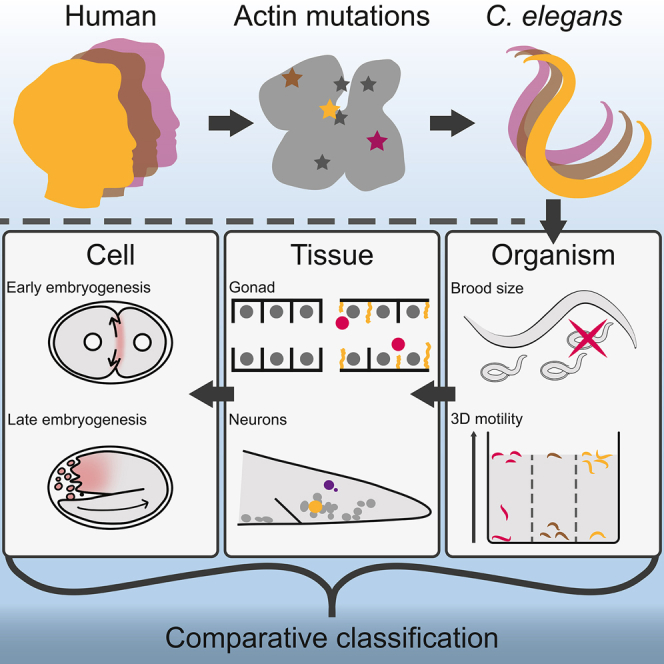
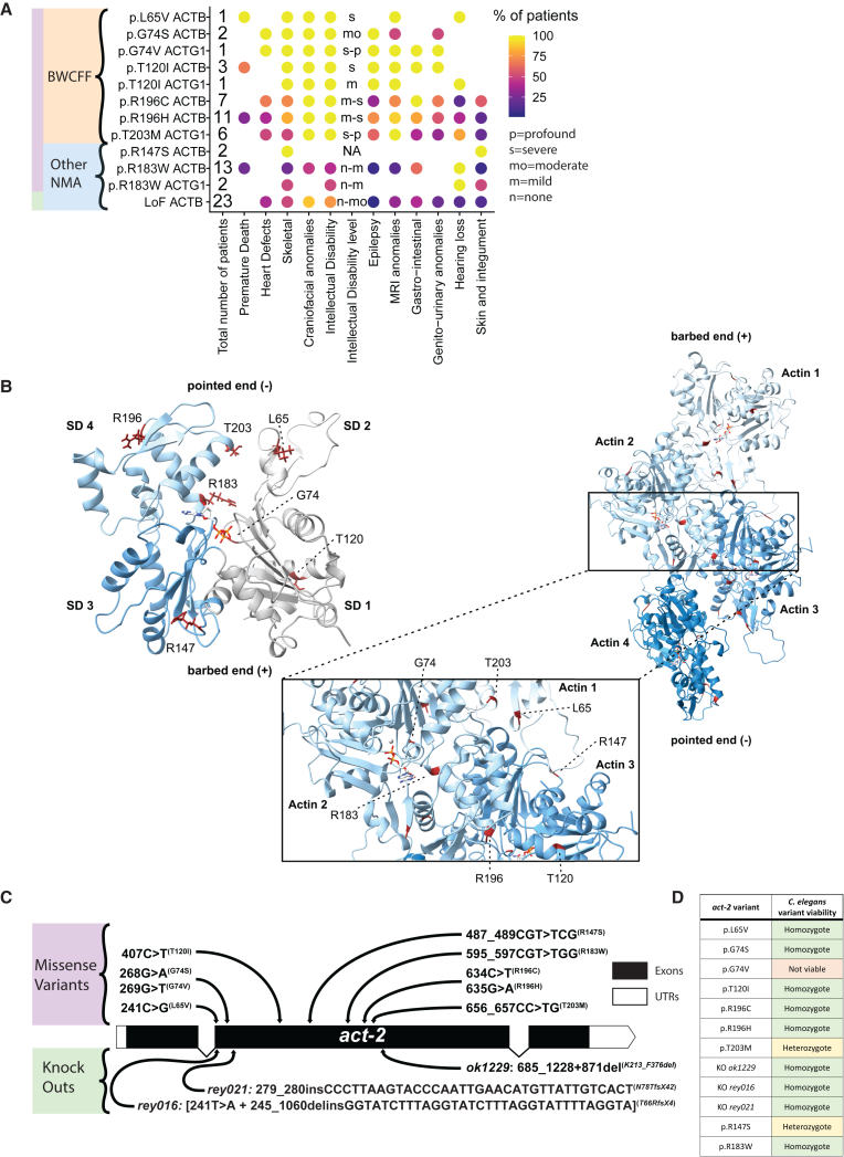
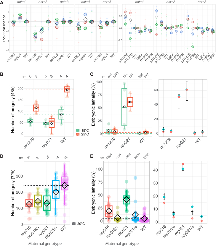
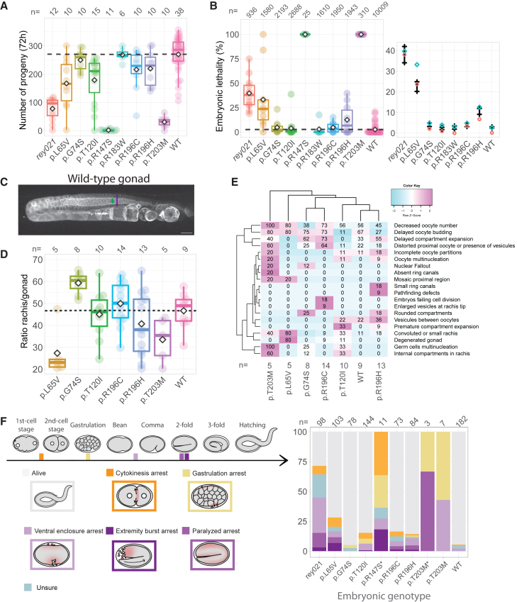
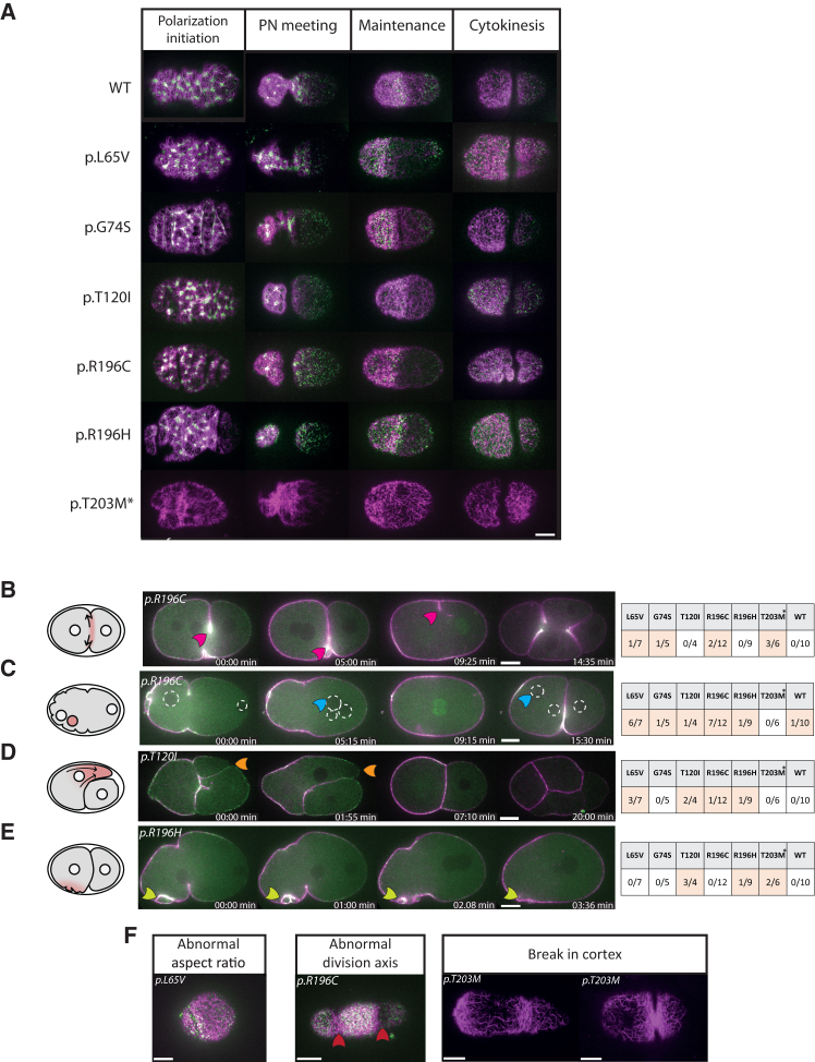
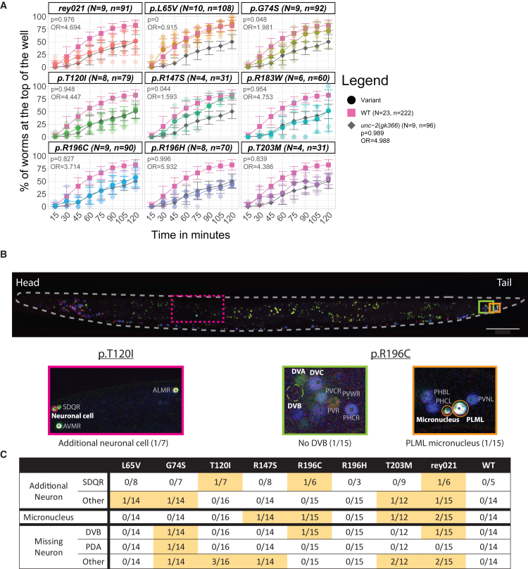
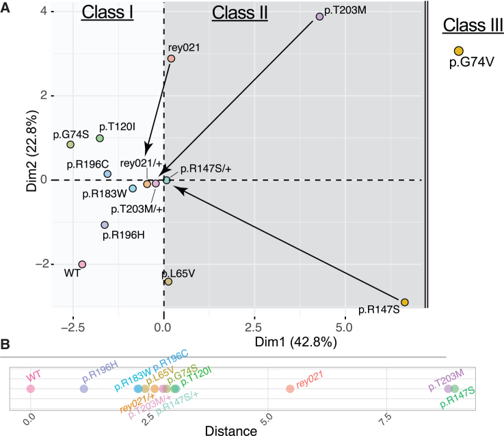
SUMMARY Actin plays a crucial role in diverse physiological processes by forming dynamic networks that determine cellular shape and mechanical properties. Non-Muscle Actinopathies (NMA) are rare diseases caused by de novo variants in human cytoskeletal β-actin ( ACTB ) and γ-actin ( ACTG1 ) genes, ranging from missense mutations to whole gene deletions. Currently, the high clinical variability and genotype-phenotype correlations in NMA remain largely unresolved. To address this concern, we inserted nine mutations identified in patients in the C. elegans cytoplasmic actin orthologue act-2 and performed a quantitative multiscale characterization of these animal models. We uncovered various perturbations including micro-scale actin network defects, cell-scale abnormalities, morphogenesis failure, and weaker behavioural phenotypes. Notably, the severity of the observed defects correlates with the severity of patients’ symptoms. Thus, we provide evidence that such C. elegans models are relevant to investigate the mechanisms underlying NMA physiopathology and could ultimately be used to screen for therapeutic strategies.
Overview
The claims and statistics of this paper haven’t been checked yet.
Run the check — free, no account needed. It reads the open-access text and checks the sources it cites.
Integrity signals
Registry
Cites
Statistics
Figures
Verification
Claims the paper makes
The claims and statistics of this paper haven’t been checked yet.
Run the check — free, no account needed. It reads the open-access text and checks the sources it cites.
Self-check
Percentages, sums and reported differences re-computed from the paper’s own numbers.
The self-check needs the paper’s full text.
Citations
Cites
Highly accurate protein structure prediction with AlphaFold
Jumper, J., Evans, R., Pritzel, A., Green, T., Figurnov, M., Ronneberger, O., Tunyasuvunakool, K., Bates, R., Žídek, A., Potapenko, A., et al. (2021) · Nature · doi:10.1038/s41586-021-03819-2 · Cited by 43,764
THE GENETICS OF CAENORHABDITIS ELEGANS
Brenner, S. (1974) · Genetics · doi:10.1093/genetics/77.1.71 · Cited by 15,081
SWISS-MODEL: homology modelling of protein structures and complexes
Waterhouse, A., Bertoni, M., Bienert, S., Studer, G., Tauriello, G., Gumienny, R., Heer, F. T., de Beer, T. A. P., Rempfer, C., Bordoli, L., et al. (2018) · Nucleic Acids Research · doi:10.1093/nar/gky427 · Cited by 13,067
UCSF ChimeraX: Structure visualization for researchers, educators, and developers
Pettersen, E. F., Goddard, T. D., Huang, C. C., Meng, E. C., Couch, G. S., Croll, T. I., Morris, J. H., & Ferrin, T. E. (2021) · Protein Science · doi:10.1002/pro.3943 · Cited by 9,514
Actin Dynamics, Architecture, and Mechanics in Cell Motility
Blanchoin, L., Boujemaa-Paterski, R., Sykes, C., & Plastino, J. (2014) · Physiological Reviews · doi:10.1152/physrev.00018.2013 · Cited by 1,457
Dominguez, R., & Holmes, K. C. (2011) · Annual Review of Biophysics · doi:10.1146/annurev-biophys-042910-155359 · Cited by 1,211
Blebs lead the way: how to migrate without lamellipodia
Charras, G., & Paluch, E. (2008) · Nature Reviews Molecular Cell Biology · doi:10.1038/nrm2453 · Cited by 769
Forcing cells into shape: the mechanics of actomyosin contractility
Murrell, M., Oakes, P. W., Lenz, M., & Gardel, M. L. (2015) · Nature Reviews Molecular Cell Biology · doi:10.1038/nrm4012 · Cited by 687
Paix, A., Folkmann, A., Rasoloson, D., & Seydoux, G. (2015) · Genetics · doi:10.1534/genetics.115.179382 · Cited by 675
The structure of crystalline profilin–β-actin
Schutt, C. E., Myslik, J. C., Rozycki, M. D., Goonesekere, N. C. W., & Lindberg, U. (1993) · Nature · doi:10.1038/365810a0 · Cited by 664
Ferguson, E. L., & Horvitz, H. R. (1985) · Genetics · doi:10.1093/genetics/110.1.17 · Cited by 541
The genetics of feeding in Caenorhabditis elegans.
Avery, L. (1993) · Genetics · doi:10.1093/genetics/133.4.897 · Cited by 506
Caenorhabditis elegans morphogenesis: The role of the cytoskeleton in elongation of the embryo
Priess, J. R., & Hirsh, D. I. (1986) · Developmental Biology · doi:10.1016/0012-1606(86)90358-1 · Cited by 419
Hoogewijs, D., Houthoofd, K., Matthijssens, F., Vandesompele, J., & Vanfleteren, J. R. (2008) · BMC Molecular Biology · doi:10.1186/1471-2199-9-9 · Cited by 400
Large-Scale Screening for Targeted Knockouts in the Caenorhabditis elegans Genome
The C. elegans Deletion Mutant Consortium (2012) · G3 Genes|Genomes|Genetics · doi:10.1534/g3.112.003830 · Cited by 372
β-Actin specifically controls cell growth, migration, and the G-actin pool
Bunnell, T. M., Burbach, B. J., Shimizu, Y., & Ervasti, J. M. (2011) · Molecular Biology of the Cell · doi:10.1091/mbc.e11-06-0582 · Cited by 320
Biochemical and mechanical regulation of actin dynamics
Lappalainen, P., Kotila, T., Jegou, A. G., & Romet‐Lemonne, G. (2022) · Nature Reviews Molecular Cell Biology · doi:10.1038/s41580-022-00508-4 · Cited by 314
A database of Caenorhabditis elegans behavioral phenotypes
Yemini, E., Jucikas, T., Grundy, L. J., Brown, A. E. X., & Schafer, W. R. (2013) · Nature Methods · doi:10.1038/nmeth.2560 · Cited by 310
NeuroPAL: A Multicolor Atlas for Whole-Brain Neuronal Identification in C. elegans
Yemini, E., Lin, A., Nejatbakhsh, A., Varol, E., Sun, R., Mena, G. E., Samuel, A. D. T., Paninski, L., Venkatachalam, V., & Hobert, O. (2020) · Cell · doi:10.1016/j.cell.2020.12.012 · Cited by 310
De novo mutations in the actin genes ACTB and ACTG1 cause Baraitser-Winter syndrome
Rivière, J., van Bon, B. W. M., Hoischen, A., Kholmanskikh, S. S., O’Roak, B. J., Gilissen, C., Gijsen, S. J., Sullivan, C. T., Christian, S. L., Abdul-Rahman, O. A., et al. (2012) · Nature Genetics · doi:10.1038/ng.1091 · Cited by 307
β- and γ-cytoplasmic actins display distinct distribution and functional diversity
Dugina, V. B., Zwaenepoel, I., Gabbiani, G., Clément, S., & Chaponnier, C. (2009) · Journal of Cell Science · doi:10.1242/jcs.041970 · Cited by 228
Architecture and Connectivity Govern Actin Network Contractility
Ennomani, H., Letort, G., Guérin, C., Martiel, J., Cao, W., Nédélec, F., Cruz, E. M. D. L., Théry, M., & Blanchoin, L. (2016) · Current Biology · doi:10.1016/j.cub.2015.12.069 · Cited by 222
Rose, L. S., & Gönczy, P. (2014) · WormBook · doi:10.1895/wormbook.1.30.2 · Cited by 217
Modeling human diseases in Caenorhabditis elegans
Markaki, M., & Tavernarakis, N. (2010) · Biotechnology Journal · doi:10.1002/biot.201000183 · Cited by 214
Green, R. A., Kao, H.-L., Audhya, A., Arur, S., Mayers, J. R., Fridolfsson, H. N., Schulman, M., Schloissnig, S., Niessen, S., Laband, K., et al. (2011) · Cell · doi:10.1016/j.cell.2011.03.037 · Cited by 207
Cortical flow aligns actin filaments to form a furrow
Reymann, A.-C., Staniscia, F., Erzberger, A., Salbreux, G., & Grill, S. W. (2016) · eLife · doi:10.7554/elife.17807 · Cited by 194
Self-citationBuilding Distinct Actin Filament Networks in a Common Cytoplasm
Michelot, A., & Drubin, D. G. (2011) · Current Biology · doi:10.1016/j.cub.2011.06.019 · Cited by 183
SETD3 is an actin histidine methyltransferase that prevents primary dystocia
Wilkinson, A. W., Diep, J., Dai, S., Liu, S., Ooi, Y. S., Song, D., Li, T., Horton, J. R., Zhang, X., Liu, C., et al. (2018) · Nature · doi:10.1038/s41586-018-0821-8 · Cited by 177
Baraitser–Winter cerebrofrontofacial syndrome: delineation of the spectrum in 42 cases
Verloès, A., Donato, N. D., Masliah‐Planchon, J., Jongmans, M. C. J., Abdul-Rahman, O. A., Albrecht, B., Allanson, J. E., Brunner, H. G., Bertola, D. R., Chassaing, N., et al. (2014) · European Journal of Human Genetics · doi:10.1038/ejhg.2014.95 · Cited by 168
Reconsidering an active role for G-actin in cytoskeletal regulation
Skruber, K., Read, T., & Vitriol, E. A. (2018) · Journal of Cell Science · doi:10.1242/jcs.203760 · Cited by 143
ACTB Loss-of-Function Mutations Result in a Pleiotropic Developmental Disorder
Cuvertino, S., Stuart, H. M., Chandler, K., Roberts, N. A., Armstrong, R., Bernardini, L., Bhaskar, S. S., Callewaert, B. L., Clayton‐Smith, J., Hernando‐Davalillo, C., et al. (2017) · The American Journal of Human Genetics · doi:10.1016/j.ajhg.2017.11.006 · Cited by 138
Bending forces and nucleotide state jointly regulate F-actin structure
Reynolds, M. J., Hachicho, C., Carl, A. G., Gong, R., & Alushin, G. M. (2022) · Nature · doi:10.1038/s41586-022-05366-w · Cited by 117
Procaccio, V., Salazar, G., Ono, S., Styers, M. L., Gearing, M., Dávila, A., Jimenez, R., Juncos, J. L., Gutekunst, C., Meroni, G., et al. (2006) · The American Journal of Human Genetics · doi:10.1086/504271 · Cited by 116
ACT-5 Is an Essential Caenorhabditis elegans Actin Required for Intestinal Microvilli Formation
MacQueen, A. J., Baggett, J. J., Perumov, N., Bauer, R., Januszewski, T., Schriefer, L. A., & Waddle, J. A. (2005) · Molecular Biology of the Cell · doi:10.1091/mbc.e04-12-1061 · Cited by 110
Ono, K., Parast, M. M., Alberico, C., Benian, G. M., & Ono, S. (2003) · Journal of Cell Science · doi:10.1242/jcs.00421 · Cited by 108
Schafer, W. R. (2005) · WormBook · doi:10.1895/wormbook.1.38.1 · Cited by 106
Delayed embryonic development and impaired cell growth and survival in Actg1 null mice
Bunnell, T. M., & Ervasti, J. M. (2010) · Cytoskeleton · doi:10.1002/cm.20467 · Cited by 98
Roles of the actin cytoskeleton in aging and age-associated diseases
Lai, W., & Wong, W. (2020) · Ageing Research Reviews · doi:10.1016/j.arr.2020.101021 · Cited by 90
Cells Lacking β-Actin are Genetically Reprogrammed and Maintain Conditional Migratory Capacity*
Tondeleir, D., Lambrechts, A., Müller, M., Jonckheere, V., Doll, T., Vandamme, D., Bakkali, K., Waterschoot, D., Lemaistre, M., Debeir, O., et al. (2012) · Molecular & Cellular Proteomics · doi:10.1074/mcp.m111.015099 · Cited by 88
Rapid RNA analysis of individual Caenorhabditis elegans
Ly, K. N., Reid, S. J., & Snell, R. G. (2015) · MethodsX · doi:10.1016/j.mex.2015.02.002 · Cited by 87
PLK-1 asymmetry contributes to asynchronous cell division ofC. elegansembryos
Budirahardja, Y., & Gönczy, P. (2008) · Development · doi:10.1242/dev.019075 · Cited by 87
Wild-type and mutant actin genes in Caenorhabditis elegans
Krause, M., Wild, M. A., Rosenzweig, B., & Hirsh, D. (1989) · Journal of Molecular Biology · doi:10.1016/0022-2836(89)90503-2 · Cited by 85
Actin Mutations and Their Role in Disease
Parker, F., Baboolal, T. G., & Peckham, M. (2020) · International Journal of Molecular Sciences · doi:10.3390/ijms21093371 · Cited by 85
The interplay of stiffness and force anisotropies drives embryo elongation
Vuong-Brender, T. T. K., Amar, M. B., Pontabry, J., & Labouesse, M. (2017) · eLife · doi:10.7554/elife.23866 · Cited by 84
Variants in exons 5 and 6 of ACTB cause syndromic thrombocytopenia
Latham, S. L., Ehmke, N., Reinke, P. Y. A., Taft, M. H., Eicke, D., Reindl, T., Stenzel, W., Lyons, M. J., Friez, M. J., Lee, J. A., et al. (2018) · Nature Communications · doi:10.1038/s41467-018-06713-0 · Cited by 71
Postzygotic Mutations in Beta-Actin Are Associated with Becker’s Nevus and Becker’s Nevus Syndrome
Cai, E. D., Sun, B. K., Chiang, A., Rogers, A., Bernet, L. L., Cheng, B., Teng, J. M. C., Rieger, K. E., & Sarin, K. Y. (2017) · Journal of Investigative Dermatology · doi:10.1016/j.jid.2017.03.017 · Cited by 66
Vedula, P., Kurosaka, S., Leu, N. A., Wolf, Y. I., Shabalina, S. A., Wang, J., Sterling, S., Dong, D. W., & Kashina, A. S. (2017) · eLife · doi:10.7554/elife.31661 · Cited by 62
Structural insights into actin isoforms
Arora, A. S., Huang, H.-L., Singh, R., et al. (2023) · eLife · doi:10.7554/elife.82015 · Cited by 57
C. elegans Embryonic Morphogenesis
Vuong-Brender, T. T. K., Yang, X., & Labouesse, M. (2016) · Current topics in developmental biology/Current Topics in Developmental Biology · doi:10.1016/bs.ctdb.2015.11.012 · Cited by 55
Bernadskaya, Y. Y., Wallace, A. G., Nguyen, J., Mohler, W. A., & Soto, M. C. (2012) · PLoS Genetics · doi:10.1371/journal.pgen.1002863 · Cited by 53
Transcriptional adaptation in Caenorhabditis elegans
Serobyan, V., Kontarakis, Z., El-Brolosy, M. A., Welker, J. M., Tolstenkov, O. O., Saadeldein, A. M., Retzer, N., GOTTSCHALK, A. R., Wehman, A. M., & Stainier, D. Y. R. (2020) · eLife · doi:10.7554/elife.50014 · Cited by 52
Baraitser–Winter cerebrofrontofacial syndrome
Yates, T. M., Turner, C. L., Firth, H. V., Berg, J., & Pilz, D. T. (2016) · Clinical Genetics · doi:10.1111/cge.12864 · Cited by 50
Nance, J. (2005) · WormBook · doi:10.1895/wormbook.1.23.1 · Cited by 49
Molecular diversity of combined and complex dystonia: insights from diagnostic exome sequencing
Zech, M., Jech, R., Wagner, M., Mantel, T., Boesch, S., Nocker, M., Jochim, A., Berutti, R., Havránková, P., Fečíková, A., et al. (2017) · Neurogenetics · doi:10.1007/s10048-017-0521-9 · Cited by 49
Morphogenetic degeneracies in the actomyosin cortex
Naganathan, S. R., Fürthauer, S., Rodriguez, J., Fievet, B. T., Jülicher, F., Ahringer, J., Cannistraci, C. V., & Grill, S. W. (2018) · eLife · doi:10.7554/elife.37677 · Cited by 48
Willis, J. H., Munro, E., Lyczak, R., & Bowerman, B. A. (2006) · Molecular Biology of the Cell · doi:10.1091/mbc.e05-09-0886 · Cited by 45
Ono, S. (2014) · The Anatomical Record · doi:10.1002/ar.22965 · Cited by 44
Essential nucleotide- and protein-dependent functions of Actb /β-actin
Patrinostro, X., Roy, P., Lindsay, A., Chamberlain, C. M., Sundby, L. J., Starker, C. G., Voytas, D. F., Ervasti, J. M., & Perrin, B. J. (2018) · Proceedings of the National Academy of Sciences · doi:10.1073/pnas.1807895115 · Cited by 44
Molecular mechanisms of inorganic-phosphate release from the core and barbed end of actin filaments
Oosterheert, W., Blanc, F. E. C., Roy, A., Belyy, A., Sanders, M. B., Hofnagel, O., Hummer, G., Bieling, P., & Raunser, S. (2023) · Nature Structural & Molecular Biology · doi:10.1038/s41594-023-01101-9 · Cited by 44
Actomyosin Tube Formation in Polar Body Cytokinesis Requires Anillin in C. elegans
Dorn, J. F., Zhang, L., Paradis, V., Edoh-Bedi, D., Jusu, S., Maddox, P. S., & Maddox, A. S. (2010) · Current Biology · doi:10.1016/j.cub.2010.10.030 · Cited by 43
Aggregation of actin and cofilin in identical twins with juvenile‐onset dystonia
Gearing, M., Juncos, J. L., Procaccio, V., Gutekunst, C., Marino‐Rodriguez, E. M., Gyure, K. A., Ono, S., Santoianni, R. A., Krawiecki, N. S., Wallace, D. C., et al. (2002) · Annals of Neurology · doi:10.1002/ana.10319 · Cited by 41
Landbl, C. P., Krause, M., Waterston, R., & Hirsh, D. (1984) · Journal of Molecular Biology · doi:10.1016/0022-2836(84)90024-x · Cited by 41
Pluronic gel-based burrowing assay for rapid assessment of neuromuscular health in C. elegans
Lesanpezeshki, L., Hewitt, J. E., Laranjeiro, R., Antebi, A., Driscoll, M., Szewczyk, N. J., Bławzdziewicz, J., Lacerda, C. M. R., & Vanapalli, S. A. (2019) · Scientific Reports · doi:10.1038/s41598-019-51608-9 · Cited by 39
Insights into the effects of disease‐causing mutations in human actins
Rubenstein, P. A., & Wen, K. (2014) · Cytoskeleton · doi:10.1002/cm.21169 · Cited by 37
Skogseid, I. M., Røsby, O., Konglund, A. E., Connelly, J. P., Nedregaard, B., Jablonski, G. E., Kvernmo, N., Stray‐Pedersen, A., & Glover, J. C. (2018) · Journal of Neurodevelopmental Disorders · doi:10.1186/s11689-018-9235-z · Cited by 30
Tailoring Common Hydrogels into 3D Cell Culture Templates
Pasturel, A., Strale, P., & Studer, V. (2020) · Advanced Healthcare Materials · doi:10.1002/adhm.202000519 · Cited by 28
Actin-based forces driving embryonic morphogenesis in Caenorhabditis elegans
Marston, D. J., & Goldstein, B. (2006) · Current Opinion in Genetics & Development · doi:10.1016/j.gde.2006.06.002 · Cited by 26
Defect-buffering cellular plasticity increases robustness of metazoan embryogenesis
Xiao, L., Fan, D., Qi, H., Cong, Y., & Du, Z. (2022) · Cell Systems · doi:10.1016/j.cels.2022.07.001 · Cited by 24
Duan, Y., Li, L., Panzade, G., Piton, A., Zinovyeva, A., & Ambros, V. (2024) · Proceedings of the National Academy of Sciences · doi:10.1073/pnas.2308255121 · Cited by 23
WormAtlas Embryo Handbook - Introduction to C. elegans Embryo Anatomy
Hall, D. H., Herndon, L. A., & Altun, Z. F. (2017) · doi:10.3908/wormatlas.4.1 · Cited by 20
Dystonia‐deafness syndrome caused by a β‐actin gene mutation and response to deep brain stimulation
Eggink, H., van Egmond, M. E., Verschuuren‐Bemelmans, C. C., Schönherr, M. C., de Koning, T. J., Oterdoom, D. L. M., van Dijk, J. M. C., & Tijssen, M. A. J. (2016) · Movement Disorders · doi:10.1002/mds.26842 · Cited by 19
A high-content imaging approach to profile C. elegans embryonic development
Wang, S., Ochoa, S. D., Khaliullin, R. N., Gerson‐Gurwitz, A., Hendel, J. M., Zhao, Z., Biggs, R. J., Chisholm, A. D., Desai, A. B., Oegema, K., et al. (2019) · Development · doi:10.1242/dev.174029 · Cited by 19
Riva, C., Hajdůšková, M., Gally, C., Suman, S. K., Ahier, A., & Jarriault, S. (2022) · Cell Reports · doi:10.1016/j.celrep.2022.111365 · Cited by 19
Acute myeloid leukemia in Baraitser–Winter cerebrofrontofacial syndrome
Cianci, P., Fazio, G., Casagranda, S., Spinelli, M., Rizzari, C., Cazzaniga, G., & Selicorni, A. (2016) · American Journal of Medical Genetics Part A · doi:10.1002/ajmg.a.38057 · Cited by 18
Expanding the Phenotype of Dystonia‐Deafness Syndrome Caused by ACTB Gene Mutation
de Freitas, J. L., Vale, T. C., Barsottini, O. G. P., & Pedroso, J. L. (2019) · Movement Disorders Clinical Practice · doi:10.1002/mdc3.12854 · Cited by 18
Greve, J. N., Schwäbe, F. V., Pokrant, T., Faix, J., Donato, N. D., Taft, M. H., & Manstein, D. J. (2022) · European Journal of Cell Biology · doi:10.1016/j.ejcb.2022.151216 · Cited by 18
Self-citationConboy, E. E., Vairo, F. P. E., Waggoner, D., Ober, C., Das, S., Dhamija, R., Klee, E. W., & Pichurin, P. N. (2017) · Case Reports in Genetics · doi:10.1155/2017/9184265 · Cited by 17
Posttranscriptional and Posttranslational Regulation of Actin
Rodriguez, A., & Kashina, A. S. (2018) · The Anatomical Record · doi:10.1002/ar.23958 · Cited by 16
Actomyosin clusters as active units shaping living matter
Kruse, K., Berthoz, R., Barberi, L., Reymann, A.-C., & Riveline, D. (2024) · Current Biology · doi:10.1016/j.cub.2024.08.043 · Cited by 16
Self-citationVontell, R., Supramaniam, V. G., Davidson, A., Thornton, C., Marnerides, A., Holder‐Espinasse, M., Lillis, S., Yau, S., Jansson, M., Hagberg, H., et al. (2019) · Frontiers in Physiology · doi:10.3389/fphys.2019.00623 · Cited by 15
Bauer, J., Poupart, V., Goupil, E., Nguyen, K. C. Q., Hall, D. H., & Labbé, J.-C. (2021) · Development · doi:10.1242/dev.199633 · Cited by 15
Greve, J. N., Marquardt, A., Heiringhoff, R. S., Reindl, T., Thiel, C., Donato, N. D., Taft, M. H., & Manstein, D. J. (2023) · eLife · doi:10.7554/elife.93013 · Cited by 15
Self-citationBiochemical and cell biological analysis of actin in the nematode Caenorhabditis elegans
Ono, S., & Pruyne, D. (2011) · Methods · doi:10.1016/j.ymeth.2011.09.008 · Cited by 14
Becker’s Nevus and Lethal Beta-Actin Mutations
Happle, R. (2017) · Journal of Investigative Dermatology · doi:10.1016/j.jid.2017.04.003 · Cited by 13
Chacon-Camacho, O. F., Barragán-Arévalo, T., Villarroel, C. E., et al. (2020) · European Journal of Medical Genetics · doi:10.1016/j.ejmg.2020.103877 · Cited by 11
Delattre, M., & Goehring, N. W. (2021) · Current topics in developmental biology/Current Topics in Developmental Biology · doi:10.1016/bs.ctdb.2020.12.006 · Cited by 11
Welker, J. M., Serobyan, V., Esfahani, E. Z., & Stainier, D. Y. R. (2023) · PLoS Genetics · doi:10.1371/journal.pgen.1010806 · Cited by 11
Polubothu, S., Abdin, D., Barysch, M. J., Thomas, A. C., Bulstrode, N., Evans, R. D., Šolman, L., Obwegeser, J. A., Hennekam, R. C. M., Weibel, L., et al. (2020) · British Journal of Dermatology · doi:10.1111/bjd.19339 · Cited by 10
Architecture of the cortical actomyosin network driving apical constriction in C. elegans
Zhang, P., Medwig-Kinney, T. N., & Goldstein, B. (2023) · The Journal of Cell Biology · doi:10.1083/jcb.202302102 · Cited by 10
Wnt signaling polarizes cortical actin polymerization to increase daughter cell asymmetry
Chai, Y., Tian, D., Zhu, Z., Jiang, Y., Huang, S., Wu, D., Ou, G., & Li, W. (2022) · Cell Discovery · doi:10.1038/s41421-022-00376-4 · Cited by 9
Specialization of actin isoforms derived from the loss of key interactions with regulatory factors
Sanders, M. B., Toret, C. P., Guillotin, A., Antkowiak, A., Vannier, T., Robinson, R. C., & Michelot, A. (2022) · The EMBO Journal · doi:10.15252/embj.2021107982 · Cited by 9
Automated profiling of gene function during embryonic development
Green, R. A., Khaliullin, R. N., Zhao, Z., Ochoa, S. D., Hendel, J. M., Chow, T.-L., Moon, H., Biggs, R. J., Desai, A. B., & Oegema, K. (2024) · Cell · doi:10.1016/j.cell.2024.04.012 · Cited by 9
Greve, J. N., & Manstein, D. J. (2025) · FEBS Journal · doi:10.1111/febs.70018 · Cited by 9
Self-citationHayashi, Y., Ono, K., & Ono, S. (2019) · F1000Research · doi:10.12688/f1000research.18476.1 · Cited by 7
Automated cell lineage reconstruction using label-free 4D microscopy
Waliman, M., Johnson, R. L., Natesan, G., Peinado, N., Tan, S., Santella, A., Hong, R. L., & Shah, P. K. (2024) · Genetics · doi:10.1093/genetics/iyae135 · Cited by 7
Flynn, J. R., & McNally, F. J. (2017) · Molecular Biology of the Cell · doi:10.1091/mbc.e17-01-0056 · Cited by 6
Time for rethinking the different β‐actin transgenic mouse models?
Vanslembrouck, B., Ampè, C., & van Hengel, J. (2020) · Cytoskeleton · doi:10.1002/cm.21647 · Cited by 5
Genomic and biological panoramas of non-muscle actinopathiesPreprint
Donato, N. D., Thom, A., Rump, A., Greve, J. N., Kropp, M., Cadiñanos, J., Calabro, S., Cathey, S. S., Chung, B. H., Cope, H., et al. (2024) · medRxiv · doi:10.1101/2024.08.21.24310320 · Cited by 4
Self-citationPacquelet, A., Jousseaume, M., Etienne, J., & Michaux, G. (2019) · Current Biology · doi:10.1016/j.cub.2019.09.013 · Cited by 3
Marquardt, A., Münchhoff, M. S., Krohn, J., et al. (2025) · FEBS Letters · doi:10.1002/1873-3468.70088 · Cited by 3
Quintin, S., Wang, S., Pontabry, J., Bender, A., Robin, F., Hyenne, V., Landmann, F., Gally, C., Oegema, K., & Labouesse, M. (2018) · Development · doi:10.1242/dev.167262 · Cited by 2
Self-citationGreve, J. N., Marquardt, A., Heiringhoff, R. S., Reindl, T., Thiel, C., Donato, N. D., Taft, M. H., & Manstein, D. J. (2023) · eLife · doi:10.7554/elife.93013.1 · Cited by 1
Self-citationGreve, J. N., & Manstein, D. J. (2024) · bioRxiv (Cold Spring Harbor Laboratory) · doi:10.1101/2024.03.20.585892 · Cited by 1
Self-citation
Metadata
- Authors
- Hecquet, T., Arbogast, N., Suhner, D., Goetz, A., Amann, G., Yürekli, S., Marangoni, F., Quintin, S., Greve, J. N., Di Donato, N., & Reymann, A.-C.
- Affiliations
- Centre National de la Recherche Scientifique (FR)
- Medizinische Hochschule Hannover (DE)
- Corresponding author
- Reymann, Anne-Cécile
- Venue
- iScience
- Publisher
- Elsevier BV
- Published
- 2025-11
- Volume
- 28
- Issue
- 11
- Pages
- 113652
- ISSN
- 2589-0042 (ISSN-L)
- PMC
- PMC12549390
- OpenAlex
- W4415648457
- Cited by
- 4
- References
- 103
- Citations by year
- 2026: 3 · 2025: 1
- FWCI
- 2.00
- Type
- journal article
- Language
- en
- Open access
- gold
- doi.org
- Licence
- cc-by-nc-nd
- www.elsevier.com/tdm/userlicense/1.0/ (tdm)
- www.elsevier.com/legal/tdmrep-license (tdm)
- creativecommons.org/licenses/by-nc-nd/4.0/ (vor)
- Locations
- iScience (publishedVersion, cc-by-nc-nd)
- bioRxiv (Cold Spring Harbor Laboratory) (acceptedVersion, cc-by-nc-nd)
- Europe PMC (PubMed Central) (publishedVersion, cc-by-nc-nd)
- PubMed (publishedVersion)
- HAL (submittedVersion, other-oa)
- HAL (submittedVersion, cc-by-nc-nd)
- Funders
- E-Rare
- Bundesministerium für Forschung, Technologie und Raumfahrt (ANR-19-RAR4-0001-03, 01GM1922A, 01GM1922B)
- Université de Strasbourg (ANR-10-IDEX-0002, ANR-20-SFRI-0012, ANR-17-EURE-0023)
- Medizinischen Hochschule Hannover
- Agence Nationale de la Recherche (ANR-10-INBS-04)
- Institut National de la Santé et de la Recherche Médicale
- Centre National de la Recherche Scientifique
- Horizon 2020 Framework Programme (825575)
- Deutsche Forschungsgemeinschaft (DI 2170/5-1, DI 2170/3-1)
- Fondation Maladies Rares (SAM-2022-121409)
- Else Kröner-Fresenius-Stiftung (2020_EKES.04)
- National Institutes of Health (P40 OD010440)
- Topics
- Life Sciences › Biochemistry, Genetics and Molecular Biology › Cell Biology › Cellular Mechanics and Interactions
- Life Sciences › Biochemistry, Genetics and Molecular Biology › Molecular Biology › Muscle Physiology and Disorders
- Life Sciences › Biochemistry, Genetics and Molecular Biology › Molecular Biology › Protein Structure and Dynamics
- Keywords
- non-muscle actin, actinopathy, multiscale characterization, therapeutic screening
About these checks
Registry, retraction, Cites and statistics results are computed, not judged, with one exception: a printed reference that no index lists is read once by a model before it counts as a citation error. Claims are extracted by a model and kept only when their quote appears verbatim in the text; each cited source is then checked, with a model’s help, for whether it says what the claim relies on. Those verdicts can be wrong — read the claim’s quote and the cited source before relying on one.
Statistics and arithmetic are recomputed, not judged.
Confidence bands are shown only for fields calibrated on our annotated test set. None is calibrated yet, so no band is shown.
What counts as a claim ›How we measure confidence ›How we check this ›
Are you an author, or do you see a problem with this page? Request a review
Data sources
Bibliographic data: Semantic Scholar, OpenAlex, Crossref.
Contains information from the Semantic Scholar Academic Graph Datasets, made available under the ODC Attribution License (ODC-BY 1.0).
Semantic Scholar’s data is described in: Kinney, R., et al. (2023). The Semantic Scholar Open Data Platform. arXiv:2301.10140.
