Student or educator? Sign up with your school email and get 6 months of Pro free.
Cited by 1interferon and immune responsesCytokine Signaling Pathways and InteractionsProtein Structure and Dynamics

The DOI matches its Crossref record; 45 of the 45 references it cites were found in a registry or an index.

Designing monomeric IFNγ: The significance of domain-swapped dimer structure in IFNγ immune responses

Open access
Goto, Yota, Miyafusa, Takamitsu, Honda, Shinya·2024·Journal of Biological Chemistry

Published version of a preprint

Abstract

Interferon (IFN) γ can initiate immune responses by inducing the expression of major histocompatibility complex molecules, suggesting its potential for cancer immunotherapy. However, it also has an immunosuppressive function that limits its application as a therapeutic agent. IFNγ has a characteristic domain-swapped dimer structure with two of the six α-helices exchanged with each other. As we hypothesized that the contrasting functions of IFNγ could be attributed to its unique domain-swapped structure, we designed monomeric IFNγ by transforming the domain-swapped dimer structure of WT IFNγ. We conjectured the evolution of this domain-swapped dimer and hypothesized that the current IFNγ structure emerged through shortening of the loop structure at the base of the swapped domain and the accumulation of hydrophobic amino acids at the newly generated interface during domain-swapping. We then designed and generated a stable monomeric IFNγ by retracing this evolutionary process, complementing the lost loop structure with a linker, and replacing the accumulated hydrophobic amino acids with hydrophilic ones. We determined that the designed variant was a monomer based on molecular size and number of epitopes and exhibited activity in cell-based assays. Notably, the monomeric IFNγ showed a qualitatively similar balance between immunostimulatory and immunosuppressive gene expression as WT IFNγ. This study demonstrates that the structural format of IFNγ affects the strength of its activity rather than regulating the fate of downstream gene expression.

Overview

The claims and statistics of this paper haven’t been checked yet.

Run the check — free, no account needed. It reads the open-access text and checks the sources it cites.

A few free checks a day · papers already verified open instantly · results are public

Integrity signals

Registry

Crossref · 10.1016/j.jbc.2024.107464

Cites

45 of 45 references verified0 citation errors · 0 not checkable

Statistics

Figures

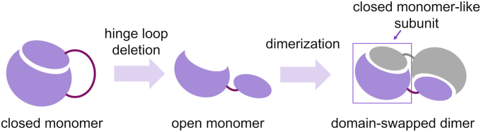
Figure 1 Hypothetical molecular evolution of domain-swapped dimers. It is believed that the open monomer evolved from the closed monomer through shortening of the hinge loop and that the formation of a domain-swapped dimer is completed by the dimerization of the open monomers. Although the domain-swapped dimer is composed of two open monomers, it can also be described as comprising two closed monomer-like subunits.
Figure 1 Hypothetical molecular evolution of domain-swapped dimers. It is believed that the open monomer evolved from the closed monomer through shortening of the hinge loop and that the formation of a domain-swapped dimer is completed by the dimerization of the open monomers. Although the domain-swapped dimer is composed of two open monomers, it can also be described as comprising two closed monomer-like subunits.CC BY-NC-ND · PubMed Central
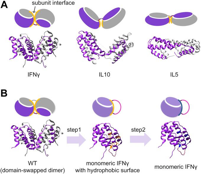
Figure 2 Proteins with domain-swapped dimers and the approach to monomerization.A, structures of IFNγ (PDB: 1FG9), IL10 (PDB: 6X93), and IL5 (PDB: 1HUL). Monomerized variants of IL10 and IL5 engineered via linker insertion have been previously reported (35, 36, 37). B, two-step approach for designing monomeric IFNγ. Step 1: A swapped domain can be structurally rearranged from an open monomer form to a closed monomer structure via linker insertion. Hydrophobic amino acids in the subunit interface are depicted in yellow. Step 2: By replacing the hydrophobic amino acids (yellow) with hydrophilic amino acids (blue), the solubility of the monomer will be increased. IFN, interferon; IL, interleukin.
Figure 2 Proteins with domain-swapped dimers and the approach to monomerization.A, structures of IFNγ (PDB: 1FG9), IL10 (PDB: 6X93), and IL5 (PDB: 1HUL). Monomerized variants of IL10 and IL5 engineered via linker insertion have been previously reported (35, 36, 37). B, two-step approach for designing monomeric IFNγ. Step 1: A swapped domain can be structurally rearranged from an open monomer form to a closed monomer structure via linker insertion. Hydrophobic amino acids in the subunit interface are depicted in yellow. Step 2: By replacing the hydrophobic amino acids (yellow) with hydrophilic amino acids (blue), the solubility of the monomer will be increased. IFN, interferon; IL, interleukin.CC BY-NC-ND · PubMed Central
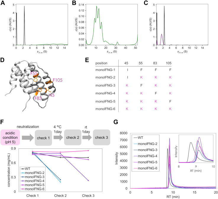
Figure 3 Characterization of IFNγ variants.A, sedimentation coefficient distribution of 1.2 mg/ml of WT (black) and monoIFNG-1 (green) measured at 20 °C and pH 5.0. B, sedimentation coefficient distribution of 1.2 mg/ml of monoIFNG-1 measured at 20 °C and pH 5.0 (enlarged figure). C, sedimentation coefficient distributions of 1.2 mg/ml of WT (black) and monoIFNG-5 (purple) measured at 20 °C and pH 5.0. D, four hydrophobic amino acids (yellow) examined in the subunit interface. E, names of the variants in which the hydrophobic amino acid was replaced with lysine. F, evaluation of the aggregation propensity of variants under neutral conditions. The concentration of the sample in supernatant was calculated from the absorbance (A280) at every checkpoint. G, analytical SEC results of variants using HPLC measured at pH 5.0. IFN, interferon; SEC, size-exclusion chromatography.
Figure 3 Characterization of IFNγ variants.A, sedimentation coefficient distribution of 1.2 mg/ml of WT (black) and monoIFNG-1 (green) measured at 20 °C and pH 5.0. B, sedimentation coefficient distribution of 1.2 mg/ml of monoIFNG-1 measured at 20 °C and pH 5.0 (enlarged figure). C, sedimentation coefficient distributions of 1.2 mg/ml of WT (black) and monoIFNG-5 (purple) measured at 20 °C and pH 5.0. D, four hydrophobic amino acids (yellow) examined in the subunit interface. E, names of the variants in which the hydrophobic amino acid was replaced with lysine. F, evaluation of the aggregation propensity of variants under neutral conditions. The concentration of the sample in supernatant was calculated from the absorbance (A280) at every checkpoint. G, analytical SEC results of variants using HPLC measured at pH 5.0. IFN, interferon; SEC, size-exclusion chromatography.CC BY-NC-ND · PubMed Central
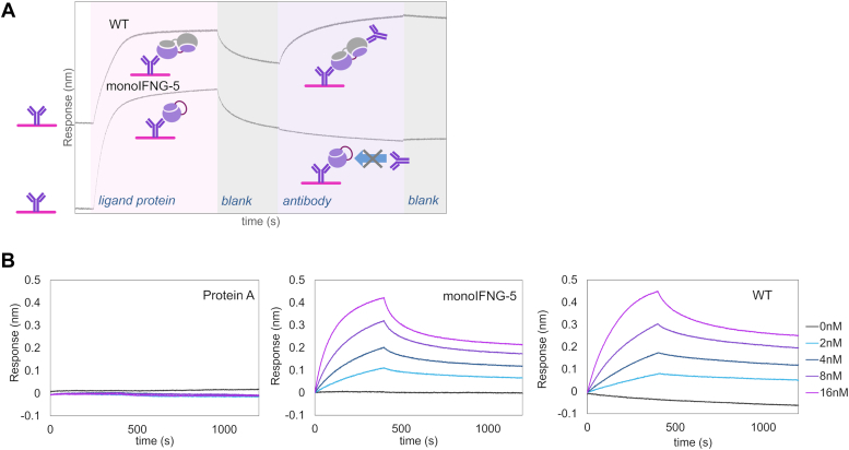
Figure 4 BLI binding assays of monomeric IFNγ.A, epitope binning assay of WT and monoIFNG-5. Biosensors immobilized with anti-IFNγ antibody were immersed into WT- or monoIFNG-5–containing solutions. Subsequently, the sensors were immersed in the same anti-IFNγ antibody solution. Diagrams illustrate the possible binding of analytes to anti-IFNγ antibody. BLI, biolayer interferometry; IFN, interferon. B, Representative sensorgrams of BLI measurements examining the interactions of IFNγR1 with protein A (left; control), monoIFNG-5 (center), and WT (right). Biosensors immobilized with IFNγR1 were immersed into solutions containing a series of analyte concentrations.
Figure 4 BLI binding assays of monomeric IFNγ.A, epitope binning assay of WT and monoIFNG-5. Biosensors immobilized with anti-IFNγ antibody were immersed into WT- or monoIFNG-5–containing solutions. Subsequently, the sensors were immersed in the same anti-IFNγ antibody solution. Diagrams illustrate the possible binding of analytes to anti-IFNγ antibody. BLI, biolayer interferometry; IFN, interferon. B, Representative sensorgrams of BLI measurements examining the interactions of IFNγR1 with protein A (left; control), monoIFNG-5 (center), and WT (right). Biosensors immobilized with IFNγR1 were immersed into solutions containing a series of analyte concentrations.CC BY-NC-ND · PubMed Central
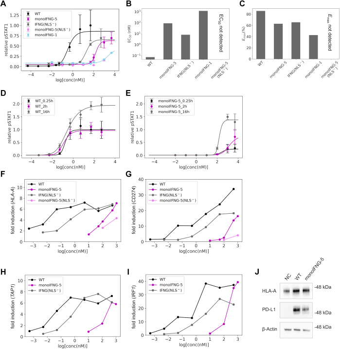
Figure 5 Upstream and downstream signaling pathway analyses upon the stimulation of IFNγ variants.A, STAT1 phosphorylation activity assay. HeLa cells were treated with WT or variants for 0.25 h. Phosphorylated STAT1 (pSTAT1) was measured using AlphaLISA. Data represent the mean ± SD of n = 3 biological replicates, except for monoIFNG-1. Data for monoIFNG-1 represent the mean ± SD of n = 2 biological replicates. Solid lines were calculated via four-parameter logistic curve fitting. B, EC50 (nM) of STAT1 phosphorylation by the variants obtained from the fitting calculation. C, relative Emax (%) of STAT1 phosphorylation by the variants. The values were normalized to measurements at 50 nM WT stimulation. D and E, time dependence of STAT1 phosphorylation by WT (D) and monoIFNG-5 (E). HeLa cells were treated with WT or monoIFNG-5 for 0.25, 2, or 16 h. pSTAT1 was measured using AlphaLISA. Data represent the mean ± SD of n = 3 biological replicates. F–I, fold induction of target genes HLA-A (F), CD274 (G), TAP1 (H), and IRF1 (I) compared to the unstimulated state (NC). HeLa cells were treated with WT and variants for 48 h, whereafter mRNA was extracted from each sample. Gene expression levels of the target genes and the reference 18S rRNA were quantified based on the amount of corresponding mRNA determined via RT-qPCR. Data represent values obtained from a single biological replicate (n = 1). J, Western blotting analysis of HLA-A, PD-L1, and β-actin in cell lysates. HeLa cells were treated with 500 nM of WT or 1000 nM of monoIFNG-5 for 48 h, and the cell lysates were analyzed. HLA, Human leukocyte antigen; IFN, interferon; PD-L1, programmed death-ligand 1; RT-qPCR, quantitative reverse transcription PCR.
Figure 5 Upstream and downstream signaling pathway analyses upon the stimulation of IFNγ variants.A, STAT1 phosphorylation activity assay. HeLa cells were treated with WT or variants for 0.25 h. Phosphorylated STAT1 (pSTAT1) was measured using AlphaLISA. Data represent the mean ± SD of n = 3 biological replicates, except for monoIFNG-1. Data for monoIFNG-1 represent the mean ± SD of n = 2 biological replicates. Solid lines were calculated via four-parameter logistic curve fitting. B, EC50 (nM) of STAT1 phosphorylation by the variants obtained from the fitting calculation. C, relative Emax (%) of STAT1 phosphorylation by the variants. The values were normalized to measurements at 50 nM WT stimulation. D and E, time dependence of STAT1 phosphorylation by WT (D) and monoIFNG-5 (E). HeLa cells were treated with WT or monoIFNG-5 for 0.25, 2, or 16 h. pSTAT1 was measured using AlphaLISA. Data represent the mean ± SD of n = 3 biological replicates. F–I, fold induction of target genes HLA-A (F), CD274 (G), TAP1 (H), and IRF1 (I) compared to the unstimulated state (NC). HeLa cells were treated with WT and variants for 48 h, whereafter mRNA was extracted from each sample. Gene expression levels of the target genes and the reference 18S rRNA were quantified based on the amount of corresponding mRNA determined via RT-qPCR. Data represent values obtained from a single biological replicate (n = 1). J, Western blotting analysis of HLA-A, PD-L1, and β-actin in cell lysates. HeLa cells were treated with 500 nM of WT or 1000 nM of monoIFNG-5 for 48 h, and the cell lysates were analyzed. HLA, Human leukocyte antigen; IFN, interferon; PD-L1, programmed death-ligand 1; RT-qPCR, quantitative reverse transcription PCR.CC BY-NC-ND · PubMed Central
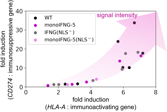
Figure 6 Relationship between immunostimulatory and immunosuppressive gene expression upon stimulation with IFNγ variants. The expression balance of two contrastive genes did not depend on the structural formats of the ligands but rather on their intracellular signal intensity. IFN, interferon.
Figure 6 Relationship between immunostimulatory and immunosuppressive gene expression upon stimulation with IFNγ variants. The expression balance of two contrastive genes did not depend on the structural formats of the ligands but rather on their intracellular signal intensity. IFN, interferon.CC BY-NC-ND · PubMed Central

Verification

Claims the paper makes

The claims and statistics of this paper haven’t been checked yet.

Run the check — free, no account needed. It reads the open-access text and checks the sources it cites.

A few free checks a day · papers already verified open instantly · results are public

Self-check

Percentages, sums and reported differences re-computed from the paper’s own numbers.

The self-check needs the paper’s full text.

Citations

Cites

Show all 45 references

Metadata

Authors
Goto, Y., Miyafusa, T., & Honda, S.
Affiliations
National Institute of Advanced Industrial Science and Technology (JP)
Corresponding author
Honda, Shinya
Venue
Journal of Biological Chemistry
Publisher
Elsevier BV
Published
2024-07
Volume
300
Issue
7
Pages
107464
ISSN
0021-9258 (ISSN-L)
OpenAlex
W4399601161
Cited by
1
References
45
Citations by year
2025: 1
FWCI
0.17
Type
journal article
Language
en
Open access
Licence
Locations
  • Journal of Biological Chemistry (publishedVersion, cc-by-nc-nd)
  • bioRxiv (Cold Spring Harbor Laboratory) (acceptedVersion)
  • PubMed Central (publishedVersion, cc-by-nc-nd)
  • PubMed (publishedVersion)
Topics
  • Life Sciences › Immunology and Microbiology › Immunology › interferon and immune responses
  • Life Sciences › Immunology and Microbiology › Immunology › Cytokine Signaling Pathways and Interactions
  • Life Sciences › Biochemistry, Genetics and Molecular Biology › Molecular Biology › Protein Structure and Dynamics
Keywords
interferon-gamma, domain-swapped dimer, protein structure design, cancer immunotherapy, immunosuppression, MHC class I expression, protein evolution, hydrophobic amino acids, linker design
MeSH terms
Protein Domains, Animals, Humans, Interferon-gamma, Mice, Protein Multimerization

About these checks

Registry, retraction, Cites and statistics results are computed, not judged, with one exception: a printed reference that no index lists is read once by a model before it counts as a citation error. Claims are extracted by a model and kept only when their quote appears verbatim in the text; each cited source is then checked, with a model’s help, for whether it says what the claim relies on. Those verdicts can be wrong — read the claim’s quote and the cited source before relying on one.

Statistics and arithmetic are recomputed, not judged.

Confidence bands are shown only for fields calibrated on our annotated test set. None is calibrated yet, so no band is shown.

What counts as a claim ›How we measure confidence ›How we check this ›

Are you an author, or do you see a problem with this page? Request a review

How to cite

Cite CoChat for the checks on this page, and the paper for its own findings.

This page
CoChat. (2026). Citation check: Designing monomeric IFNγ: The significance of domain-swapped dimer structure in IFNγ immune responses. https://app.cochat.ai/papers/10.1016/j.jbc.2024.107464
The paper
Goto, Y., Miyafusa, T., & Honda, S. (2024). Designing monomeric IFNγ: The significance of domain-swapped dimer structure in IFNγ immune responses. Journal of Biological Chemistry. https://doi.org/10.1016/j.jbc.2024.107464

Data sources

Bibliographic data: Semantic Scholar, OpenAlex, Crossref.

Contains information from the Semantic Scholar Academic Graph Datasets, made available under the ODC Attribution License (ODC-BY 1.0).

Semantic Scholar’s data is described in: Kinney, R., et al. (2023). The Semantic Scholar Open Data Platform. arXiv:2301.10140.