Student or educator? Sign up with your school email and get 6 months of Pro free.
Cited by 6Enzyme Production and CharacterizationPolysaccharides and Plant Cell WallsSeaweed-derived Bioactive Compounds

The DOI matches its Crossref record; 57 of the 57 references it cites were found in a registry or an index.

The complete λ-carrageenan depolymerization cascade from a marine Pseudoalteromonad revealed by structural analysis of the enzymes

Open access

Published version of a preprint

Abstract

Carrageenans are a complex group of polysaccharides derived from the cell walls of red macroalgae. They are an abundant, yet recalcitrant, nutrient source for most marine heterotrophic bacteria. Some member species of the Pseudoalteromonas genus are effective at metabolizing carrageenan. However, the enzymatic pathway for λ-carrageenan, one of the most sulfated naturally occurring polysaccharides, remains unknown. Using detailed structural analysis by X-ray crystallography, we reveal the sophisticated and cyclic enzymatic cascade deployed by Pseudoalteromonas distincta (strain U2A) to utilize λ-carrageenan. The cascade incorporates 10 glycoside hydrolases and five sulfatases that are specific for λ-carrageenan and cooperate to completely deconstruct this polysaccharide, thus yielding galactose monosaccharides for subsequent energy production. The detailed molecular understanding of λ-carrageenan depolymerisation provided includes structural evidence for a lesser described sulfatase catalytic mechanism and elucidation of a distinct catabolic cascade that is unique from previously described carrageenan metabolic pathways. This insight also holds potential for the application of enzymatic logic in the generation of high-value products from abundant natural biopolymers, such as carrageenans.

Overview

The claims and statistics of this paper haven’t been checked yet.

Run the check — free, no account needed. It reads the open-access text and checks the sources it cites.

A few free checks a day · papers already verified open instantly · results are public

Integrity signals

Registry

Crossref · 10.1016/j.jbc.2025.110719

Cites

57 of 57 references verified0 citation errors · 0 not checkable

Statistics

Figures

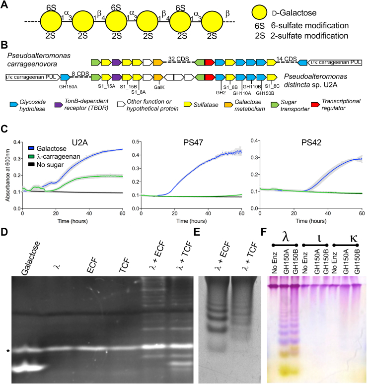
Figure 1 Pseudoalteromonas distincta U2A λ-CarPUL organization and λ-carrageenan growth experiments.A, schematic of the typical structure of λ-carrageenan. B, schematic of the additional λ-CarPUL segment in the Pseudoalteromonas distincta U2A (U2A) CarPUL that is syntenic to a region of the Pseudoalteromonas carrageenovora 9T CarPUL and is hypothesised to confer λ-carrageenan metabolism. Each gene is represented by an arrow, which are colour coded according to putative function. C, carrageenan growth experiments of isolated Pseudoalteromonas sp where growth on λ-carrageenan is exhibited by U2A exclusively. Gray bars indicate the error from n = 4 independent experiments. FACE (D) and C-PAGE (E) metabolic localization experiments showing different λ-carrageenan depolymerisation profiles by U2A extracellular fractions (ECF or cleared culture supernatant) and total cell fractions (TCF, lysed cells removed from culture medium). F, U2A C-PAGE carrageenan activity experiment showing different depolymerisation profiles of GH150A and GH150B exclusively on λ-carrageenan. The asterisk in panel B indicates the free label ANTS.
Figure 1 Pseudoalteromonas distincta U2A λ-CarPUL organization and λ-carrageenan growth experiments.A, schematic of the typical structure of λ-carrageenan. B, schematic of the additional λ-CarPUL segment in the Pseudoalteromonas distincta U2A (U2A) CarPUL that is syntenic to a region of the Pseudoalteromonas carrageenovora 9T CarPUL and is hypothesised to confer λ-carrageenan metabolism. Each gene is represented by an arrow, which are colour coded according to putative function. C, carrageenan growth experiments of isolated Pseudoalteromonas sp where growth on λ-carrageenan is exhibited by U2A exclusively. Gray bars indicate the error from n = 4 independent experiments. FACE (D) and C-PAGE (E) metabolic localization experiments showing different λ-carrageenan depolymerisation profiles by U2A extracellular fractions (ECF or cleared culture supernatant) and total cell fractions (TCF, lysed cells removed from culture medium). F, U2A C-PAGE carrageenan activity experiment showing different depolymerisation profiles of GH150A and GH150B exclusively on λ-carrageenan. The asterisk in panel B indicates the free label ANTS.CC BY · PubMed Central
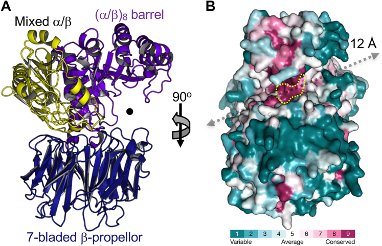
Figure 2 Structural modelling of GH150A and GH150B with AlphaFold 2.A, cartoon representation of the GH150A AlphaFold two model as representative of the overall fold of both GH150A and GH150B (and other GH150 enzymes). The structure is coloured and labelled according to the three identified domains. B, consurf analysis revealing conserved deep cleft running the width of the protein that is created by the two lobes. The length (approximately 12 Å) and location of the cleft is indicated by a grey arrow. The most conserved patch, which presumably comprised the catalytic machinery or other functionally relevant residues, is highlighted by the yellow dotted line.
Figure 2 Structural modelling of GH150A and GH150B with AlphaFold 2.A, cartoon representation of the GH150A AlphaFold two model as representative of the overall fold of both GH150A and GH150B (and other GH150 enzymes). The structure is coloured and labelled according to the three identified domains. B, consurf analysis revealing conserved deep cleft running the width of the protein that is created by the two lobes. The length (approximately 12 Å) and location of the cleft is indicated by a grey arrow. The most conserved patch, which presumably comprised the catalytic machinery or other functionally relevant residues, is highlighted by the yellow dotted line.CC BY · PubMed Central
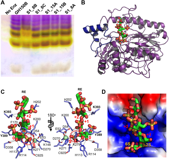
Figure 3 Characterization of S1_8B.A, screening of the five sulfatases in the U2A λ-CarPUL by C-PAGE to identify the λ-neocarrageenoligosaccharide specific exo-galactose-2[,6]-sulfate-2-O-sulfohydrolase. The substrates used for this screen was a pool of oligosaccharides produced by the depolymerization of λ-carrageenan by GH150B. S1_8B was the only sulfatase that showed activity on these oligosaccharides. B, overlay of the apo X-ray crystallographic structure of S1_8B (purple) with the inactive C92S structure bound to λ-NC4 revealing the typical two-domain alkaline phosphatase fold of the S1 family. The structure of the complex was nearly identical to that of the apo structure and only the loop that was disordered in the apo structure is shown in blue for the complex. C, interactions between residues on the dynamic active site-adjacent loop and λ-NC4. The 2S of the non-reducing end G2,6S engages with the catalytic machinery in the S-subsite. D, surface electrostatics show the catalytic machinery in the S-subsite is buried in the base of a positively charged active site pocket that is complementary to the negatively charged l-NC4 substrate.
Figure 3 Characterization of S1_8B.A, screening of the five sulfatases in the U2A λ-CarPUL by C-PAGE to identify the λ-neocarrageenoligosaccharide specific exo-galactose-2[,6]-sulfate-2-O-sulfohydrolase. The substrates used for this screen was a pool of oligosaccharides produced by the depolymerization of λ-carrageenan by GH150B. S1_8B was the only sulfatase that showed activity on these oligosaccharides. B, overlay of the apo X-ray crystallographic structure of S1_8B (purple) with the inactive C92S structure bound to λ-NC4 revealing the typical two-domain alkaline phosphatase fold of the S1 family. The structure of the complex was nearly identical to that of the apo structure and only the loop that was disordered in the apo structure is shown in blue for the complex. C, interactions between residues on the dynamic active site-adjacent loop and λ-NC4. The 2S of the non-reducing end G2,6S engages with the catalytic machinery in the S-subsite. D, surface electrostatics show the catalytic machinery in the S-subsite is buried in the base of a positively charged active site pocket that is complementary to the negatively charged l-NC4 substrate.CC BY · PubMed Central
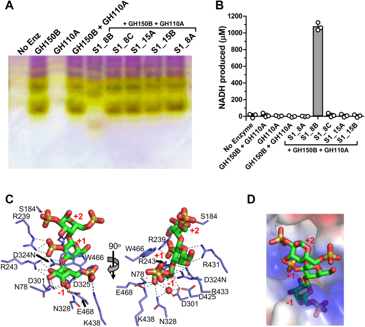
Figure 4 Characterisation of GH110A.A, C-PAGE screening for GH110A and sulfatase activities in combination on GH150B produced λ-carrageenan oligosaccharides. B, galactose release assays confirm monosaccharide release from assays containing GH150B, S1_8B and GH110A indicating removal of G6S from the λ-neocarrageenoligosaccharides. Gray bars indicate the mean values of triplicate independent measurements and the error bars the standard deviation. Closed circles show individual data points. C, X-ray crystallographic structure of GH110A D324N mutant showing the active site bound to λ-NC4 that was pre-treated with S1_8B. D, surface electrostatics show complementary charge of the +1 and +2 subsites to the edge of the glycan region that presents the sugars with 2S modifications.
Figure 4 Characterisation of GH110A.A, C-PAGE screening for GH110A and sulfatase activities in combination on GH150B produced λ-carrageenan oligosaccharides. B, galactose release assays confirm monosaccharide release from assays containing GH150B, S1_8B and GH110A indicating removal of G6S from the λ-neocarrageenoligosaccharides. Gray bars indicate the mean values of triplicate independent measurements and the error bars the standard deviation. Closed circles show individual data points. C, X-ray crystallographic structure of GH110A D324N mutant showing the active site bound to λ-NC4 that was pre-treated with S1_8B. D, surface electrostatics show complementary charge of the +1 and +2 subsites to the edge of the glycan region that presents the sugars with 2S modifications.CC BY · PubMed Central
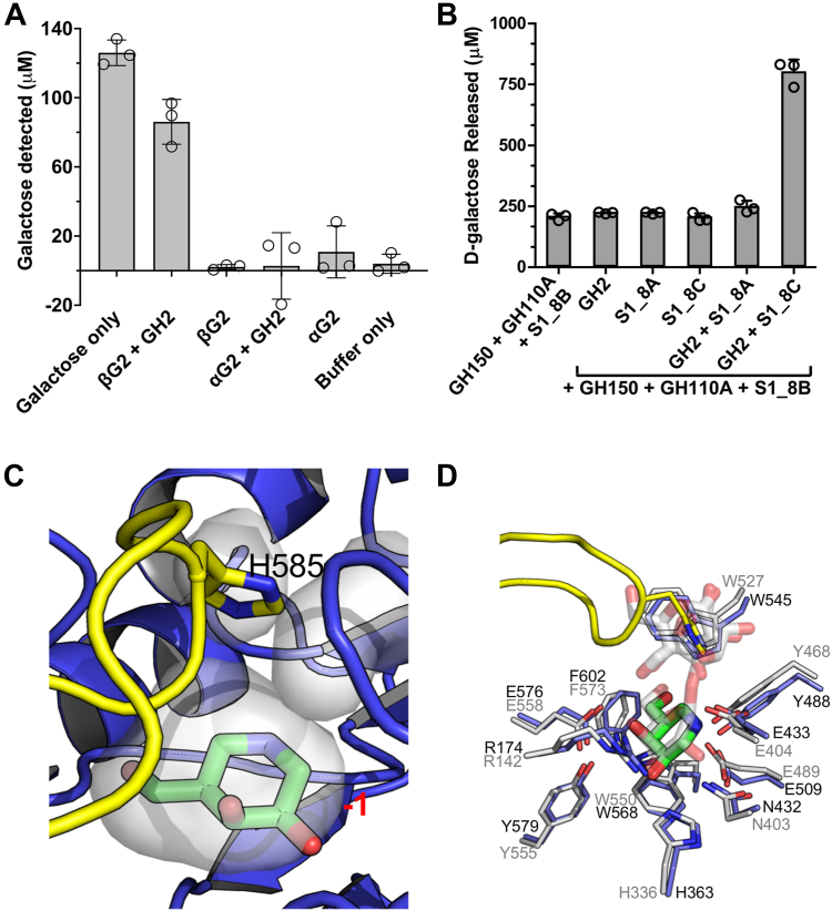
Figure 5 Release of unsulfated galactose by S1_8C and GH2.A, GH2 is a β-galactosidase that shows activity on Galβ1–4Gal in a galactose release assay. B, In galactose release assays GH2 releases monosaccharide from oligosaccharides produced from λ-carrageenan treated with a combination of GH150B, S1_8B and GH110A but only when S1_8C is included in assays i.e., S1_8C and GH2 work together to remove a monosaccharides from carrageenoligosaccharides with a terminal G2S at the non-reducing end. In panels (A) and (B) gray bars indicate the mean values of triplicate independent measurements and the error bars the standard deviation. Closed circles show individual data points. C, GH2 crystallographic structure showing the −1 subsite bound to inhibitor galactoisofagomine (GIF) with capping loop residue H585 at its apex immediately over the actives site. The −1 subsite of GH2 is contoured very closely around the GIF molecule such that a sulfated galactose residue could not be accommodated. D, superimposition of the GH2 structure and β-galactosidase II from Bacillus circulans (PDB ID 7CWD).
Figure 5 Release of unsulfated galactose by S1_8C and GH2.A, GH2 is a β-galactosidase that shows activity on Galβ1–4Gal in a galactose release assay. B, In galactose release assays GH2 releases monosaccharide from oligosaccharides produced from λ-carrageenan treated with a combination of GH150B, S1_8B and GH110A but only when S1_8C is included in assays i.e., S1_8C and GH2 work together to remove a monosaccharides from carrageenoligosaccharides with a terminal G2S at the non-reducing end. In panels (A) and (B) gray bars indicate the mean values of triplicate independent measurements and the error bars the standard deviation. Closed circles show individual data points. C, GH2 crystallographic structure showing the −1 subsite bound to inhibitor galactoisofagomine (GIF) with capping loop residue H585 at its apex immediately over the actives site. The −1 subsite of GH2 is contoured very closely around the GIF molecule such that a sulfated galactose residue could not be accommodated. D, superimposition of the GH2 structure and β-galactosidase II from Bacillus circulans (PDB ID 7CWD).CC BY · PubMed Central
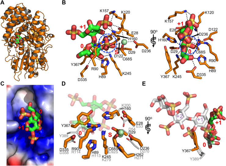
Figure 6 Characterization of S1_8C.A, x-ray crystallographic structure of S1_8C. B, the S1_8C C68S structure bound to λ-C3 which makes extensive interactions with the active site, specifically with the 2S group of the G2S residue engaging the catalytic machinery. C, solvent accessible surface showing a blind-pocket active site architecture. Superimposition of S1_8C and S1_8B showing (D) conservation of the S and 0 subsites and (E) differences in +1 and +2 subsites associated with the different trajectories of substrates when bound in active sites.
Figure 6 Characterization of S1_8C.A, x-ray crystallographic structure of S1_8C. B, the S1_8C C68S structure bound to λ-C3 which makes extensive interactions with the active site, specifically with the 2S group of the G2S residue engaging the catalytic machinery. C, solvent accessible surface showing a blind-pocket active site architecture. Superimposition of S1_8C and S1_8B showing (D) conservation of the S and 0 subsites and (E) differences in +1 and +2 subsites associated with the different trajectories of substrates when bound in active sites.CC BY · PubMed Central
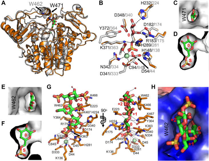
Figure 7 Structural analysis of 6S sulfatases S1_15A and S1_15B.A, superimposition of S1_15A (orange) and S1_15B (gray) X-ray crystallographic structures showing structural variation at the entrance to the active site at tryptophans W471 and W462. B) Both enzymes interact with G6S in an almost identical manner consistent with the specific interactions of galactose-6-sulfate-6-O-sulfohydrolases. C and D, In S1_15A the G6S residue is nearly completely enclosed in the active site by virtue of the α-helix containing W471 closing in over the active site (E) and (F). In contrast, the analogous helix in S1_15B is retracted from the entrance to the active site providing an opening into the pocket. In neither case was there room to accommodate a 2S modification on the galactose in the 0-subsite. G, S1_15B C84S in complex with λ-NC4 pretreated with S1_8B showing the poise of the terminal G6S in the active site was the same as that observed in the monosaccharide G6S complex. H, W462, which lines the entrance to the active site, packs against the pyranose ring of the G2S residue in the +1 subsite guiding the oligosaccharide into the base of the active site pocket.
Figure 7 Structural analysis of 6S sulfatases S1_15A and S1_15B.A, superimposition of S1_15A (orange) and S1_15B (gray) X-ray crystallographic structures showing structural variation at the entrance to the active site at tryptophans W471 and W462. B) Both enzymes interact with G6S in an almost identical manner consistent with the specific interactions of galactose-6-sulfate-6-O-sulfohydrolases. C and D, In S1_15A the G6S residue is nearly completely enclosed in the active site by virtue of the α-helix containing W471 closing in over the active site (E) and (F). In contrast, the analogous helix in S1_15B is retracted from the entrance to the active site providing an opening into the pocket. In neither case was there room to accommodate a 2S modification on the galactose in the 0-subsite. G, S1_15B C84S in complex with λ-NC4 pretreated with S1_8B showing the poise of the terminal G6S in the active site was the same as that observed in the monosaccharide G6S complex. H, W462, which lines the entrance to the active site, packs against the pyranose ring of the G2S residue in the +1 subsite guiding the oligosaccharide into the base of the active site pocket.CC BY · PubMed Central
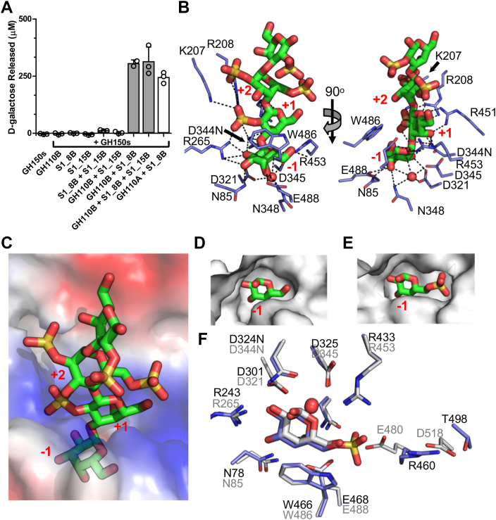
Figure 8 Characterization of GH110B.A, galactose release assay showing GH110B requires S1_8B and S1_15B pre-treatment of oligosaccharides before it could remove monosaccharides. Grey bars indicate the mean values of triplicate independent measurements, and the error bars the standard deviation. Closed circles show individual data points. B, GH110B in complex with λ-NC4 that was pretreated with S1_8B and S1_15B to fully desulfate the non-reducing terminal galactose. The recognition of the sugars involved a complex network of hydrogen bonds, including specific interactions with 2S modifications on the monosaccharides occupying the +1 and +2 subsites. C, the unsulfated non-reducing terminal galactose is bound in −1 subsite at the bottom of a deep pocket. D, the surface of the −1 subsite closely complements the unsulfated galactose which is in contrast to (E) the −1 subsite of GH110A which accommodates the 6S of G6S. F, superimposition of GH110A and GH110B showing nearly identical −1 subsites apart from the region surrounding the 6S group of G6S.
Figure 8 Characterization of GH110B.A, galactose release assay showing GH110B requires S1_8B and S1_15B pre-treatment of oligosaccharides before it could remove monosaccharides. Grey bars indicate the mean values of triplicate independent measurements, and the error bars the standard deviation. Closed circles show individual data points. B, GH110B in complex with λ-NC4 that was pretreated with S1_8B and S1_15B to fully desulfate the non-reducing terminal galactose. The recognition of the sugars involved a complex network of hydrogen bonds, including specific interactions with 2S modifications on the monosaccharides occupying the +1 and +2 subsites. C, the unsulfated non-reducing terminal galactose is bound in −1 subsite at the bottom of a deep pocket. D, the surface of the −1 subsite closely complements the unsulfated galactose which is in contrast to (E) the −1 subsite of GH110A which accommodates the 6S of G6S. F, superimposition of GH110A and GH110B showing nearly identical −1 subsites apart from the region surrounding the 6S group of G6S.CC BY · PubMed Central
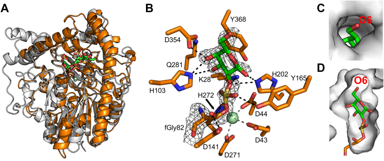
Figure 9 Characterization of monosaccharide specific 2S sulfatase S1_8A.A, superimposition of S1_8A and S1_8B showing overall structural fold conservation. B, contiguous electron density between catalytically matured wild-type S1_8A catalytic residue fGly82 and bound G2S moiety modelled as covalent diester sulfo-galactose species, which is a proposed reaction intermediate. C and D, binding of substrate results in movement of an active-site loop that causes the active site to be enclosed to the extent that only a monosaccharide could be accommodated during catalysis.
Figure 9 Characterization of monosaccharide specific 2S sulfatase S1_8A.A, superimposition of S1_8A and S1_8B showing overall structural fold conservation. B, contiguous electron density between catalytically matured wild-type S1_8A catalytic residue fGly82 and bound G2S moiety modelled as covalent diester sulfo-galactose species, which is a proposed reaction intermediate. C and D, binding of substrate results in movement of an active-site loop that causes the active site to be enclosed to the extent that only a monosaccharide could be accommodated during catalysis.CC BY · PubMed Central
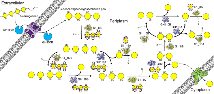
Figure 10 Schematic of λ-carrageenan metabolism by Pseudoalteromonas distincta U2A. Depolymerization is initiated by the activity of the GH150 enzymes that produce a series of λ-neocarrageenoligosaccharides (joined yellow circles with sulfates indicated) with even numbered degrees of polymerization. λ-neocarrageenoligosaccharide breakdown in the periplasm proceeds via a cyclical process whereby the non-reducing end monosaccharides are sequentially removed by GH110A and GH110B and GH2. However, as each non-reducing terminal residue is revealed it must first be prepared for removal by specific sulfatase processing. GH110A requires removal of the 2-sulfate by S1_8 sulfatases. While this desulfation is sufficient to enable GH110B activity we suggest its optimal activity occurs after additional removal of the 6-sulfate by sulfatases from the S1_15 family. GH2 is active only when the terminal β-linked galactose is fully desulfated by the removal of the 2-sulfate group by S1_8C. The process of removing sulfates from the products of the cascade, namely the G6S released by GH110B and G2S left from the final hydrolysis step, to completely reduce λ-carrageenan to galactose (free yellow circle) is accomplished by sulfatases specific to G6S and G2S, i.e., S1_15A and S1_8A. Enzyme structures and schematics of the active sites representing the specificity of substrate recognition are provided for each reaction step.
Figure 10 Schematic of λ-carrageenan metabolism by Pseudoalteromonas distincta U2A. Depolymerization is initiated by the activity of the GH150 enzymes that produce a series of λ-neocarrageenoligosaccharides (joined yellow circles with sulfates indicated) with even numbered degrees of polymerization. λ-neocarrageenoligosaccharide breakdown in the periplasm proceeds via a cyclical process whereby the non-reducing end monosaccharides are sequentially removed by GH110A and GH110B and GH2. However, as each non-reducing terminal residue is revealed it must first be prepared for removal by specific sulfatase processing. GH110A requires removal of the 2-sulfate by S1_8 sulfatases. While this desulfation is sufficient to enable GH110B activity we suggest its optimal activity occurs after additional removal of the 6-sulfate by sulfatases from the S1_15 family. GH2 is active only when the terminal β-linked galactose is fully desulfated by the removal of the 2-sulfate group by S1_8C. The process of removing sulfates from the products of the cascade, namely the G6S released by GH110B and G2S left from the final hydrolysis step, to completely reduce λ-carrageenan to galactose (free yellow circle) is accomplished by sulfatases specific to G6S and G2S, i.e., S1_15A and S1_8A. Enzyme structures and schematics of the active sites representing the specificity of substrate recognition are provided for each reaction step.CC BY · PubMed Central

Verification

Claims the paper makes

The claims and statistics of this paper haven’t been checked yet.

Run the check — free, no account needed. It reads the open-access text and checks the sources it cites.

A few free checks a day · papers already verified open instantly · results are public

Self-check

Percentages, sums and reported differences re-computed from the paper’s own numbers.

The self-check needs the paper’s full text.

Citations

Cites

Show all 57 references

Metadata

Authors
Vickers, C. J., Hettle, A. G., Hobbs, J. K., Shapiro-Ward, S., Pluvinage, B., Medley, B. J., McGuire, B. E., Mihalynuk, L., Nitin, N., Zandberg, W. F., & Boraston, A. B.
Affiliations
  • University of Victoria (CA)
  • University of St Andrews (GB)
  • University of British Columbia (CA)
Corresponding author
Boraston, Alisdair B.
Venue
Journal of Biological Chemistry
Publisher
Elsevier BV
Published
2025-10
Volume
301
Issue
10
Pages
110719
ISSN
0021-9258 (ISSN-L)
OpenAlex
W4414245781
Cited by
6
References
57
Citations by year
2026: 5 · 2025: 1
FWCI
2.52
Type
journal article
Language
en
Open access
Licence
Locations
  • Journal of Biological Chemistry (publishedVersion, cc-by)
  • bioRxiv (Cold Spring Harbor Laboratory) (acceptedVersion, cc-by)
  • Europe PMC (PubMed Central) (publishedVersion, cc-by)
  • PubMed (publishedVersion)
  • St Andrews Research Repository (St Andrews Research Repository) (acceptedVersion, cc-by)
Funders
Natural Sciences and Engineering Research Council of Canada (FRN 04355)
Topics
  • Life Sciences › Biochemistry, Genetics and Molecular Biology › Biotechnology › Enzyme Production and Characterization
  • Life Sciences › Agricultural and Biological Sciences › Plant Science › Polysaccharides and Plant Cell Walls
  • Life Sciences › Agricultural and Biological Sciences › Aquatic Science › Seaweed-derived Bioactive Compounds
Keywords
lambda-carrageenan, sulfated polysaccharides, glycoside hydrolases, sulfatase, marine bacteria, enzyme cascade
MeSH terms
Bacterial Proteins, Carrageenan, Glycoside Hydrolases, Sulfatases, Crystallography, X-Ray, Pseudoalteromonas, Polymerization

About these checks

Registry, retraction, Cites and statistics results are computed, not judged, with one exception: a printed reference that no index lists is read once by a model before it counts as a citation error. Claims are extracted by a model and kept only when their quote appears verbatim in the text; each cited source is then checked, with a model’s help, for whether it says what the claim relies on. Those verdicts can be wrong — read the claim’s quote and the cited source before relying on one.

Statistics and arithmetic are recomputed, not judged.

Confidence bands are shown only for fields calibrated on our annotated test set. None is calibrated yet, so no band is shown.

What counts as a claim ›How we measure confidence ›How we check this ›

Are you an author, or do you see a problem with this page? Request a review

How to cite

Cite CoChat for the checks on this page, and the paper for its own findings.

This page
CoChat. (2026). Citation check: The complete λ-carrageenan depolymerization cascade from a marine Pseudoalteromonad revealed by structural analysis of the enzymes. https://app.cochat.ai/papers/10.1016/j.jbc.2025.110719
The paper
Vickers, C. J., Hettle, A. G., Hobbs, J. K., Shapiro-Ward, S., Pluvinage, B., Medley, B. J., McGuire, B. E., Mihalynuk, L., Nitin, N., Zandberg, W. F., & Boraston, A. B. (2025). The complete λ-carrageenan depolymerization cascade from a marine Pseudoalteromonad revealed by structural analysis of the enzymes. Journal of Biological Chemistry. https://doi.org/10.1016/j.jbc.2025.110719

Data sources

Bibliographic data: Semantic Scholar, OpenAlex, Crossref.

Contains information from the Semantic Scholar Academic Graph Datasets, made available under the ODC Attribution License (ODC-BY 1.0).

Semantic Scholar’s data is described in: Kinney, R., et al. (2023). The Semantic Scholar Open Data Platform. arXiv:2301.10140.