

The DOI matches its Crossref record; 1 of the 1 references it cites were found in a registry or an index.
Uptake of different crystal structures of TiO2 nanoparticles by Caco-2 intestinal cells
Overview
The claims and statistics of this paper haven’t been checked yet.
Run the check — free, no account needed. It reads the open-access text and checks the sources it cites.
Integrity signals
Registry
Cites
Statistics
Figures
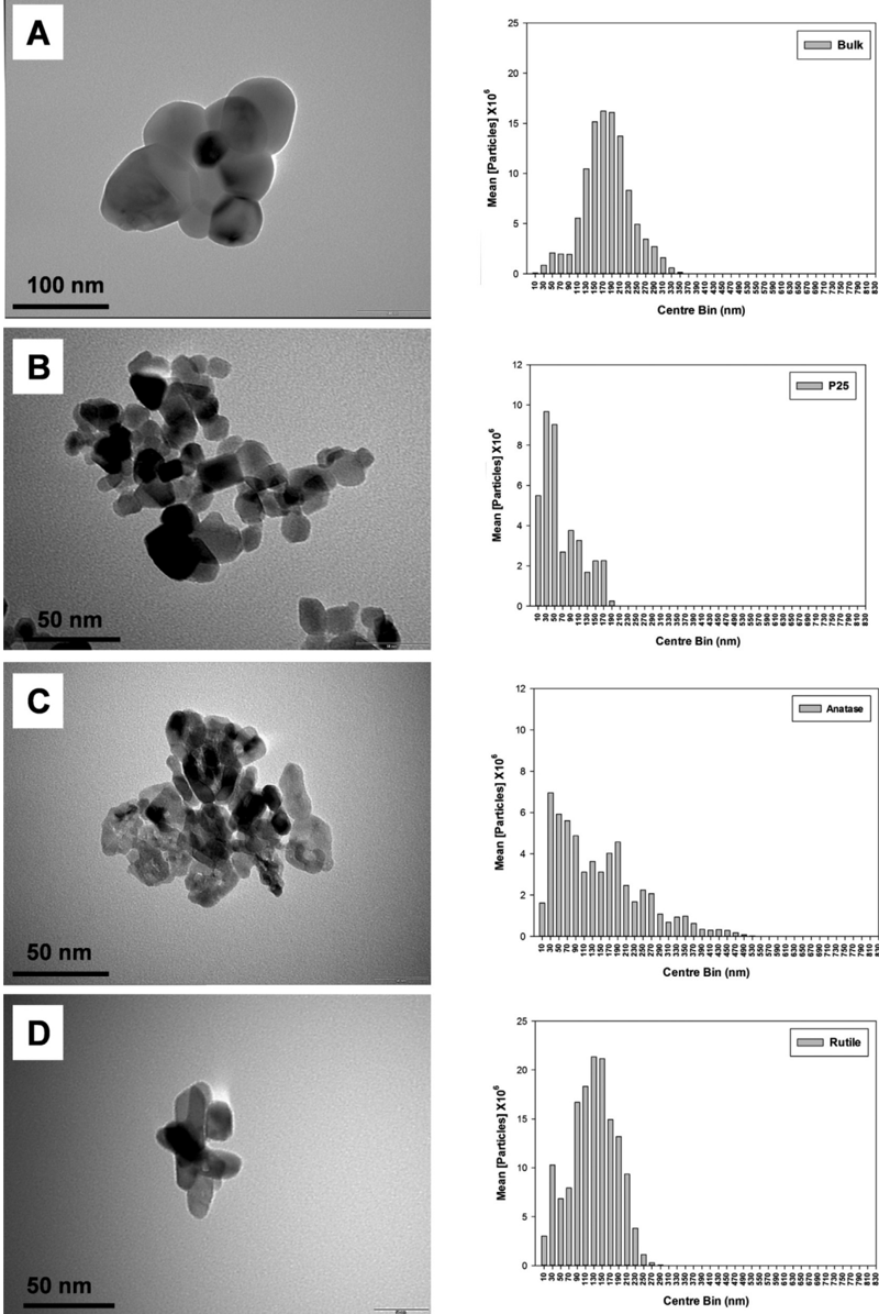
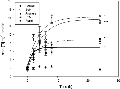
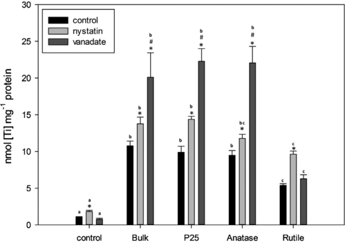
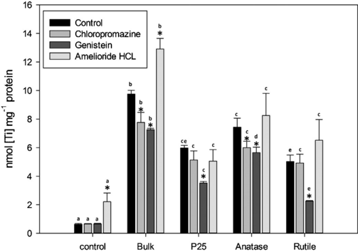
Verification
Claims the paper makes
The claims and statistics of this paper haven’t been checked yet.
Run the check — free, no account needed. It reads the open-access text and checks the sources it cites.
Claims other papers make about this one
Each is checked against this paper’s own text: does it say what the citing paper says it does?
In the citing paper
“the gut is able to differentiate crystal structures of the same chemical substance (e.g., TiO2 materials [250]).”
From Particle toxicology and health - where are we? (2019) · cites this paper as [250]
Self-check
Percentages, sums and reported differences re-computed from the paper’s own numbers.
The self-check needs the paper’s full text.
Citations
Cites
Shannon, R. D. (1976) · Acta Crystallographica Section A · doi:10.1107/s0567739476001551 · Cited by 66,145
Research works printed but not linked: 69
- Aitken, 2006, Hull Manufacture and use of nanomaterials: current status in the UK and global trends, Occupat. Med.-Oxford, 56, 300, 10.1093/occmed/kql051 · DOI
- Akiyama, 1987, Genistein, a specific inhibitor of Tyrosine-specific protein kinases, J. Biol. Chem., 262, 5592, 10.1016/S0021-9258(18)45614-1 · DOI
- Al-Jubory, 2012, Uptake of TiO2 nanoparticles across the isolated perfused intestine of rainbow trout: nystatin, vanadate, and novel gas-sensitive components, Nanotoxicology, 1
- Arrendondo, 2000, Regulation of copper uptake and transport in intestinal monolayers by acute and chronic copper exposure, Biochim. Biophys. Acta, 1474, 169, 10.1016/S0304-4165(00)00015-5 · DOI
- Arrendondo, 2006, Inhibition of iron and copper uptake by iron, copper and zinc, Biol. Res., 39, 95
- Au, 2000, Caco-2 cells can be used ti assess human iron bioavailability from semipurified meal, J. Nutr., 130, 1329, 10.1093/jn/130.5.1329 · DOI
- Baro, 2001, Lipid rafts required for GLUT4 internalization in adipose cells, PNAS, 98, 12050, 10.1073/pnas.211341698 · DOI
- Bermudez, 2004, Pulmonary responses of mice, rats, and hamsters to subchronic inhalation of ultrafine titanium dioxide particles, Toxicol. Sci., 77, 347, 10.1093/toxsci/kfh019 · DOI · Paper page
- Bockmann, 2000, Blood titanium levels before and after oral administration titanium dioxide, Pharmazie, 55, 140
- Bouwmeester, 2009, Review of health safety aspects of nanotechnologies in food production, Regul. Toxicol. Pharmacol., 53, 52, 10.1016/j.yrtph.2008.10.008 · DOI
- Brookes, 2006, Modulation of iron transport proteins in human colorectal carcinogenesis, Gut, 55, 1449, 10.1136/gut.2006.094060 · DOI · Paper page
- Burke, 2005, Sodium-sensitive and -insensitive copper accumulation by isolated intestinal cells of rainbow trout Oncorhynchus mykiss, J. Exp. Biol., 208, 391, 10.1242/jeb.01379 · DOI
- Bury, 2010, Copper and iron uptake in teleost fish, vol. 2, 107
- Cantley, 1978, Vanadate inhibits the red cell (Na+, K+) ATPase from the cytoplasmic side, Nature, 272, 552, 10.1038/272552a0 · DOI
- Chaudry, 2008, Applications and implications of nanotechnologies for the food sector, Food Addit. Contam., Part A 25, 241, 10.1080/02652030701744538 · DOI · Paper page
- Chen, 2007, Titanium dioxide nanomaterials: synthesis, properties, modifications, and applications, Chem. Rev., 107, 2891, 10.1021/cr0500535 · DOI
- Chen, 2011, Cholesterol sequestration by nystatin enhances the uptake and activity of endostatin in endothelium via regulating distinct endocytic pathways, Blood, 117, 6392, 10.1182/blood-2010-12-322867 · DOI
- Chen, 2006, Acute toxicological effects of copper nanoparticles in vivo, Toxicol. Lett., 163, 109, 10.1016/j.toxlet.2005.10.003 · DOI
- Dameron, 1998, Mechanisms for protection against copper toxicity, Am. J. Clin. Nutr., 67, 1091, 10.1093/ajcn/67.5.1091S · DOI
- Ferin, 1985, Biological effects and toxicity assessment of titanium dioxides: anatase and rutile, Am. Ind. Hyg. Assoc. J., 46, 69, 10.1080/15298668591394419 · DOI · Paper page
- Dharmsathaphorn, 1990, Established intestinal cell lines as model systems for electrolyte transport studies, Methods Enzymol., 192, 354, 10.1016/0076-6879(90)92082-O · DOI
- Ferin, 1992, Pulmonary retention of ultrafine and fine particles in rats, Am. J. Respir. Cell. Mol. Biol., 6, 535, 10.1165/ajrcmb/6.5.535 · DOI
- Garnett, 2006, Nanomedicines and nanotoxicology: some physiological principles, Occup. Med. (Lond.), 56, 307, 10.1093/occmed/kql052 · DOI
- Handy, 2007, Ecotoxicity of nanomaterials to fish: Challenges for ecotoxicity testing, Integr. Environ. Assess. Manage, 3, 458, 10.1002/ieam.5630030316 · DOI
- Handy, 2003, Chronic effects of copper exposure versus endocrine toxicity: two sides of the same toxicological process, Comp. Biochem. Physiol., Part A: Mol. Integr. Physiol., 135, 25, 10.1016/S1095-6433(03)00018-7 · DOI
- Handy, 2000, Mechanisms of gastrointestinal copper absorption in the African walking catfish: copper dose-effects and a novel anion-dependent pathway in the intestine, J. Exp. Biol., 203, 2365, 10.1242/jeb.203.15.2365 · DOI
- Handy, 2002, Sodium-dependent copper uptake across epithelia: a review of rationale with experimental evidence from gill and intestine, Biochim. Biophys. Acta, 1566, 104, 10.1016/S0005-2736(02)00590-4 · DOI
- Handy, 1996, Na-dependent regulation of intracellular free magnesium concentration in isolated rat ventricular myocytes, J. Mol. Cell Cardiol., 28, 1641, 10.1006/jmcc.1996.0154 · DOI
- Iverson, 2011, Endocytosis and intracellular transport of nanoparticles: Present knowledge and need for future studies, Nano Today, 6, 176, 10.1016/j.nantod.2011.02.003 · DOI
- Jani, 1994, Titanium dioxide (rutile) particle uptake from the rat GI tract and translocation to systemic organs after oral administration, Int. J. Pharm., 105, 157, 10.1016/0378-5173(94)90461-8 · DOI
- Juttner, 1997, Characterization of Mg2+ transport in brush border membrane vesicles of rabbit ileum studied with mag-fura-2, Biochim. Biophys. Acta, 1370, 51, 10.1016/S0005-2736(97)00242-3 · DOI
- Kaplan, 2009, Copper transport in mammalian cells: Special care for metal with special needs, J. Biol. Chem., 284, 25461, 10.1074/jbc.R109.031286 · DOI
- Kirchhausen, 2009, Imaging endocytic clathrin structures in living cells, Trends Cell Biol., 19, 596, 10.1016/j.tcb.2009.09.002 · DOI
- Kobayashi, 2009, Comparative pulmonary toxicity study of nano-TiO(2) particles of different sizes and agglomerations in rats: different short- and long-term post-instillation results, Toxicology, 264, 110, 10.1016/j.tox.2009.08.002 · DOI
- Koeneman, 2010, Toxicity and cellular responses of intestinal cells exposed to titanium dioxide, Cell Biol. Toxicol., 26, 225, 10.1007/s10565-009-9132-z · DOI
- Lewis, 1977, Nystatin as a probe for investigating the electrical properties of a tight epithelium, J. Cell Biol., 70, 427
- Liu, 2006, Experimental study on toxicity of nanosized titanium dioxide, Modern Prevent. Med., 33, 1211
- Linder, 2006, Vesicular transport of Fe and interaction with other metal ions in polarized Caco-2 Cell monolayers, Biol. Res., 39, 143, 10.4067/S0716-97602006000100016 · DOI
- Lomer, 2000, Determination of titanium dioxide in foods using inductively coupled plasma optical emission spectrometry, Roy. Soc. Ch., 125, 2339
- Moriya, 2005, Vesicular transport and apotransferrin in intestinal iron absorbtion, as shown in the Caco-2 cell model, Am. J. Physiol., 290, 301
- Nabi, 2003, Caveolae/raft-dependent endocytosis, J. Cell. Biol., 161, 673, 10.1083/jcb.200302028 · DOI
- Panessa-Warren, 2006, Biological cellular response to carbon nanoparticle toxicity, J. Phys.-Condens. Matter., 18, 2185, 10.1088/0953-8984/18/33/S34 · DOI
- Peterson, 1993, An in vitro model for the analysis of intestinal brush border assembly I. Ultrastructural analysis of cell contact induced brush border assembly in Caco-2BBe cells, J. Cell Sci., 105, 445, 10.1242/jcs.105.2.445 · DOI
- Peterson, 1993, An in vitro model for the analysis of intestinal brush border assembly II. Changes in expression and localization of brush border proteins during cell contact induced brush border assembly in Caco-2BBe cells, J. Cell Sci., 105, 461, 10.1242/jcs.105.2.461 · DOI
- Ramsden, 2009, Dietary exposure to titanium dioxide nanoparticles in rainbow trout, (Oncorhynchus mykiss): no effect on growth, but subtle biochemical disturbances in the brain, Ecotoxicology, 18, 939, 10.1007/s10646-009-0357-7 · DOI
- Razani, 2002, Caveolae from cell biology to animal physiology, Pharmacol. Rev., 54, 431, 10.1124/pr.54.3.431 · DOI
- Romani, 1992, Regulation of cell magnesium, Arch. Biochem. Biophys., 298, 1, 10.1016/0003-9861(92)90086-C · DOI
- Ryabchikova, 2010, The crystalline forms of titanium dioxide nanoparticles affect their interactions with individual cells, J. Med. CBR Def., 8, 1
- Sayes, 2006, Correlating nanoscale titania structure with toxicity: A cytotoxicity and inflammatory response study with human dermal fibroblasts and human lung epithelial cells, Toxicol. Sci., 92, 174, 10.1093/toxsci/kfj197 · DOI
- Schmidt, 2009, Aqueous long- term solubility of titania nanoparticles and titanium (IV) hydrolysis in a sodium chloride system studied by adsorbative stripping voltammetry, J. Sol. Chem., 38, 1267, 10.1007/s10953-009-9445-9 · DOI
- Shaw, 2011, Physiological effects of nanoparticles on fish: A comparison of nanometals versus metal ions, Environ. Int., 37, 1083, 10.1016/j.envint.2011.03.009 · DOI
- Silva, 2006, Competitive binding of cholesterol and ergosterol to the polyene antibiotic Nystatin. A fluorescence study, Biophys. J., 90, 3625, 10.1529/biophysj.105.075408 · DOI
- Simon, 2011, Titanium dioxide nanoparticles induced intracellular calcium homeostasis modification in primary keratinocytes. Towards an in vitro explanation of titanium dioxide nanoparticles toxicity, Nanotoxicology, 5, 125, 10.3109/17435390.2010.502979 · DOI
- Singh, 2007, Endocytosis, oxidative stress and IL-8 expression in human lung epithelial cells upon treatment with fine and ultrafine TiO2: Role of the specific surface area and of surface methylation of the particles, Toxicol. Appl. Pharmacol., 222, 131, 10.1016/j.taap.2007.05.001 · DOI
- Sonnier, 2011, TNF-(induced vectorial secretion of IL-8 corresponds to development of transepithelial electrical resistance in Caco-2 cells (2011), Gastroenterology, 140, 1044, 10.1016/S0016-5085(11)64340-0 · DOI
- Tiede, 2008, Detection and characterization of engineered nanoparticles in food and the environment, Food Addit. Contam., Part A 25, 795, 10.1080/02652030802007553 · DOI
- Trouiller, 2009, Titanium dioxide nanoparticles induce DNA damage and genetic instability in vivo in mice, Cancer Res., 69, 8784, 10.1158/0008-5472.CAN-09-2496 · DOI
- Van der Ma, 2007, Cellular uptake of cationic polymer- DNA complexes via caveolae plays a pivotal role in gene transfection in COS-7 cells, Pharm. Res., 24, 1590, 10.1007/s11095-007-9287-3 · DOI
- Wang, 2006, Acute toxicity of nano - and micro-scaled zinc powder in adult healthy mice, Toxicol. Lett., 161, 115, 10.1016/j.toxlet.2005.08.007 · DOI
- Wang, 1993, Mis-assembly of clathrin lattices on endosomes reveals a regulatory switch for coated pit formation, J. Cell. Biol., 123, 1107, 10.1083/jcb.123.5.1107 · DOI
- Wang, 2007, Acute toxicity and biodistribution of different sized titanium dioxide particles in mice after oral administration, Toxicol. Lett., 168, 176, 10.1016/j.toxlet.2006.12.001 · DOI
- Wang, 2008, Time-dependent translocation and potential impairment on central nervous system by intranasally instilled TiO2 nanoparticles, Toxicology, 254, 82, 10.1016/j.tox.2008.09.014 · DOI
- Wang, 2003, Phosphatidylinositol 4 phosphate regulates targeting of clathrin adaptor AP-1 complexes to the golgi, Cell, 114, 299, 10.1016/S0092-8674(03)00603-2 · DOI
- Warheit, 2007, Pulmonary toxicity study in rats with three forms of ultrafine-TiO2 particles: Differential responses related to surface properties, Toxicology, 230, 90, 10.1016/j.tox.2006.11.002 · DOI
- Weir, 2012, Titanium dioxide nanoparticles in food and personal care products, Environ. Sci. Technol., 46, 2242, 10.1021/es204168d · DOI
- Wodnicka, 1994, Tyrosine phosphorylation is involved in reorganization of the actin cytoskeleton in response to serum or LPA stimulation, J. Cell Sci., 107, 3643, 10.1242/jcs.107.12.3643 · DOI
- Yamashita, 2002, New and better protocols for short term Caco-2 cell culture system, J. Pharm. Cell Sci., 91, 669, 10.1002/jps.10050 · DOI
- Zerounian, 2002, Effects of copper and ceruloplasmin on iron transport in the Caco-2 cell intestinal model, J. Nutr. Biochem., 13, 138, 10.1016/S0955-2863(01)00205-4 · DOI
- Zhang, 2009, Mechanisms of quantum dot nanoparticle cellular uptake, Toxicol. Sci., 110, 138, 10.1093/toxsci/kfp087 · DOI
Cited by
Sorted by citations
Particle toxicology and health - where are we?
Riediker, M., Zink, D., Kreyling, W., Oberdörster, G., Elder, A., Graham, U., Lynch, I., Duschl, A., Ichihara, G., Ichihara, S., et al. (2019) · Particle and Fibre Toxicology · doi:10.1186/s12989-019-0302-8 · Cited by 237
Self-citation
Metadata
- Authors
- Gitrowski, C., Al-Jubory, A. R., & Handy, R. D.
- Venue
- Toxicology Letters
- Publisher
- Elsevier BV
- Published
- 2014-05
- Volume
- 226
- Issue
- 3
- Pages
- 264-276
- ISSN
- 0378-4274
- Cited by
- 72
- References
- 1
- Language
- en
About these checks
Registry, retraction, Cites and statistics results are computed, not judged, with one exception: a printed reference that no index lists is read once by a model before it counts as a citation error. Claims are extracted by a model and kept only when their quote appears verbatim in the text; each cited source is then checked, with a model’s help, for whether it says what the claim relies on. Those verdicts can be wrong — read the claim’s quote and the cited source before relying on one.
Statistics and arithmetic are recomputed, not judged.
Confidence bands are shown only for fields calibrated on our annotated test set. None is calibrated yet, so no band is shown.
What counts as a claim ›How we measure confidence ›How we check this ›
Are you an author, or do you see a problem with this page? Request a review
