

The DOI matches its Crossref record; 143 of the 143 references it cites were found in a registry or an index.
Cardiac macrophage biology in the steady-state heart, the aging heart, and following myocardial infarction
Open accessAbstract
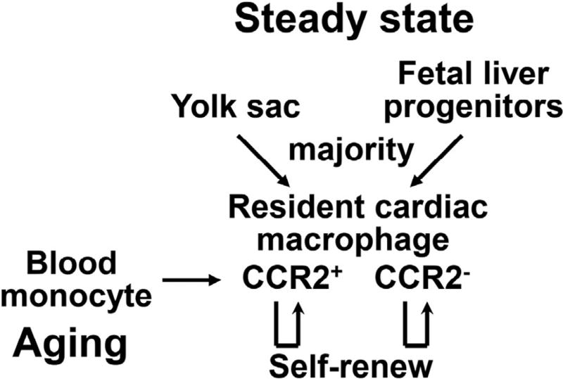
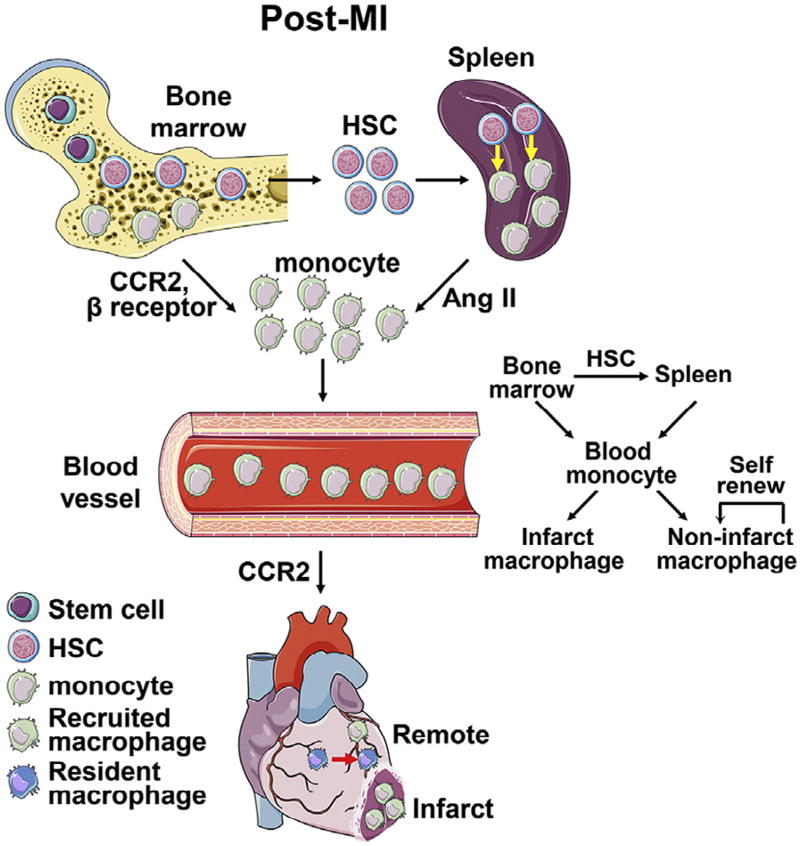
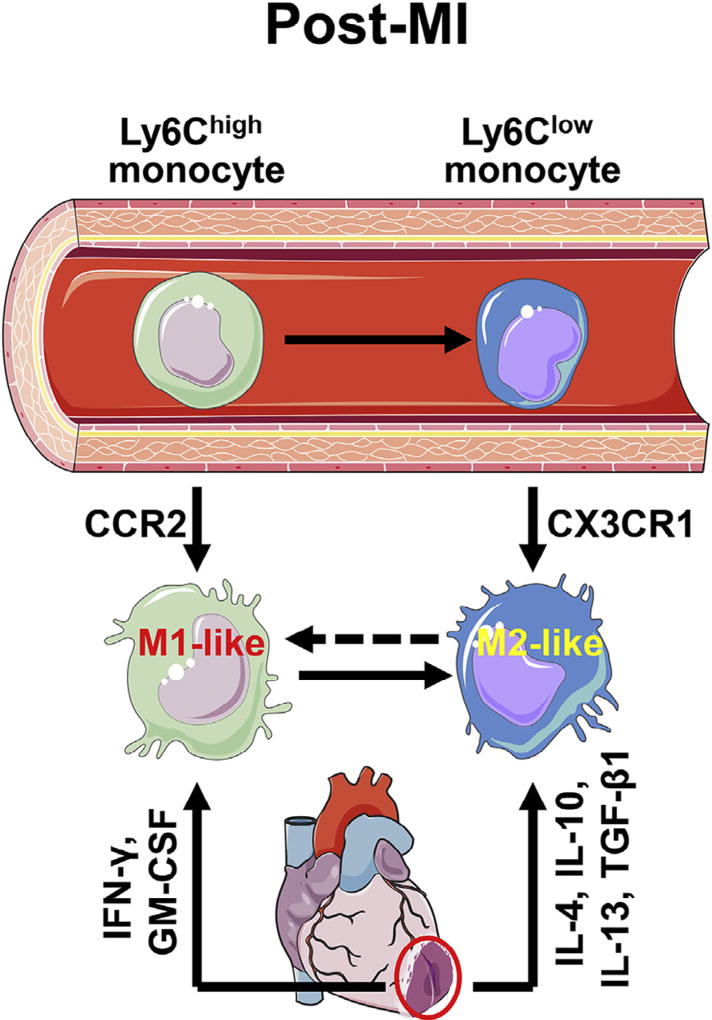
Macrophages play critical roles in homeostatic maintenance of the myocardium under normal conditions and in tissue repair after injury. In the steady-state heart, resident cardiac macrophages remove senescent and dying cells and facilitate electrical conduction. In the aging heart, the shift in macrophage phenotype to a proinflammatory subtype leads to inflammaging. Following myocardial infarction (MI), macrophages recruited to the infarct produce both proinflammatory and anti-inflammatory mediators (cytokines, chemokines, matrix metalloproteinases, and growth factors), phagocytize dead cells, and promote angiogenesis and scar formation. These diverse properties are attributed to distinct macrophage subtypes and polarization status. Infarct macrophages exhibit a proinflammatory M1 phenotype early and become polarized toward an anti-inflammatory M2 phenotype later post-MI. Although this classification system is oversimplified and needs to be refined to accommodate the multiple different macrophage subtypes that have been recently identified, general concepts on macrophage roles are independent of subtype classification. This review summarizes current knowledge about cardiac macrophage origins, roles, and phenotypes in the steady state, with aging, and after MI, as well as highlights outstanding areas of investigation.
Overview
The claims and statistics of this paper haven’t been checked yet.
Run the check — free, no account needed. It reads the open-access text and checks the sources it cites.
Integrity signals
Registry
Cites
Statistics
Figures
Verification
Claims the paper makes
The claims and statistics of this paper haven’t been checked yet.
Run the check — free, no account needed. It reads the open-access text and checks the sources it cites.
Claims other papers make about this one
Each is checked against this paper’s own text: does it say what the citing paper says it does?
The source backs part of the claim, not all of it — or backs it for a different population, condition or comparison.
In the citing paper
“However, in the aging heart, macrophages recruited to the site of infarction exhibit a pro-inflammatory M1 phenotype initially, but subsequently transition to an anti-inflammatory M2 phenotype after myocardial infarction (MI).”
From Inflammation and aging: signaling pathways and intervention therapies (2023) · cites this paper as [143]
Experimental — accuracy not yet measuredThe cited source says what the claim attributes to it.
In the citing paper
“This phenotypic switch promotes angiogenesis and scar formation, aiding in the recovery process.143”
From Inflammation and aging: signaling pathways and intervention therapies (2023) · cites this paper as [143]
Self-check
Percentages, sums and reported differences re-computed from the paper’s own numbers.
The self-check needs the paper’s full text.
Citations
Cites
1 retracted
Yin, H., Li, P., Hu, F., Wang, Y., Chai, X., & Zhang, Y. (2014) · Molecular Medicine Reports · doi:10.3892/mmr.2014.2051 · Cited by 42
RetractedThe chemokine system in diverse forms of macrophage activation and polarization
MANTOVANI, A., SICA, A., SOZZANI, S., ALLAVENA, P., VECCHI, A., & LOCATI, M. (2004) · Trends in Immunology · doi:10.1016/j.it.2004.09.015 · Cited by 6,534
Macrophage plasticity and polarization: in vivo veritas
Sica, A., & Mantovani, A. (2012) · Journal of Clinical Investigation · doi:10.1172/jci59643 · Cited by 6,335
Cytokine-mediated regulation of CXCR4 expression in human neutrophils
Nagase, H., Miyamasu, M., Yamaguchi, M., Imanishi, M., Tsuno, N. H., Matsushima, K., Yamamoto, K., Morita, Y., & Hirai, K. (2002) · Journal of Leukocyte Biology · doi:10.1189/jlb.71.4.711 · Cited by 4,713
Synaptic Pruning by Microglia Is Necessary for Normal Brain Development
Paolicelli, R. C., Bolasco, G., Pagani, F., Maggi, L., Scianni, M., Panzanelli, P., Giustetto, M., Ferreira, T. A., Guiducci, E., Dumas, L., et al. (2011) · Science · doi:10.1126/science.1202529 · Cited by 4,244
Fate Mapping Reveals Origins and Dynamics of Monocytes and Tissue Macrophages under Homeostasis
Yona, S., Kim, K.-W., Wolf, Y., Mildner, A., Varol, D., Breker, M., Strauss-Ayali, D., Viukov, S., Guilliams, M., Misharin, A., et al. (2013) · Immunity · doi:10.1016/j.immuni.2012.12.001 · Cited by 3,245
Macrophage activation and polarization
Martinez, F. O. (2008) · Frontiers in Bioscience · doi:10.2741/2692 · Cited by 3,160
Development of Monocytes, Macrophages, and Dendritic Cells
Geissmann, F., Manz, M. G., Jung, S., Sieweke, M. H., Merad, M., & Ley, K. (2010) · Science · doi:10.1126/science.1178331 · Cited by 3,049
Sugiyama, T., Kohara, H., Noda, M., & Nagasawa, T. (2006) · Immunity · doi:10.1016/j.immuni.2006.10.016 · Cited by 2,419
Nahrendorf, M., Swirski, F. K., Aikawa, E., Stangenberg, L., Wurdinger, T., Figueiredo, J.-L., Libby, P., Weissleder, R., & Pittet, M. J. (2007) · The Journal of Experimental Medicine · doi:10.1084/jem.20070885 · Cited by 2,300
Research works printed but not linked: 1
- Pawelec, 1999, Immunosenescence: impact in the young as well as the old?, Mech Ageing Dev, 108, 1
Cited by
Sorted by citations
Inflammation and aging: signaling pathways and intervention therapies
Li, X., Li, C., Zhang, W., Wang, Y., Qian, P., & Huang, H. (2023) · Signal Transduction and Targeted Therapy · doi:10.1038/s41392-023-01502-8 · Cited by 1,443
Bajpai, G., Bredemeyer, A., Li, W., Zaitsev, K., Koenig, A. L., Lokshina, I., Mohan, J., Ivey, B., Hsiao, H.-M., Weinheimer, C., et al. (2019) · Circulation Research · doi:10.1161/circresaha.118.314028 · Cited by 751
Macrophages in inflammation, repair and regeneration
Oishi, Y., & Manabe, I. (2018) · International Immunology · doi:10.1093/intimm/dxy054 · Cited by 770
Obesity, Hypertension, and Cardiac Dysfunction
Mouton, A. J., Li, X., Hall, M. E., & Hall, J. E. (2020) · Circulation Research · doi:10.1161/circresaha.119.312321 · Cited by 561
Self-citationWang, L., Peng, Y., Zhou, B., Song, J., Li, Z., Zhang, M., Guo, G., Wang, Y., Chen, X., Han, L., et al. (2020) · Nature Cell Biology · doi:10.1038/s41556-019-0446-7 · Cited by 405
Metadata
- Authors
- Ma, Y., Mouton, A. J., & Lindsey, M. L.
- Affiliations
- University of Mississippi Medical Center (US)
- Corresponding author
- Lindsey, Merry L.
- Venue
- Translational Research
- Publisher
- Elsevier BV
- Published
- 2018-01
- Volume
- 191
- Pages
- 15-28
- ISSN
- 1878-1810 (ISSN-L)
- 1931-5244
- PMC
- PMC5846093
- OpenAlex
- W2763341318
- Cited by
- 405
- References
- 143
- Citations by year
- 2026: 34 · 2025: 27 · 2024: 64 · 2023: 73 · 2022: 49 · 2021: 50 · 2020: 55 · 2019: 32 · 2018: 21
- FWCI
- 4.19
- Type
- journal article
- Language
- en
- Open access
- hybrid
- doi.org
- Licence
- Locations
- Translational research (publishedVersion, cc-by-nc-nd)
- PubMed (publishedVersion)
- PubMed Central (acceptedVersion, cc-by-nc-nd)
- Funders
- American Heart Association (15SDG22930009)
- National Institutes of Health (GM104357, GM114833, HL051971, HL129823, HL075360, HL105324, GM115428)
- Biomedical Laboratory Research and Development, VA Office of Research and Development (5I01BX000505)
- Topics
- Health Sciences › Medicine › Cardiology and Cardiovascular Medicine › Cardiac Fibrosis and Remodeling
- Life Sciences › Immunology and Microbiology › Immunology › Atherosclerosis and Cardiovascular Diseases
- Life Sciences › Biochemistry, Genetics and Molecular Biology › Cell Biology › Mesenchymal stem cell research
- Keywords
- cardiac macrophages, macrophage polarization, M1/M2 macrophages, myocardial infarction, cardiac aging, inflammaging, tissue homeostasis, phagocytosis, angiogenesis, scar formation, macrophage origin, macrophage phenotype
- MeSH terms
- Aging, Heart, Humans, Macrophages, Myocardial Infarction, Myocardium, Biomarkers
About these checks
Registry, retraction, Cites and statistics results are computed, not judged, with one exception: a printed reference that no index lists is read once by a model before it counts as a citation error. Claims are extracted by a model and kept only when their quote appears verbatim in the text; each cited source is then checked, with a model’s help, for whether it says what the claim relies on. Those verdicts can be wrong — read the claim’s quote and the cited source before relying on one.
Statistics and arithmetic are recomputed, not judged.
Confidence bands are shown only for fields calibrated on our annotated test set. None is calibrated yet, so no band is shown.
What counts as a claim ›How we measure confidence ›How we check this ›
Are you an author, or do you see a problem with this page? Request a review
