

The DOI matches its Crossref record; 41 of the 41 references it cites were found in a registry or an index.
Culturing of ‘unculturable’ human microbiota reveals novel taxa and extensive sporulation
Open accessAbstract
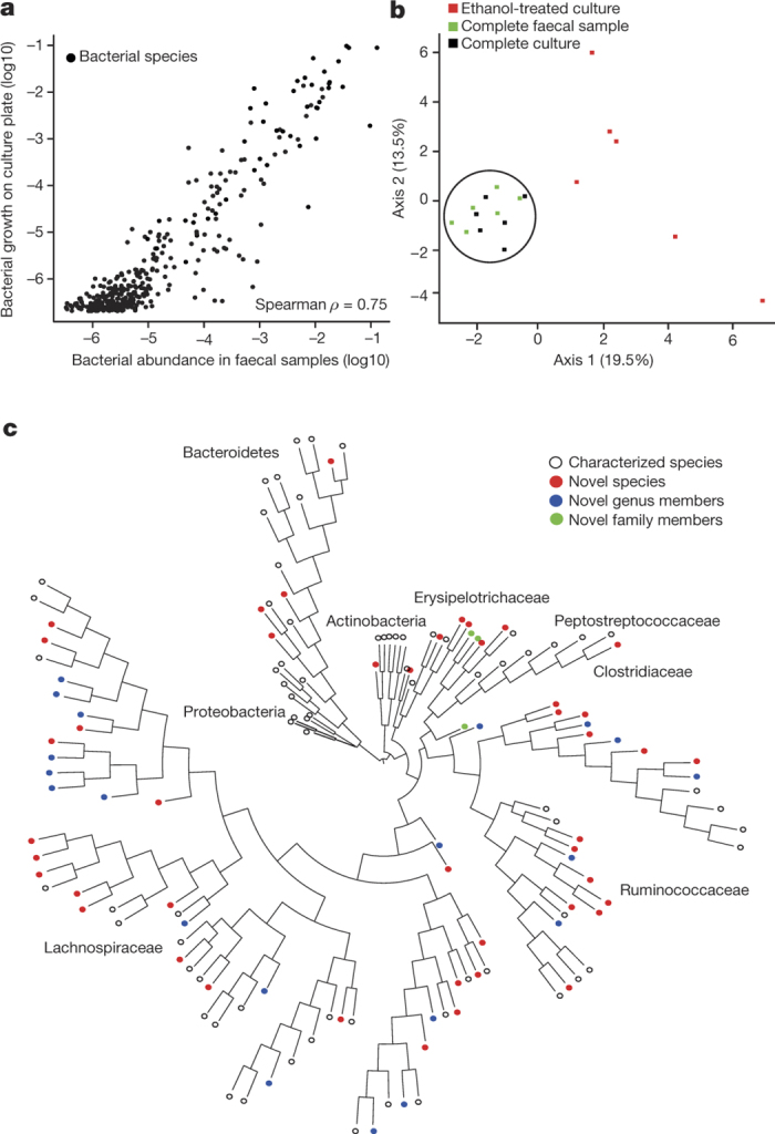
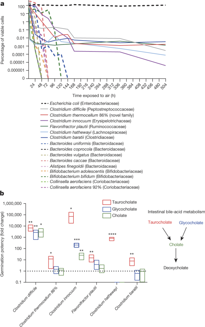
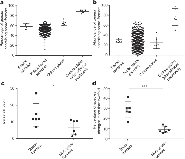
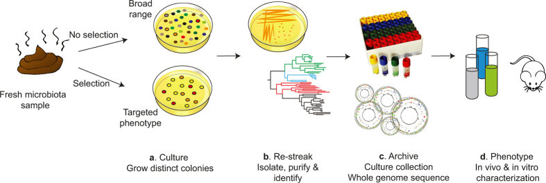
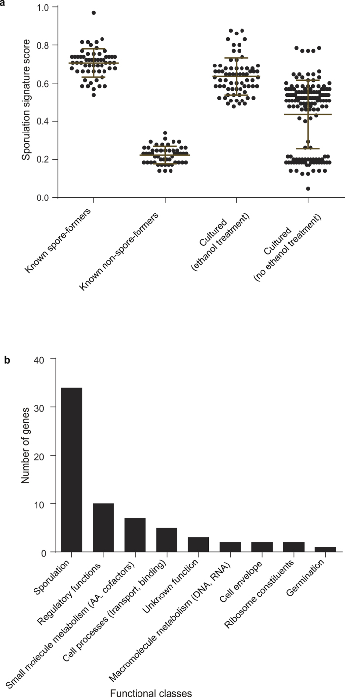
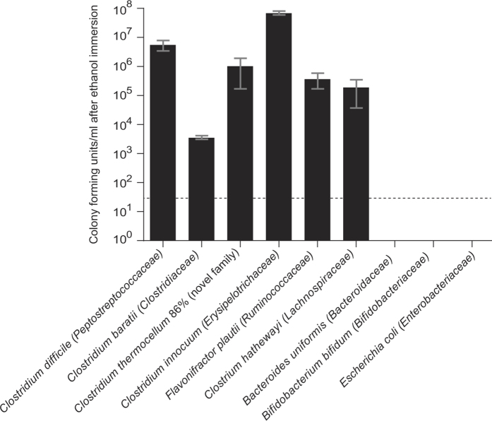
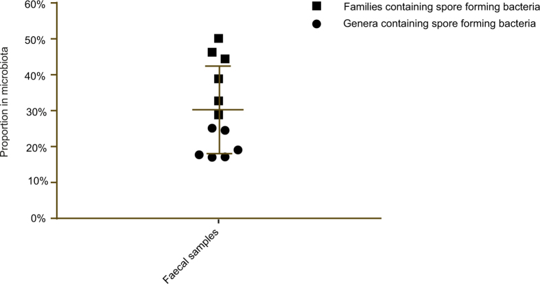
Our intestinal microbiota harbours a diverse bacterial community required for our health, sustenance and wellbeing. Intestinal colonization begins at birth and climaxes with the acquisition of two dominant groups of strict anaerobic bacteria belonging to the Firmicutes and Bacteroidetes phyla. Culture-independent, genomic approaches have transformed our understanding of the role of the human microbiome in health and many diseases. However, owing to the prevailing perception that our indigenous bacteria are largely recalcitrant to culture, many of their functions and phenotypes remain unknown. Here we describe a novel workflow based on targeted phenotypic culturing linked to large-scale whole-genome sequencing, phylogenetic analysis and computational modelling that demonstrates that a substantial proportion of the intestinal bacteria are culturable. Applying this approach to healthy individuals, we isolated 137 bacterial species from characterized and candidate novel families, genera and species that were archived as pure cultures. Whole-genome and metagenomic sequencing, combined with computational and phenotypic analysis, suggests that at least 50-60% of the bacterial genera from the intestinal microbiota of a healthy individual produce resilient spores, specialized for host-to-host transmission. Our approach unlocks the human intestinal microbiota for phenotypic analysis and reveals how a marked proportion of oxygen-sensitive intestinal bacteria can be transmitted between individuals, affecting microbiota heritability.
Overview
The claims and statistics of this paper haven’t been checked yet.
Run the check — free, no account needed. It reads the open-access text and checks the sources it cites.
Integrity signals
Registry
Cites
Statistics
Figures
Verification
Claims the paper makes
The claims and statistics of this paper haven’t been checked yet.
Run the check — free, no account needed. It reads the open-access text and checks the sources it cites.
Claims other papers make about this one
Each is checked against this paper’s own text: does it say what the citing paper says it does?
The cited source says what the claim attributes to it.
In the citing paper
“Many more species continue to be cultured as laboratory-based efforts have become more high-throughput 43,44 .”
From Human microbiome myths and misconceptions (2023) · cites this paper as [44]
Self-check
Percentages, sums and reported differences re-computed from the paper’s own numbers.
The self-check needs the paper’s full text.
Citations
Cites
Basic local alignment search tool
Altschul, S. F., Gish, W., Miller, W., Myers, E. W., & Lipman, D. J. (1990) · Journal of Molecular Biology · doi:10.1006/jmbi.1990.9999 · Cited by 98,669
Fast gapped-read alignment with Bowtie 2
Langmead, B., & Salzberg, S. L. (2012) · Nature Methods · doi:10.1038/nmeth.1923 · Cited by 56,400
Naive Bayesian Classifier for Rapid Assignment of rRNA Sequences into the New Bacterial Taxonomy
Wang, Q., Garrity, G. M., Tiedje, J. M., & Cole, J. R. (2007) · Applied and Environmental Microbiology · doi:10.1128/aem.00062-07 · Cited by 20,145
Schloss, P. D., Westcott, S. L., Ryabin, T., Hall, J. R., Hartmann, M., Hollister, E. B., Lesniewski, R. A., Oakley, B. B., Parks, D. H., Robinson, C. J., et al. (2009) · Applied and Environmental Microbiology · doi:10.1128/aem.01541-09 · Cited by 19,549
FastTree 2 – Approximately Maximum-Likelihood Trees for Large Alignments
Price, M. N., Dehal, P. S., & Arkin, A. P. (2010) · PLoS ONE · doi:10.1371/journal.pone.0009490 · Cited by 14,894
A human gut microbial gene catalogue established by metagenomic sequencing
MetaHIT Consortium, Qin, J., Li, R., Raes, J., Arumugam, M., Burgdorf, K. S., Manichanh, C., Nielsen, T., Pons, N., Levenez, F., et al. (2010) · Nature · doi:10.1038/nature08821 · Cited by 11,624
Kozich, J. J., Westcott, S. L., Baxter, N. T., Highlander, S. K., & Schloss, P. D. (2013) · Applied and Environmental Microbiology · doi:10.1128/aem.01043-13 · Cited by 7,246
ARB: a software environment for sequence data
Ludwig, W. (2004) · Nucleic Acids Research · doi:10.1093/nar/gkh293 · Cited by 6,714
Diversity, stability and resilience of the human gut microbiota
Lozupone, C. A., Stombaugh, J. I., Gordon, J. I., Jansson, J. K., & Knight, R. (2012) · Nature · doi:10.1038/nature11550 · Cited by 5,533
Kraken: ultrafast metagenomic sequence classification using exact alignments
Wood, D. E., & Salzberg, S. L. (2014) · Genome Biology · doi:10.1186/gb-2014-15-3-r46 · Cited by 4,521
Cited by
Sorted by citations
Items 901–1,000 of 1,000
Ren, Q., Li, Y., Duan, M., Li, J., Shi, F., Zhou, Y., Hu, W., Mao, J., & Li, X. (2025) · Frontiers in Microbiology · doi:10.3389/fmicb.2025.1515865 · Cited by 2
Potential pitfalls in claiming novel taxa
Abdugheni, R., & Liu, S. (2025) · iMeta · doi:10.1002/imt2.70036 · Cited by 2
Jensen, R., McKenney, E. A., Beasley, J. C., Cloete, C. C., Melton, M. H., & Lafferty, D. J. R. (2025) · PLoS ONE · doi:10.1371/journal.pone.0333639 · Cited by 2
Do we really understand how faecal microbiota transplantation works?
Quraishi, M. N., McNally, A., & van Schaik, W. (2019) · EBioMedicine · doi:10.1016/j.ebiom.2019.03.024 · Cited by 2
Hologenomics: The Interaction Between Host, Microbiome and Diet
Marsh, A. J., & Azcárate-Peril, M. A. (2020) · Elsevier eBooks · doi:10.1016/b978-0-08-100596-5.22701-7 · Cited by 2
Camarinha‐Silva, A., & Fricke, W. F. (2018) · Springer eBooks · doi:10.1007/978-3-319-90545-7_4 · Cited by 2
Microbiome: Current Status and Future Applications
Ramos-Jiménez, R. G., & Morowitz, M. J. (2019) · Success in academic surgery · doi:10.1007/978-3-030-14644-3_14 · Cited by 2
The role of the microbiome in precision medicine
Pincelli, J. V., Vittorelli, L. O., Stabellini, N., & Pinho, J. R. R. (2019) · Elsevier eBooks · doi:10.1016/b978-0-12-819178-1.00002-2 · Cited by 2
Вячеславович, К. И., Litvinenko, I., Родионов, Г. Г., Игнатьевич, Ш. И., & Светкина, Е. В. (2019) · Human Physiology · doi:10.1134/s036211971908005x · Cited by 2
Adhikari, B., & Kwon, Y. M. (2020) · BMC Research Notes · doi:10.1186/s13104-020-05113-2 · Cited by 2
Blanco, J. M., Borghese, F., McHugh, N. J., Kelleher, P., Sengupta, R., Marchesi, J. R., & Abraham, S. (2020) · BMC Rheumatology · doi:10.1186/s41927-020-00155-2 · Cited by 2
Methods in Computational Biology
Carlson, R. P., & Sauro, H. M. (2019) · MDPI eBooks · doi:10.3390/books978-3-03921-164-7 · Cited by 2
Tejesvi, M. V., Tapiainen, T., Vänni, P., Uhari, M. K., Suokas, M., Lantto, U., Koivunen, P. T., & Renko, M. (2021) · Frontiers in Cellular and Infection Microbiology · doi:10.3389/fcimb.2020.616814 · Cited by 2
Helicobacter pylori-induced gastric carcinogenesis
Noto, J. M., & Ferrero, R. L. (2021) · Elsevier eBooks · doi:10.1016/b978-0-323-85563-1.00004-6 · Cited by 2
Wongkuna, S., Ghimire, S., Janvilisri, T., Doerner, K., Chankhamhaengdecha, S., & Scaria, J. (2020) · F1000Research · doi:10.12688/f1000research.25823.2 · Cited by 2
Tiny Drosophila intestinal stem cells, big power
Zhai, J., Li, W., Liu, X., Wang, D., Zhang, D., Liu, Y., Liang, X., & Chen, Z. (2022) · Cell Biology International · doi:10.1002/cbin.11911 · Cited by 2
Vandamme, P. A. R., Peeters, C., Hettiarachchi, A., Cnockaert, M. C., & Carlier, A. L. (2023) · Systematic and Applied Microbiology · doi:10.1016/j.syapm.2023.126405 · Cited by 2
Predicting evolution in diverse communities
Barraclough, T. G. (2019) · Oxford University Press eBooks · doi:10.1093/oso/9780198749745.003.0009 · Cited by 2
Castaneda, E. U., Brady, J. A., & Speshock, J. L. (2019) · bioRxiv (Cold Spring Harbor Laboratory) · doi:10.1101/734145 · Cited by 2
A regulatory protein that represses sporulation in Clostridioides difficilePreprint
Martins, D., Mendes, A. L., Antunes, J. S., Henriques, A. O., & Serrano, M. (2020) · bioRxiv (Cold Spring Harbor Laboratory) · doi:10.1101/2020.02.25.964569 · Cited by 2
Wu, C., Xu, W., Yu, J., Li, Z., Zhang, Y., Zhang, F., Yang, Y., Wang, Q., Yong, X., & Zhao, B. (2021) · Research Square · doi:10.21203/rs.3.rs-989302/v1 · Cited by 2
Takada, T., Kurakawa, T., Tsuji, H., & Nomoto, K. (2019) · Bergey's Manual of Systematics of Archaea and Bacteria · doi:10.1002/9781118960608.gbm01639 · Cited by 2
Limited vertical transmission of gut bacteria in commercial chickensPreprint
Shterzer, N., Rothschild, N., Sbehat, Y., Dayan, J., Eytan, D., Uni, Z., & Mills, E. (2022) · Research Square · doi:10.21203/rs.3.rs-2132073/v1 · Cited by 2
Host microbiome determines host specificity of the human whipworm, Trichuris trichiuraPreprint
Stark, K. A., Mkandawire, T. T., Tolley, C., Thompson, S., Fortune‐Grant, R., Brandt, C., Clare, S., Lawley, T. D., Cantacessi, C., Almeida, A., et al. (2025) · bioRxiv (Cold Spring Harbor Laboratory) · doi:10.1101/2025.05.09.653168 · Cited by 2
Self-citationConvergent genomic evolution of human gut bacteria along an industrialization gradient
Rühlemann, M. C., Ma, Y., Szánthó, L. L., Waschina, S., Moitinho-Silva, L., Mews, L. C., Faus-Camarena, J., Jebens, H., Costa, J., Juimo, V., et al. (2026) · Cell · doi:10.1101/2025.10.20.683395 · Cited by 3
Afouda, P., Dubourg, G., Cadoret, F., Fournier, P., & Raoult, D. (2017) · New Microbes and New Infections · doi:10.1016/j.nmni.2017.10.011 · Cited by 1
Altmann, D. M. (2018) · Immunology · doi:10.1111/imm.12975 · Cited by 1
Visualization of Respiratory Commensal Bacteria in Context of Their Natural Host Environment
Bonifacio, J. P. P., & Schmolke, M. (2021) · Frontiers in Microbiology · doi:10.3389/fmicb.2021.678389 · Cited by 1
Oprea, M. M., Cristea, D., Dinu, S., Ciontea, S. A., Bojinca, V. C., Predețeanu, D., Balanescu, A. R., & Usein, C. R. (2022) · Revista română de medicină de laborator · doi:10.2478/rrlm-2022-0004 · Cited by 1
Jõers, A., Putrinš, M., Kaldalu, N., Luidalepp, H., & Tenson, T. (2019) · Springer eBooks · doi:10.1007/978-3-030-25241-0_9 · Cited by 1
Tanaka, M., & Nakayama, J. (2022) · KAGAKU TO SEIBUTSU · doi:10.1271/kagakutoseibutsu.60.79 · Cited by 1
Comtet-Marre, S., Chakoory, O., Rochette, E., Gallot, D., Merlin, É., Pons, M., & Peyret, P. (2024) · Cahiers de Nutrition et de Diététique · doi:10.1016/j.cnd.2024.04.006 · Cited by 2
Suggestions for prophylactic ileostomy after rectal cancer surgery
Suo, H.-Y., Li, R.-J., & Song, X.-Q. (2024) · Asian Journal of Surgery · doi:10.1016/j.asjsur.2024.07.291 · Cited by 1
Sánchez, M. C., Hernández, P., Velapatiño, Á., Cuba, E., Ciudad, M. J., & Yurrita, L. C. (2024) · Antibiotics · doi:10.3390/antibiotics13111000 · Cited by 1
Mobile genetic elements: the hidden puppet masters underlying infant gut microbiome assembly?
Kreuze, K., Friman, V., & Vatanen, T. (2024) · Microbiome Research Reports · doi:10.20517/mrr.2024.51 · Cited by 1
Funahashi, K., Lee, C. G., Sugitate, K., Kagata, N., Fukuda, N. N., Song, I., Ishii, C., Hirayama, A., & Fukuda, S. (2024) · Gut Microbes Reports · doi:10.1080/29933935.2024.2429408 · Cited by 1
A high-throughput anaerobic method for viability assays
El-Fateh, M., Meyer, C. T., Chatterjee, A., & Zhao, X. (2025) · Microbiology Spectrum · doi:10.1128/spectrum.02706-24 · Cited by 1
A Novel Therapeutic Strategy for Parkinson's Disease based on the Gut Microbiota: A Rreview Article
Kardan, R., Nazari, M., Hemmati, J., Ahmadi, A., & Ashab, M. (2024) · Scientific Journal of Kurdistan University of Medical Sciences · doi:10.61186/sjku.29.3.11 · Cited by 1
Uncovering the Single Amino-Acid Polymorphisms of the Human Gut Ecosystem
Xue, K., Wang, P., Lin, Q., Xie, J., Li, C., & Yan, Z. (2025) · Journal of Proteome Research · doi:10.1021/acs.jproteome.5c00108 · Cited by 1
Liu, X., Zhu, L., Jin, Q., Peng, H., & Zhu, T. (2025) · Food Science & Nutrition · doi:10.1002/fsn3.70978 · Cited by 1
Tian, R., Hu, X., Yu, L., Tian, F., Zhao, J., Zhai, Q., & Chen, W. (2025) · Food Science and Human Wellness · doi:10.26599/fshw.2025.9250793 · Cited by 1
Integrating theory and machine learning to reveal determinants of plasmid copy number
Shahzadi, I., Xue, W., Ullah, H. U., Maddamsetti, R., You, L., & Wang, T. (2026) · Nature Communications · doi:10.1038/s41467-026-72303-0 · Cited by 1
Hassan, M., Elenany, Y. E., Hassan, A. A.-M., & Ahmed, N. (2025) · Probiotics and Antimicrobial Proteins · doi:10.1007/s12602-025-10817-7 · Cited by 2
Plomp, N., Gaćeša, R., Slager, J., Samsom, J. N., Faber, K. N., Jonkers, I. H., Withoff, S., Wijmenga, C., Weersma, R. K., & Harmsen, H. J. M. (2025) · Scientific Reports · doi:10.1038/s41598-025-29138-4 · Cited by 1
Bacterial Nutrient Acquisition and Its Function in Diseases
Soni, V., & Upadhyay, A. (2026) · Springer eBooks · doi:10.1007/978-3-031-46895-7_5 · Cited by 1
Yamamoto, E. M., Hisatomi, A., Miwa, K., Sakurai, N., Koizumi, A., Ohkuma, M., Tsuchihashi, H., & Sakamoto, M. (2026) · INTERNATIONAL JOURNAL OF SYSTEMATIC AND EVOLUTIONARY MICROBIOLOGY · doi:10.1099/ijsem.0.007011 · Cited by 1
Lu, J., Chen, X., Huang, J., Ye, K., Wu, Z., Chen, X., Zhao, X., Li, Z., Chi, H., Liu, C., et al. (2026) · Journal of Future Foods · doi:10.1016/j.jfutfo.2026.01.009 · Cited by 1
Ceylani, T., Teker, H. T., Önlü, H., Ünver, T., Allahverdi, H., Sahin, E., & Atalan, E. (2026) · BMC Genomics · doi:10.1186/s12864-026-12650-w · Cited by 1
Minty, M., Lê, S., Addi, M., Canceill, T., Thomas, C., Loubières, P., Burcelin, R., & Blasco‐Baqué, V. (2026) · Reviews in Endocrine and Metabolic Disorders · doi:10.1007/s11154-026-10017-w · Cited by 1
Wu, R., Wen, T., Shang, N., Xie, P., Wang, Z., Li, H., Li, S., & Zhang, D. (2026) · iMeta · doi:10.1002/imt2.70118 · Cited by 1
Medium-aware multi-task learning for robust bacterial identification using Raman spectroscopy
Shi, W., Xie, H., Song, X., Shi, L., Bi, C., & Li, B. (2026) · Spectrochimica Acta Part A Molecular and Biomolecular Spectroscopy · doi:10.1016/j.saa.2026.127831 · Cited by 1
Pilosibacter rotomagensis sp. nov., a Butyrate-Producing Bacterium Isolated from Human Faeces
Gloanec, N., Huré, M., Bailly, L., Petit, É., Loutelier‐Bourhis, C., Goux, D., Coëffier, M., & Ribet, D. (2026) · Current Microbiology · doi:10.1007/s00284-026-04906-1 · Cited by 1
Developing spore-forming gut bacteria as model organisms
Faherty-McGrath, L. B., van Sinderen, D., & Browne, H. P. (2026) · Trends in Microbiology · doi:10.1016/j.tim.2026.04.006 · Cited by 1
Self-citationSpagnuolo, M. S., Petecca, N., Palma, F. D., Troise, A. D., Porzio, A. D., Barrella, V., Saggese, A., Pascale, S. D., Stefano, M. D., Scaloni, A., et al. (2026) · Molecular Medicine · doi:10.1186/s10020-026-01479-4 · Cited by 1
Tuncay, K., & Erkuş, F. Ş. (2026) · One Health Microbiology & Infection · doi:10.66585/ohmi.2026.2.0015 · Cited by 1
Volatile fatty acids production from straw and algae by rumen microorganisms: the role of pH
Gu, Z., Yan, Y., Liu, T., Xing, M., Mustafa, A. M., & Feng, L. (2026) · Biomass and Bioenergy · doi:10.1016/j.biombioe.2026.109661 · Cited by 1
Xue, N.-Y., Feng, Q., Liu, D., Cai, W., Zhou, Y., Hou, Z., Xu, J., & Tao, J. (2026) · Microorganisms · doi:10.3390/microorganisms14081659 · Cited by 1
La gastroenterologia verso il 2020
Koch, M. (2016) · Recenti Progressi in Medicina · doi:10.1701/2296.24678 · Cited by 1
Molecular Pathology of Pediatric Oculoplastic Disorders
Lee, V., Milman, T., & Eagle, R. C. (2017) · Springer eBooks · doi:10.1007/978-3-319-60814-3_16 · Cited by 1
Development of Gut Microbiota during Infancy: Important Event Effecting the Life-Span Health Status
Makino, H., & Matsuki, T. (2018) · KAGAKU TO SEIBUTSU · doi:10.1271/kagakutoseibutsu.56.279 · Cited by 1
Species boundaries and contemporary evolution
Barraclough, T. G. (2019) · Oxford University Press eBooks · doi:10.1093/oso/9780198749745.003.0007 · Cited by 1
Determinants of the Gut Microbiota
Muthaiyan, A. (2020) · Springer eBooks · doi:10.1007/978-3-030-47384-6_2 · Cited by 1
(2019) · Вестник Российского государственного медицинского университета · doi:10.24075/vrgmu.2019.048 · Cited by 2
A New Technique for Use in Culturing Prokaryotes Comprising the Mouse Intestinal Microbiome
Castaneda, E. U., Carroll, K., Speshock, J. L., & Brady, J. A. (2023) · Advances in Microbiology · doi:10.4236/aim.2023.133008 · Cited by 1
Ferenec, D. (2022) · Acta Clinica Croatica · doi:10.20471/acc.2022.61.03.17 · Cited by 1
Wills, C. J. (2024) · Oxford University Press eBooks · doi:10.1093/oso/9780192887573.003.0001 · Cited by 1
Gtari, M., Maaoui, R., Ghodhbane‐Gtari, F., Slama, K. B., & Sbissi, I. (2024) · Frontiers in Microbiology · doi:10.3389/fmicb.2024.1367490 · Cited by 1
Kogoj, R., Bosilj, M., Šturm, A. C., Korva, M., Smrdel, K. S., Kvas, E., Pirš, M., Lepen, L., & Triglav, T. (2025) · Journal of Biomedical Informatics · doi:10.1016/j.jbi.2025.104841 · Cited by 1
Habitat characteristic of macrozoobenthos in Naborsahan River of Toba Lake, North Sumatra, Indonesia
Basyuni, M., Lubis, M. S., & Suryanti, A. (2018) · IOP Conference Series Earth and Environmental Science · doi:10.1088/1755-1315/122/1/012106 · Cited by 1
An Overview of the Human Microbiome
Ishiguro, E. E., Haskey, N., & Campbell, K. (2018) · Elsevier eBooks · doi:10.1016/b978-0-12-810541-2.00001-4 · Cited by 1
Ovallath, S., & Sulthana, B. (2018) · Annals of Movement Disorders · doi:10.4103/aomd.aomd_4_18 · Cited by 1
Barraclough, T. G. (2019) · Oxford University Press eBooks · doi:10.1093/oso/9780198749745.003.0005 · Cited by 1
Species and speciation without sex
Barraclough, T. G. (2019) · Oxford University Press eBooks · doi:10.1093/oso/9780198749745.003.0006 · Cited by 1
Species interactions and contemporary evolution
Barraclough, T. G. (2019) · Oxford University Press eBooks · doi:10.1093/oso/9780198749745.003.0008 · Cited by 1
Some New Targets and Approaches to the Construction of Intended-Use Metabiotics
Ba, S., Sinitsa, A. V., Zakharchenko, M. M., & Lang, C. (2020) · Springer eBooks · doi:10.1007/978-3-030-34167-1_16 · Cited by 1
ROSENBERG, E. B. (2021) · The microbiomes of humans, animals, plants, and the environment · doi:10.1007/978-3-030-65317-0_2 · Cited by 2
Ba, S., Sinitsa, A. V., Zakharchenko, M. M., & Lang, C. (2020) · Springer eBooks · doi:10.1007/978-3-030-34167-1_17 · Cited by 1
Mycobacteria, Immunoregulation, and Autoimmunity
Rook, G. A. W. (2018) · Elsevier eBooks · doi:10.1016/b978-0-12-814603-3.00009-4 · Cited by 1
Goethem, M. W. V., Couradeau, E., & Northen, T. R. (2022) · Ecological studies · doi:10.1007/978-3-030-98415-1_2 · Cited by 1
Gut Microbiome in Health and Gastrointestinal Cancer
Lau, H. C.-H. (2023) · Springer eBooks · doi:10.1007/978-981-19-4492-5_2 · Cited by 1
Interplay between gut microbiota and antibiotics
Gonzalez, T. D. J. B. (2016) · doi:10.18174/394954 · Cited by 1
Ravi, A., Avershina, E., Angell, I. L., Ludvigsen, J., Manohar, P., Padmanaban, S., Nachimuthu, R., & Rudi, K. (2017) · bioRxiv (Cold Spring Harbor Laboratory) · doi:10.1101/191445 · Cited by 1
Electron microscopy reveals unique spore-like nano forms of Bacillus cereusPreprint
Ghosh, S., Chakraborty, B., Ghosh, S., Dey, S., Biswas, C., Chowdhury, R., Chattopadhyay, K., & Sengupta, J. (2017) · bioRxiv (Cold Spring Harbor Laboratory) · doi:10.1101/228833 · Cited by 1
Browne, H. (2018) · LSHTM Research Online (London School of Hygiene and Tropical Medicine) · doi:10.17037/pubs.04647551 · Cited by 2
Self-citationFenske, G. J., Ghimire, S., Antony, L., Christopher‐Hennings, J., & Scaria, J. (2019) · bioRxiv (Cold Spring Harbor Laboratory) · doi:10.1101/738278 · Cited by 1
Li, S., Zhang, S., Li, B., Sha, S., Kang, J., Li, P., Zhang, A., Ji, Q., Lv, Q., Zhang, X., et al. (2020) · bioRxiv (Cold Spring Harbor Laboratory) · doi:10.1101/2020.01.29.926196 · Cited by 1
Identification of Lipid Droplets in Gut MicrobiotaPreprint
Zhang, K., Zhou, C., Zhou, Z., Li, X., Li, Z., Zhang, M., Zhang, X., Zhang, C., Wei, T., Zhang, S., et al. (2020) · bioRxiv (Cold Spring Harbor Laboratory) · doi:10.1101/2020.05.06.080317 · Cited by 1
The indoor environment - a potential source for intact human-associated anaerobesPreprint
Pausan, M.-R., Blohs, M., Mahnert, A., & Moissl‐Eichinger, C. (2020) · bioRxiv (Cold Spring Harbor Laboratory) · doi:10.1101/2020.12.02.406132 · Cited by 1
Olenic, S., Heo, L., Feig, M., & Kroos, L. (2021) · bioRxiv (Cold Spring Harbor Laboratory) · doi:10.1101/2021.07.09.451828 · Cited by 1
Lawson, P. A. (2020) · Wiley eBooks · doi:10.1002/9781118960608.gbm01644 · Cited by 1
Alili, R., Belda, E., Clément, K., Le, P. T., Prifti, E., Zucker, J., & Wirth, T. (2020) · Research Square · doi:10.21203/rs.3.rs-131495/v1 · Cited by 1
Single-cell dissection of bacterial communities into functional gene carriers with ddPCRPreprint
Dutra, L. A. L., Jalasvuori, M., Franz, O., Nurminen, K., Salmi, P., Tiirola, M. A., & Penttinen, R. K. (2022) · bioRxiv (Cold Spring Harbor Laboratory) · doi:10.1101/2022.09.27.509627 · Cited by 1
Alauzet, C., Lozniewski, A., & Cailliez‐Grimal, C. (2023) · Bergey's Manual of Systematics of Archaea and Bacteria · doi:10.1002/9781118960608.gbm01733 · Cited by 1
Ammar, A., Singh, V., Ilić, S., Samiksha, F., Marsh, A. E., & Rodriguez‐Palacios, A. (2024) · bioRxiv (Cold Spring Harbor Laboratory) · doi:10.1101/2024.03.22.585885 · Cited by 1
Daele, E. V. (2024) · doi:10.18174/656497 · Cited by 1
Sokolovskaya, O. M., Uzunović, J., Peng, Y., Okumura, M., Wang, L. M., Zhou, Y., Lai, Z., Skippington, E., & Tan, M. (2024) · bioRxiv (Cold Spring Harbor Laboratory) · doi:10.1101/2024.08.02.604340 · Cited by 1
Longitudinal dynamics of ethanol-resistant microbes and sporulation in the human gutPreprint
Miklavcic, U., Mahnič, A., Shoemaker, W. R., Grilli, J., Janežič, S., & Rupnik, M. (2025) · bioRxiv (Cold Spring Harbor Laboratory) · doi:10.1101/2025.07.08.663423 · Cited by 1
Cowell, R. P., Bickart, A. C., Shashi, S. R., Vásquez, A., Brackman, L. C., Abdel-Dayem, M. A., Chen, W., Bowman, D. M., Krystofiak, E., Essalki, Y., et al. (2025) · bioRxiv (Cold Spring Harbor Laboratory) · doi:10.1101/2025.11.25.690363 · Cited by 1
Lee, J.-E., Kim, T., Park, S., Lee, J. Y., Kim, K. S., Koh, H., Lee, K., Lee, D.-W., & Kang, N. J. (2026) · bioRxiv (Cold Spring Harbor Laboratory) · doi:10.64898/2026.01.12.698921 · Cited by 1
Activation mechanism of AhR pathway in intestinal epithelial cells by commensal bacteria
Marinelli, L. (2018) · theses.fr (ABES) · doi:10.70675/fcff0336z601cz4152z97f7z6e398d94c34b · Cited by 1
Metadata
- Authors
- Browne, H. P., Forster, S. C., Anonye, B. O., Kumar, N., Neville, B. A., Stares, M. D., Goulding, D., & Lawley, T. D.
- Affiliations
- Wellcome Sanger Institute (GB)
- Corresponding author
- Lawley, Trevor D.
- ORCID
- Venue
- Nature
- Published
- 2016-05-04
- Volume
- 533
- Issue
- 7604
- Pages
- 543-546
- ISSN
- 0028-0836 (ISSN-L)
- 1476-4687
- PMC
- PMC4890681
- OpenAlex
- W2345813655
- Cited by
- 1,299
- References
- 41
- Citations by year
- 2026: 60 · 2025: 90 · 2024: 119 · 2023: 102 · 2022: 131 · 2021: 164 · 2020: 170 · 2019: 180 · 2018: 119 · 2017: 120 · 2016: 41
- FWCI
- 79.72
- Type
- journal article
- Language
- en
- Open access
- hybrid
- doi.org
- Locations
- Nature (publishedVersion, cc-by)
- PubMed Central (publishedVersion, cc-by)
- PubMed (publishedVersion)
- CLOK (University of Central Lancashire) (acceptedVersion)
- Monash University Research Portal (Monash University) (submittedVersion, other-oa)
- RePEc: Research Papers in Economics (submittedVersion)
- Funders
- Wellcome Trust (098051)
- State Government of Victoria
- Medical Research Council (098051, PF451, MR/K000551/1)
- National Health and Medical Research Council (1091097)
- Topics
- Life Sciences › Biochemistry, Genetics and Molecular Biology › Molecular Biology › Gut microbiota and health
- Physical Sciences › Environmental Science › Ecology › Microbial Community Ecology and Physiology
- Life Sciences › Biochemistry, Genetics and Molecular Biology › Molecular Biology › Genomics and Phylogenetic Studies
- Keywords
- uncultured bacteria, human gut microbiome, bacterial sporulation, spore-forming bacteria, host-to-host transmission, novel bacterial taxa, Firmicutes, Bacteroidetes, anaerobic bacteria, metagenomics, whole genome sequencing
- MeSH terms
- Gastrointestinal Microbiome, Anaerobiosis, Bacteria, Feces, Health, Humans, Oxygen, Phenotype, Phylogeny, Species Specificity, Spores, Bacterial, Bacterial Typing Techniques, Genome, Bacterial, Sequence Analysis, DNA, Cell Culture Techniques, Metagenome, Metagenomics
About these checks
Registry, retraction, Cites and statistics results are computed, not judged, with one exception: a printed reference that no index lists is read once by a model before it counts as a citation error. Claims are extracted by a model and kept only when their quote appears verbatim in the text; each cited source is then checked, with a model’s help, for whether it says what the claim relies on. Those verdicts can be wrong — read the claim’s quote and the cited source before relying on one.
Statistics and arithmetic are recomputed, not judged.
Confidence bands are shown only for fields calibrated on our annotated test set. None is calibrated yet, so no band is shown.
What counts as a claim ›How we measure confidence ›How we check this ›
Are you an author, or do you see a problem with this page? Request a review
