

The DOI matches its Crossref record; 138 of the 139 references it cites were found in a registry or an index.
Digital pathology and artificial intelligence in translational medicine and clinical practice
Open accessAbstract
Traditional pathology approaches have played an integral role in the delivery of diagnosis, semi-quantitative or qualitative assessment of protein expression, and classification of disease. Technological advances and the increased focus on precision medicine have recently paved the way for the development of digital pathology-based approaches for quantitative pathologic assessments, namely whole slide imaging and artificial intelligence (AI)-based solutions, allowing us to explore and extract information beyond human visual perception. Within the field of immuno-oncology, the application of such methodologies in drug development and translational research have created invaluable opportunities for deciphering complex pathophysiology and the discovery of novel biomarkers and drug targets. With an increasing number of treatment options available for any given disease, practitioners face the growing challenge of selecting the most appropriate treatment for each patient. The ever-increasing utilization of AI-based approaches substantially expands our understanding of the tumor microenvironment, with digital approaches to patient stratification and selection for diagnostic assays supporting the identification of the optimal treatment regimen based on patient profiles. This review provides an overview of the opportunities and limitations around implementing AI-based methods in biomarker discovery and patient selection and discusses how advances in digital pathology and AI should be considered in the current landscape of translational medicine, touching on challenges this technology may face if adopted in clinical settings. The traditional role of pathologists in delivering accurate diagnoses or assessing biomarkers for companion diagnostics may be enhanced in precision, reproducibility, and scale by AI-powered analysis tools.
Overview
The claims and statistics of this paper haven’t been checked yet.
Run the check — free, no account needed. It reads the open-access text and checks the sources it cites.
Integrity signals
Registry
Cites
Statistics
Figures
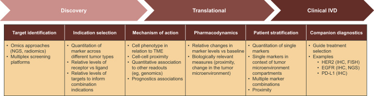
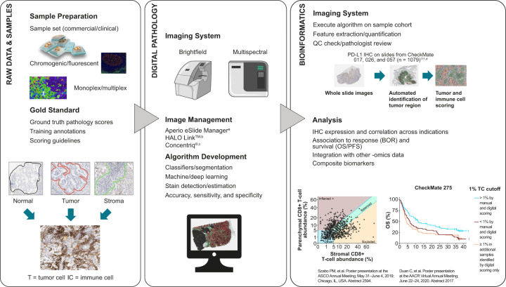
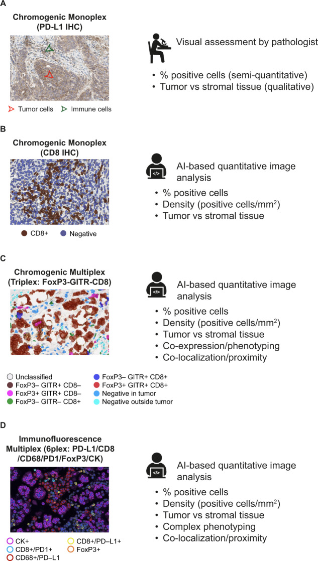
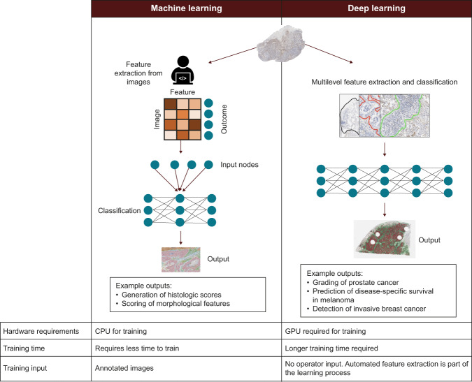
Verification
Claims the paper makes
The claims and statistics of this paper haven’t been checked yet.
Run the check — free, no account needed. It reads the open-access text and checks the sources it cites.
Claims other papers make about this one
Each is checked against this paper’s own text: does it say what the citing paper says it does?
The check wasn’t confident either way, so it gives no verdict — never disputed.
In the citing paper
“In radiology, AI can assist radiologists in improving diagnoses and enhancing patient care by analyzing chest X-rays, magnetic resonance images, and computed tomography scans (41).”
From Artificial Intelligence for Drug Discovery: Are We There Yet? (2024) · cites this paper as [41]
Experimental — accuracy not yet measured
Self-check
Percentages, sums and reported differences re-computed from the paper’s own numbers.
The self-check needs the paper’s full text.
Citations
Cites
PD-1 and PD-L1 Immune Checkpoint Blockade to Treat Breast Cancer
Hartkopf, A. D., Taran, F., Wallwiener, M., Walter, C. B., Krämer, B., Grischke, E., & Brucker, S. Y. (2016) · Breast Care · doi:10.1159/000453569 · Cited by 30,989
Adverse Renal Effects of Immune Checkpoint Inhibitors: A Narrative Review
Wanchoo, R., Karam, S., Uppal, N. N., Barta, V. S., Deray, G., Devoe, C., Launay‐Vacher, V., Jhaveri, K. D., & on behalf of Cancer and Kidney International Network Workgroup on Immune Checkpoint Inhibitors (2017) · American Journal of Nephrology · doi:10.1159/000455014 · Cited by 15,320
Pembrolizumab versus Chemotherapy for PD-L1-Positive Non-Small-Cell Lung Cancer
Reck, M., Rodríguez-Abreu, D., Robinson, A. G., Hui, R., Csőszi, T., Fülöp, A., Gottfried, M., Peled, N., Tafreshi, A., Cuffe, S., et al. (2016) · New England Journal of Medicine · doi:10.1056/nejmoa1606774 · Cited by 10,089
Nivolumab versus docetaxel in advanced nonsquamous non-small-cell lung cancer
Borghaei, H., Paz-Ares, L., Horn, L., Spigel, D. R., Steins, M., Ready, N. E., Chow, L. Q., Vokes, E. E., Felip, E., Holgado, E., et al. (2015) · New England Journal of Medicine · doi:10.1056/nejmoa1507643 · Cited by 9,335
Herbst, R. S., Baas, P., Kim, D.-W., Felip, E., Pérez-Gracia, J. L., Han, J.-Y., Molina, J., Kim, J.-H., Arvis, C. D., Ahn, M.-J., et al. (2016) · The Lancet · doi:10.1016/s0140-6736(15)01281-7 · Cited by 6,449
Pembrolizumab for the Treatment of Non–Small-Cell Lung Cancer
Garon, E. B., Rizvi, N. A., Hui, R., Leighl, N., Balmanoukian, A. S., Eder, J. P., Patnaik, A., Aggarwal, C., Gubens, M., Horn, L., et al. (2015) · New England Journal of Medicine · doi:10.1056/nejmoa1501824 · Cited by 6,198
Cortazar, P., Zhang, L., Untch, M., Mehta, K. M., Costantino, J. P., Wolmark, N., Bonnefoi, H. R., Cameron, D., Gianni, L., Valagussa, P., et al. (2014) · The Lancet · doi:10.1016/s0140-6736(13)62422-8 · Cited by 4,777
Rittmeyer, A., Barlési, F., Waterkamp, D., Park, K., Ciardiello, F., von Pawel, J., Gadgeel, S. M., Hida, T., Kowalski, D. M., Cobo, M., et al. (2016) · The Lancet · doi:10.1016/s0140-6736(16)32517-x · Cited by 4,718
Durvalumab after chemoradiotherapy in stage III non–small-cell lung cancer
Antonia, S. J., Villegas, A., Daniel, D., Vicente, D., Murakami, S., Hui, R., Yokoi, T., Chiappori, A., Lee, K. H., de Wit, M., et al. (2017) · New England Journal of Medicine · doi:10.1056/nejmoa1709937 · Cited by 4,593
Atezolizumab and nab-paclitaxel in advanced triple-negative breast cancer
Schmid, P., Adams, S., Rugo, H. S., Schneeweiss, A., Barrios, C. H., Iwata, H., Diéras, V., Hegg, R., Im, S.-A., Shaw Wright, G., et al. (2018) · New England Journal of Medicine · doi:10.1056/nejmoa1809615 · Cited by 4,350
Cited by
Sorted by citations
Items 501–593 of 593
Artificial Intelligence-Based Pathomics: Technological Features and Clinical Applications
Kang, L., Liu, L., Chen, H., Zheng, C., & Ye, T. (2025) · Bratislavské lekárske listy/Bratislava medical journal · doi:10.1007/s44411-025-00436-7
Balan, J., & Larson, N. B. (2025) · Scientific Reports · doi:10.1038/s41598-025-30947-w
Jung, M., Song, S. G., Cho, S. I., Shin, S., Lee, T., Jung, W., Lee, H., Park, J., Song, S., Park, G., et al. (2024) · Figshare · doi:10.6084/m9.figshare.25282805
Sato, N., Tsujimoto, M., Nakatsuji, M., Tsuji, H., Sugama, Y., Shimazu, K., Shimoda, M., & Ishihara, H. (2024) · Figshare · doi:10.6084/m9.figshare.26829077
Sato, N., Tsujimoto, M., Nakatsuji, M., Tsuji, H., Sugama, Y., Shimazu, K., Shimoda, M., & Ishihara, H. (2024) · Figshare · doi:10.6084/m9.figshare.26829014
Jiang, W., Wang, H., Dong, X., Zhao, Y., Long, C., Chen, D., Yan, B., Cheng, J., Lin, Z., Zhuo, S., et al. (2024) · Figshare · doi:10.6084/m9.figshare.25067167
Scalco, R., Saito, N., Beckett, L. A., Nguyen, M.-L., Huie, E. Z., Wang, H.-P., Flaherty, D., Honig, L. S., DeCarli, C. S., Rissman, R. A., et al. (2024) · Figshare · doi:10.6084/m9.figshare.26600926
Gopinath, B., & Santhi, R. (2025) · Lecture notes in networks and systems · doi:10.1007/978-981-96-9196-8_40
AI-Powered Diagnostic Tools for Menopause
Saini, A., Verma, I., & Bhatia, R. (2025) · IGI Global eBooks · doi:10.4018/979-8-3373-2970-3.ch007
Yang, Z., Li, K., Ramandi, S. G., Brassard, P., Khellaf, A., Trinh, V. Q., Zhang, J., Chen, L., Rowsell, C. H., Varma, S., et al. (2025) · Journal of Pathology Informatics · doi:10.1016/j.jpi.2025.100537 · Cited by 1
A novel approach for bubble restoration in histopathological images
Fan, Y., Hao, X., Xing, Z., Chang, H., Teng, X., Wang, L., Wang, X., Yang, Z., Lian, J., Jiang, Y., et al. (2026) · Biomedical Signal Processing and Control · doi:10.1016/j.bspc.2025.108826
An automatic, rapid and accurate method for the annotation of tumor components on whole slide images
Tang, H., Wang, X., Zhang, X., Wu, X., Tang, X., Ma, Y., Chen, Y., & yu, G. (2026) · Journal of Histotechnology · doi:10.1080/01478885.2025.2602268
Diagnosis of Endometrial Carcinoma
Hussain, N., Gahine, R., Chandrakar, J., Jain, S., & Hussain, R. (2026) · Obstetrics and gynecology. · doi:10.5772/intechopen.1012666
Intraoperative biopsy imaging of lung cancer risk
Qian, S., Yang, L., Meng, J., Zhou, L., Han, T., Chen, L., Xi, G., Jiang, R., Wang, C., Niu, B., et al. (2026) · Communications Medicine · doi:10.1038/s43856-026-01406-y
Liu, B., Wang, Q., Qin, G., Gu, Y., Ding, F., Jin, W., Hou, Q., Sun, M., & Xu, M. (2026) · Biopreservation and Biobanking · doi:10.1177/19475535261417938
Bielecki, M., Lu, F.-I., Vo, A., Rakovitch, E., Jerzak, K. J., Salgado, R., Karshafian, R., & Tran, W. T. (2026) · Therapeutic Advances in Medical Oncology · doi:10.1177/17588359261417762
Kumar, S., & Mishra, P. K. (2026) · Engineering Applications of Artificial Intelligence · doi:10.1016/j.engappai.2026.114262
梁, 长. (2026) · Advances in Clinical Medicine · doi:10.12677/acm.2026.1631112
Kawamura, K., Hiraoka, S.-I., Lee, C., Ohya, K., Akiyama, R., Uchida, S., Itakura, Y., Toyosawa, S., & Tanaka, S. (2026) · Oral Surgery Oral Medicine Oral Pathology and Oral Radiology · doi:10.1016/j.oooo.2026.03.006
Deep learning-based early screening for porcine embryos with different developmental potential
Yang, Y., Li, H., Bi, D., Deng, F., Jia, K., Qin, G., Mao, K., Wang, Y., Zhang, Z., & Zhao, J. (2026) · Theriogenology · doi:10.1016/j.theriogenology.2026.117900
Artificial Intelligence in Cardiovascular Pathology: Toward a Diagnostic Revolution
Marzullo, A., Quaranta, A., Cazzato, G., & Salzillo, C. (2026) · BioMedInformatics · doi:10.3390/biomedinformatics6020018
Sonar, P., Mathuvanti, K. R., Venkataramanan, A., Reddy, H., & Mohanty, A. (2026) · Biotechnology Letters · doi:10.1007/s10529-026-03732-6
Pye, H., Brettle, D. S., Kaye, D., Dunn, C. M., Humphries, M. P., & Treanor, D. E. (2026) · Journal of Histotechnology · doi:10.1080/01478885.2026.2643973
Conditional Generative AI in Oncology Diagnostics
Frascarelli, C., Concardi, A., Mangione, E., Negrelli, M., Porta, F. M., Tulino, M., Sorino, J., Marra, A., Fusco, N., Guerini‐Rocco, E., et al. (2026) · Applied Sciences · doi:10.3390/app16084015
Wu, B., Gu, J., & Chen, T. (2026) · International Journal of General Medicine · doi:10.2147/ijgm.s593911
Luo, Y., Xie, P., Yang, S., Qu, P., Hou, L., & Yi, Q. (2026) · iScience · doi:10.1016/j.isci.2026.116000
Megneishvili, S. R., & Евгеньевна, Ш. И. (2026) · A breakthrough in science: development strategies · doi:10.21661/r-588705
AI Regulations in Medical Devices
Khinvasara, T. (2026) · Springer handbooks · doi:10.1007/978-3-032-01636-2_40
Benimam, M. M., Frafjord, A., Corthay, A., Lagache, T., Olivo‐Marín, J., & Meas‐Yedid, V. (2026) · IEEE International Symposium on Biomedical Imaging (ISBI) · doi:10.1109/isbi61048.2026.11515781
Knowledge-Augmented Multimodal Learning for Breast Cancer Diagnosis
Li, L., Wang, L., & Hou, L. (2026) · Communications in computer and information science · doi:10.1007/978-981-95-7299-1_19
Oguzhanoglu, M. K., Uzuner, E. G., Oguzhanoglu, K., Cetin, A., Karacabey, S., & Asgarova, Z. (2026) · Frontiers in Oncology · doi:10.3389/fonc.2026.1819567
Prediction of prediabetes: Artificial neural networking
Thakur, N., Kumar, P., Sharma, P., & Kumar, R. (2026) · Elsevier eBooks · doi:10.1016/b978-0-443-36348-1.00011-6
Majeed, T., Aalam, S. W., Masoodi, T. A., Ahanger, A. B., Mishra, D., Nayyar, V., Khan, M. A., Bhat, A. A., Bhat, M. R., Macha, M. A., et al. (2026) · Head and Neck Pathology · doi:10.1007/s12105-026-01920-4
Usui, G., & Kaneda, A. (2026) · Cancer Science · doi:10.1111/cas.70432
Topology-guided hard example mining for cell detection
Çakı, O., Ünver, S., Karasayar, A. H. D., Mericoz, C. A., Bulutay, P., Kapucuoğlu, N., Eren, H., Dilbaz, O. F., Osmanli, J., Yetkili, B. S., et al. (2026) · Medical Image Analysis · doi:10.1016/j.media.2026.104155
Fernandes, G., Wang, W., Parwani, A. V., Ahmadian, S. S., Alves, M. J., Philips, J. J., & Otero, J. J. (2026) · bioRxiv (Cold Spring Harbor Laboratory) · doi:10.64898/2026.06.04.730099
Banaszek, J., Bąk, D., Barańska, K., Czajka, A., Ciesielska, D., Kleinrok, J., Pająk, W., Korolczuk, A., Mazur, M., & Rusztyn, K. (2026) · International Journal of Molecular Sciences · doi:10.3390/ijms27125244
Artificial intelligence for gastric cancer: Advances and current status in diagnosis and therapy
Chi, Z.-C. (2026) · World Chinese Journal of Digestology · doi:10.11569/wcjd.v34.i5.345
Younas, M., Mahmood, A., Zafar, M., Mohammed, N. K., Alakilli, S. Y. M., Shahin, O. R., Islam, F., & Ndagire, C. T. (2026) · Silicon · doi:10.1007/s12633-026-03705-4
Advancing metastatic prostate cancer care: best practices and future directions in molecular testing
Castro, E., Chen, J., Lowentritt, B. H., Das, P. M., Dowlati, R., & Olmos, D. (2026) · Prostate International · doi:10.1016/j.prnil.2026.06.002
Canberk, Ş., & Nosé, V. (2026) · Advances in Anatomic Pathology · doi:10.1097/pap.0000000000000544
Artificial intelligence and the future of telemonitoring in home mechanical ventilation
Crimi, C., Luján, M., & Duiverman, M. L. (2026) · ERJ Open Research · doi:10.1183/23120541.01717-2025
A Homology‐Based Application to Diagnose Colorectal Adenoma and Early‐Stage Cancer
Yokoyama, Y., Kanayama, K., Matsuda, M., Hatori, N., Asano, T., Morii, E., Yamamoto, H., & Nakane, K. (2026) · Pathology International · doi:10.1111/pin.70143
Altundağ, K. (2026) · Breast Cancer · doi:10.1007/s12282-026-01894-9
DeepPlaque: a scalable multimodal platform for Aβ pathology and cell analysis in Alzheimer’s disease
Wong, H. Y., Jin, C., Yuen, S. L., Yang, X., Gill, S. S., Xu, J., Lai, K. P. E., Fu, W., Gao, W., Wang, X., et al. (2026) · EMBO Molecular Medicine · doi:10.1038/s44321-026-00488-4
Rethinking pathology image analysis through shuffling
Liu, Z., Zhang, T., Chen, B. K., Feng, Y., Lyu, S., Lei, Y., Ying, N., Feng, Y., Zhao, Y., Zhang, P., et al. (2026) · npj Biomedical Innovations. · doi:10.1038/s44385-026-00096-4 · Cited by 1
Kuan, L., Jia, Z., 鲁海文, Yan, Z., Haihui, H., & Liu, X. (2026) · Diagnostic Pathology · doi:10.1186/s13000-026-01822-7
Artificial Intelligence Applications in Pathology
Ercan, A. T., & Güzeliş, İ. (2026) · Springer eBooks · doi:10.1007/978-3-032-24420-8_20
Stromal Laminin Subunit Alpha 3 is a Novel Prognostic Marker for Breast Cancer
Tan, Z., Liu, S., Zhao, D., Du, S., Sheng, C., Wang, H., & Lu, Z. (2026) · Clinical Medicine Insights Oncology · doi:10.1177/11795549261476199
Bolck, H. A., Migh, E., Kriston, A., Zajac, N., Kreutzer, S., Totu, T., Leary, P., Kovács, F., Rutishauser, D., Pfammatter, S., et al. (2026) · EMBO Molecular Medicine · doi:10.1038/s44321-026-00484-8
Wu, Q., Gong, Q., Yuan, L., Li, Z., Ashenberg, O., Chen, F., Xavier, R. J., & Uhler, C. (2026) · bioRxiv (Cold Spring Harbor Laboratory) · doi:10.64898/2026.07.31.741265 · Cited by 1
Wang, X., (2085), W. G., Wang, Q., He, S., Cui, S., Duan, B., Ding, G., Huang, Y., & Zou, B. (2026) · Frontiers in Immunology · doi:10.3389/fimmu.2026.1762484
Poombal, Lail, R. A., Satturwar, S., Singh, R., Ullah, E., & Parwani, A. V. (2026) · Diagnostic Pathology · doi:10.1186/s13000-026-01816-5
Su, Q., Wu, J., Li, B., Wang, Y., Xu, Y., Chen, Y., Wang, N., Hou, F., Yang, G., & Nie, P. (2026) · Translational Oncology · doi:10.1016/j.tranon.2026.102979
Baviello, C., Capuano, D. M., & Verna, R. (2026) · Life · doi:10.3390/life16081346
KOPARIR, O. F., Tarakçı, B. G., & Şengür, A. (2026) · Bioengineering · doi:10.3390/bioengineering13080937
Sulley, S., & Tranesh, G. A. (2026) · Journal of Pathology Informatics · doi:10.1016/j.jpi.2026.100710
Digital Biobanking in the Era of Artificial Intelligence
Dey, P. (2026) · Biopreservation and Biobanking · doi:10.1177/19475535261474367
Innovative pathological and therapeutic approaches for poorly cohesive gastric cancer
Muratore, M., Sullo, F. G., Bittoni, A., Cardisciani, L., Valgiusti, M., Bartolini, G., Esposito, L., Molinari, C., Câmara, R., & Passardi, A. (2026) · Frontiers in Oncology · doi:10.3389/fonc.2026.1842715
Lei, Q.-K., Wu, T.-X., Zhang, C.-L., & Zheng, Y. (2026) · Liver Research · doi:10.1016/j.livres.2026.08.001
Antibody–Drug Conjugates in Lung Cancer: Promise, Progress, and Persistent Challenges
Paliogiannis, P., Fara, G., Zinellu, A., Fois, A. G., & Palmieri, G. (2026) · Current Issues in Molecular Biology · doi:10.3390/cimb48090868
Chen, H., Tong, L., Liu, J., Liu, K., Li, X., Shi, S., Xi, S., Tian, G., Zhang, M., Zhong, D., et al. (2026) · Frontiers in Big Data · doi:10.3389/fdata.2026.1924721
Federated Learning Pipeline for Whole-Slide Image Classification in Digital Neuropathology
Cong, C., Ieva, A. D., & Liu, S. (2026) · Methods in molecular biology · doi:10.1007/978-1-0716-5440-8_13
Kalykakis, G.-E., Tarampoulous, I., Gyftodimos, K., Sepsa, A., Agrogiannis, G., Vamvakaris, G., Samaras, M. G., Spyropoulos, C., Manolis, C., Niotis, N., et al. (2026) · Bioengineering · doi:10.3390/bioengineering13091021
Mirza, K., Rao, A., Baker, J. A., Parker, R. K., Quinonez, S. C., Njenga, P., Saleh, M. N., Balis, U. J., Waljee, A. K., & Sayed, S. (2026) · BMJ Digital Health & AI · doi:10.1136/bmjdh-2026-000026
Federated learning for multi-institutional AI in healthcare via digital pathology
Soleimani, R., Azimi, M., & Talebi, N. (2026) · Biomedizinische Technik/Biomedical Engineering · doi:10.1515/bmt-2025-0489 · Cited by 2
Other applications of digital pathology
Rasool, G., Singh, R., & Bui, M. (2026) · Elsevier eBooks · doi:10.1016/b978-0-443-40383-5.00009-1
Insights into artificial intelligence-based digital pathology in hepatobiliary and pancreatic cancer
Huang, H.-Y., Kow, A. W. C., Fan, Z., & Lv, G. (2026) · Frontiers in Oncology · doi:10.3389/fonc.2026.1871770
Bülbül, M. V., & Demircan, T. (2026) · Organoids · doi:10.3390/organoids5030030
Tondon, C., Erb, G., Egelé, C., Michot, J.-P., Fetique, D., Chenard, M., & Bellocq, J. (2026) · Archiv für Pathologische Anatomie und Physiologie und für Klinische Medicin · doi:10.1007/s00428-026-04714-9
Liu, S., Wang, Z., Qian, Z., & Gao, H. (2026) · Optics & Laser Technology · doi:10.1016/j.optlastec.2026.116360
Digital twins in neuroscience: A narrative review of technical challenges for child neurology
Canepa, M. E., Villano, G., Ramenghi, L. A., di Pianella, A. V., & Striano, P. (2026) · Advanced technology in neuroscience . · doi:10.4103/atn.atn-d-26-00025
Histological Analysis of Zoonotic Diseases and Artificial Intelligence
Rustamovna, A. B., Gayratovich, E. N., Xursandov, I., Nagendra, K. V., Dharrao, D. S., Dharrao, M., Sivaraman, S. K., & Singh, A. P. (2026) · Advances in computational intelligence and robotics book series · doi:10.4018/979-8-2600-1358-8.ch013
Explainable Artificial Intelligence (AI) and Clinical Reliability in Histology
Rustamovna, A. B., Gayratovich, E. N., Xursandov, I., Nagendra, K. V., Dharrao, D. S., Dharrao, M., Sivaraman, S. K., & Singh, A. P. (2026) · Advances in computational intelligence and robotics book series · doi:10.4018/979-8-2600-1358-8.ch012
SmartHisto: Bayesian active learning for histology images
Vijendran, S., Arruda, B. L., Anderson, T. K., & Eulenstein, O. (2026) · PLoS Computational Biology · doi:10.1371/journal.pcbi.1013611
Chi, H., Liu, X., Yan, Y., Wang, L., Luo, P., Su, L., Jiang, L., Zhou, X., Huang, G., Jiang, Y., et al. (2026) · Genes & Diseases · doi:10.1016/j.gendis.2026.102480 · Cited by 1
Mishra, S., Singh, R., Zargar, N. N., & Khanuja, S. (2026) · Global Journal of Guntur Medical College · doi:10.25259/gjgmc_11_2026
Wang, Z.-Y., Liu, N., & Chen, M. (2026) · Frontiers in Molecular Biosciences · doi:10.3389/fmolb.2026.1875788
Tham, J. A., Xie, F., Chen, B., Yu, P., Wu, J., Feng, T., Tse, G. M. K., To, K. F., & Kang, W. (2026) · Intelligent Systems with Applications · doi:10.1016/j.iswa.2026.200736
Immunophenotyping by Spatial Biology
Curras-Alonso, S., Adimi, Y., Levin, C., & Delahaye, F. (2026) · Methods in molecular biology · doi:10.1007/978-1-0716-5607-5_18
Combining artificial intelligence and neuroscience to diagnose and predict neurological disorders
Rahaman, S. A., Babu, M. R., Patel, S., Kumar, R., Löebenberg, R., & Chettupalli, A. K. (2026) · Elsevier eBooks · doi:10.1016/b978-0-443-27722-1.00015-8
Park, J., Subramaniam, R., Cazzato, G., Belloni, E., Torres-Cabala, C. A., & Giubellino, A. (2026) · American Journal of Dermatopathology · doi:10.1097/dad.0000000000003450
Üstüner, E., & Öz, M. (2026) · Veterinary Research Communications · doi:10.1007/s11259-026-11567-9
Barbosa, V. M., Giraldi, J. M. M., Soares, T. T. M. S., Laurindo, A. K. S., Gentili, A., Berejuck, M. D., Macedo, D. D. J. D., von Wangenheim, A., & Sobieranski, A. C. (2026) · Proceedings - IEEE Symposium on Computer-Based Medical Systems · doi:10.1109/cbms69103.2026.00289
The accuracy–utility gap: Why AI accuracy does not guarantee clinical benefit in digital pathology
Battiato, R., Silva, F. M., Morales, M. C., Romero, D. S., & Benites, M. H. (2026) · Journal of Pathology Informatics · doi:10.1016/j.jpi.2026.100751
Boot, G. F., Masotti, C., Corrò, C., Schraepen, C., Williams, H. L., Wright, T. A., Ishaqwala, F., Steele, C. W., Rasschaert, G., Lafarge, M. W., et al. (2026) · The Journal of Pathology · doi:10.1002/path.70127
Agrawal, U. K., Jaiswal, S., & Singh, D. (2026) · Surgical and Experimental Pathology · doi:10.1186/s42047-026-00267-y
Chen, W., Chang, F., & Qi, J. (2026) · Journal of Imaging Informatics in Medicine · doi:10.1007/s10278-026-02390-4
Li, Z., Duan, X., Lian, J., Li, X., Liang, Q., Liu, X., Tan, Y., Yang, G., Du, J., 武丽娜, et al. (2026) · Frontiers in Immunology · doi:10.3389/fimmu.2026.1845251
Opportunities and challenges of artificial intelligence and digital pathology in adult CNS tumors
Lechpammer, M., Norrell, J., Parnas, J. N., Vojnic, M., & Sherman, J. H. (2026) · Journal of Neuro-Oncology · doi:10.1007/s11060-026-05833-8
Zhu, M., Aschenaki, Y., Zhang, X., Sali, R., Lin, Z., Dixon, C. M., & Zhang, D. Y. (2026) · BMJ Oncology · doi:10.1136/bmjonc-2026-001208
Xu, H., Wu, Y., Fang, H., & Gu, Z. (2026) · International Journal of Surgery · doi:10.1097/js9.0000000000005583
Metadata
- Authors
- Baxi, V., Edwards, R., Montalto, M., & Saha, S.
- Affiliations
- Bristol-Myers Squibb (United States) (US)
- PathAI (United States) (US)
- Corresponding author
- Baxi, Vipul
- Venue
- Modern Pathology
- Publisher
- Elsevier BV
- Published
- 2022-01
- Volume
- 35
- Issue
- 1
- Pages
- 23-32
- ISSN
- 0893-3952 (ISSN-L)
- PMC
- PMC8491759
- OpenAlex
- W3204013916
- Cited by
- 592
- References
- 139
- Citations by year
- 2026: 107 · 2025: 158 · 2024: 175 · 2023: 102 · 2022: 49
- FWCI
- 5.91
- Type
- journal article
- Language
- en
- Open access
- hybrid
- doi.org
- Licence
- Locations
- Modern Pathology (publishedVersion, cc-by)
- PubMed Central (publishedVersion, cc-by)
- PubMed (publishedVersion)
- Funders
- Bristol-Myers Squibb
- Topics
- Physical Sciences › Computer Science › Artificial Intelligence › AI in cancer detection
- Health Sciences › Medicine › Health Informatics › Artificial Intelligence in Healthcare and Education
- Health Sciences › Medicine › Radiology, Nuclear Medicine and Imaging › Radiomics and Machine Learning in Medical Imaging
- Keywords
- digital pathology, artificial intelligence, whole slide imaging, cancer immunotherapy, biomarker discovery, patient selection, precision medicine, tumor microenvironment, companion diagnostics, translational medicine, clinical implementation barriers
- MeSH terms
- Translational Science, Biomedical, Algorithms, Artificial Intelligence, Humans, Pathology, Practice Patterns, Physicians', Biomarkers
About these checks
Registry, retraction, Cites and statistics results are computed, not judged, with one exception: a printed reference that no index lists is read once by a model before it counts as a citation error. Claims are extracted by a model and kept only when their quote appears verbatim in the text; each cited source is then checked, with a model’s help, for whether it says what the claim relies on. Those verdicts can be wrong — read the claim’s quote and the cited source before relying on one.
Statistics and arithmetic are recomputed, not judged.
Confidence bands are shown only for fields calibrated on our annotated test set. None is calibrated yet, so no band is shown.
What counts as a claim ›How we measure confidence ›How we check this ›
Are you an author, or do you see a problem with this page? Request a review
