

The DOI matches its Crossref record; 138 of the 139 references it cites were found in a registry or an index.
Digital pathology and artificial intelligence in translational medicine and clinical practice
Open accessAbstract
Traditional pathology approaches have played an integral role in the delivery of diagnosis, semi-quantitative or qualitative assessment of protein expression, and classification of disease. Technological advances and the increased focus on precision medicine have recently paved the way for the development of digital pathology-based approaches for quantitative pathologic assessments, namely whole slide imaging and artificial intelligence (AI)-based solutions, allowing us to explore and extract information beyond human visual perception. Within the field of immuno-oncology, the application of such methodologies in drug development and translational research have created invaluable opportunities for deciphering complex pathophysiology and the discovery of novel biomarkers and drug targets. With an increasing number of treatment options available for any given disease, practitioners face the growing challenge of selecting the most appropriate treatment for each patient. The ever-increasing utilization of AI-based approaches substantially expands our understanding of the tumor microenvironment, with digital approaches to patient stratification and selection for diagnostic assays supporting the identification of the optimal treatment regimen based on patient profiles. This review provides an overview of the opportunities and limitations around implementing AI-based methods in biomarker discovery and patient selection and discusses how advances in digital pathology and AI should be considered in the current landscape of translational medicine, touching on challenges this technology may face if adopted in clinical settings. The traditional role of pathologists in delivering accurate diagnoses or assessing biomarkers for companion diagnostics may be enhanced in precision, reproducibility, and scale by AI-powered analysis tools.
Overview
The claims and statistics of this paper haven’t been checked yet.
Run the check — free, no account needed. It reads the open-access text and checks the sources it cites.
Integrity signals
Registry
Cites
Statistics
Figures
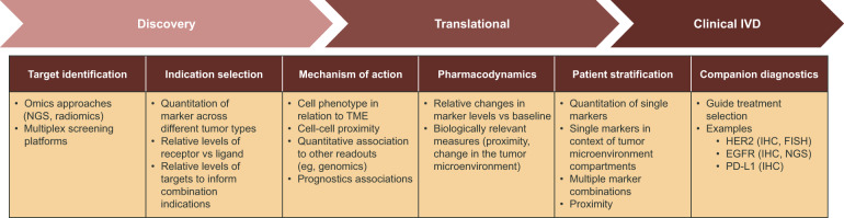
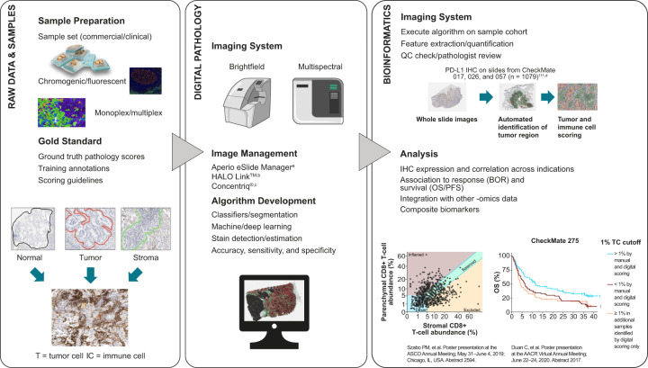
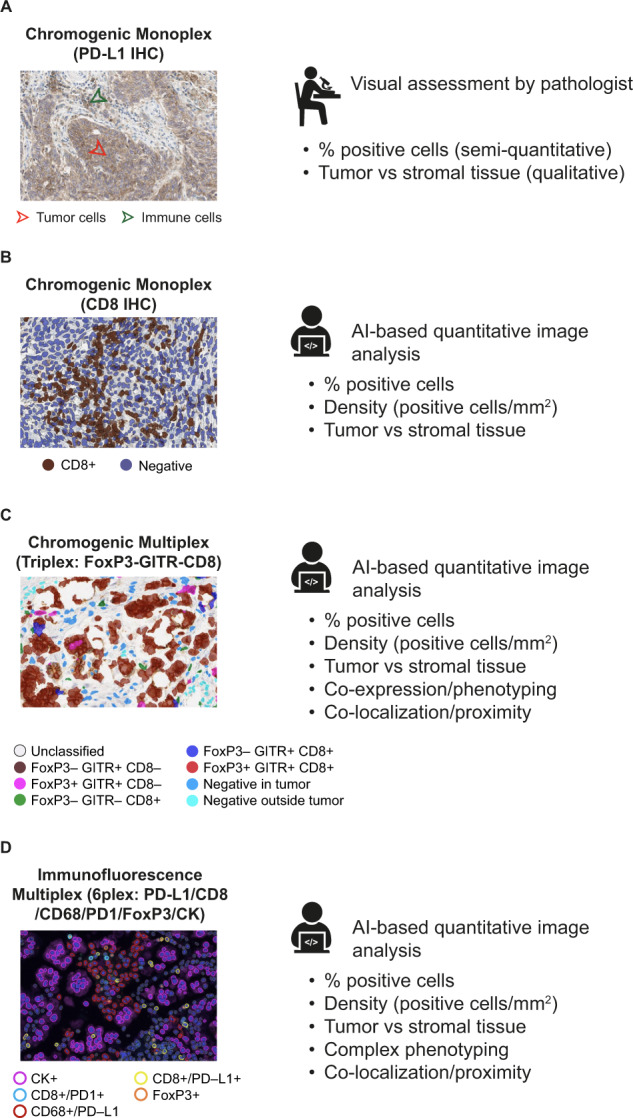
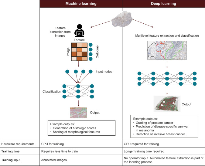
Verification
Claims the paper makes
The claims and statistics of this paper haven’t been checked yet.
Run the check — free, no account needed. It reads the open-access text and checks the sources it cites.
Claims other papers make about this one
Each is checked against this paper’s own text: does it say what the citing paper says it does?
The check wasn’t confident either way, so it gives no verdict — never disputed.
In the citing paper
“In radiology, AI can assist radiologists in improving diagnoses and enhancing patient care by analyzing chest X-rays, magnetic resonance images, and computed tomography scans (41).”
From Artificial Intelligence for Drug Discovery: Are We There Yet? (2024) · cites this paper as [41]
Experimental — accuracy not yet measured
Self-check
Percentages, sums and reported differences re-computed from the paper’s own numbers.
The self-check needs the paper’s full text.
Citations
Cites
PD-1 and PD-L1 Immune Checkpoint Blockade to Treat Breast Cancer
Hartkopf, A. D., Taran, F., Wallwiener, M., Walter, C. B., Krämer, B., Grischke, E., & Brucker, S. Y. (2016) · Breast Care · doi:10.1159/000453569 · Cited by 30,989
Adverse Renal Effects of Immune Checkpoint Inhibitors: A Narrative Review
Wanchoo, R., Karam, S., Uppal, N. N., Barta, V. S., Deray, G., Devoe, C., Launay‐Vacher, V., Jhaveri, K. D., & on behalf of Cancer and Kidney International Network Workgroup on Immune Checkpoint Inhibitors (2017) · American Journal of Nephrology · doi:10.1159/000455014 · Cited by 15,320
Pembrolizumab versus Chemotherapy for PD-L1-Positive Non-Small-Cell Lung Cancer
Reck, M., Rodríguez-Abreu, D., Robinson, A. G., Hui, R., Csőszi, T., Fülöp, A., Gottfried, M., Peled, N., Tafreshi, A., Cuffe, S., et al. (2016) · New England Journal of Medicine · doi:10.1056/nejmoa1606774 · Cited by 10,089
Nivolumab versus docetaxel in advanced nonsquamous non-small-cell lung cancer
Borghaei, H., Paz-Ares, L., Horn, L., Spigel, D. R., Steins, M., Ready, N. E., Chow, L. Q., Vokes, E. E., Felip, E., Holgado, E., et al. (2015) · New England Journal of Medicine · doi:10.1056/nejmoa1507643 · Cited by 9,335
Herbst, R. S., Baas, P., Kim, D.-W., Felip, E., Pérez-Gracia, J. L., Han, J.-Y., Molina, J., Kim, J.-H., Arvis, C. D., Ahn, M.-J., et al. (2016) · The Lancet · doi:10.1016/s0140-6736(15)01281-7 · Cited by 6,449
Pembrolizumab for the Treatment of Non–Small-Cell Lung Cancer
Garon, E. B., Rizvi, N. A., Hui, R., Leighl, N., Balmanoukian, A. S., Eder, J. P., Patnaik, A., Aggarwal, C., Gubens, M., Horn, L., et al. (2015) · New England Journal of Medicine · doi:10.1056/nejmoa1501824 · Cited by 6,198
Cortazar, P., Zhang, L., Untch, M., Mehta, K. M., Costantino, J. P., Wolmark, N., Bonnefoi, H. R., Cameron, D., Gianni, L., Valagussa, P., et al. (2014) · The Lancet · doi:10.1016/s0140-6736(13)62422-8 · Cited by 4,777
Rittmeyer, A., Barlési, F., Waterkamp, D., Park, K., Ciardiello, F., von Pawel, J., Gadgeel, S. M., Hida, T., Kowalski, D. M., Cobo, M., et al. (2016) · The Lancet · doi:10.1016/s0140-6736(16)32517-x · Cited by 4,718
Durvalumab after chemoradiotherapy in stage III non–small-cell lung cancer
Antonia, S. J., Villegas, A., Daniel, D., Vicente, D., Murakami, S., Hui, R., Yokoi, T., Chiappori, A., Lee, K. H., de Wit, M., et al. (2017) · New England Journal of Medicine · doi:10.1056/nejmoa1709937 · Cited by 4,593
Atezolizumab and nab-paclitaxel in advanced triple-negative breast cancer
Schmid, P., Adams, S., Rugo, H. S., Schneeweiss, A., Barrios, C. H., Iwata, H., Diéras, V., Hegg, R., Im, S.-A., Shaw Wright, G., et al. (2018) · New England Journal of Medicine · doi:10.1056/nejmoa1809615 · Cited by 4,350
Cited by
Sorted by citations
Serrano, D. R., Luciano, F. C., Anaya, B. J., Öngoren, B., Kara, A., Molina, G., Ramirez, B. I., Sánchez-Guirales, S. A., Simon, J. A., Tomietto, G., et al. (2024) · Pharmaceutics · doi:10.3390/pharmaceutics16101328 · Cited by 594
Artificial intelligence in histopathology: enhancing cancer research and clinical oncology
Shmatko, A., Ghaffari Laleh, N., Gerstung, M., & Kather, J. N. (2022) · Nature Cancer · doi:10.1038/s43018-022-00436-4 · Cited by 446
Multimodal data fusion for cancer biomarker discovery with deep learning
Steyaert, S., Pizurica, M., Nagaraj, D., Khandelwal, P., Hernandez-Boussard, T., Gentles, A. J., & Gevaert, O. (2023) · Nature Machine Intelligence · doi:10.1038/s42256-023-00633-5 · Cited by 402
High-throughput proteomics: a methodological mini-review
Cui, M., Cheng, C., & Zhang, L. (2022) · Laboratory Investigation · doi:10.1038/s41374-022-00830-7 · Cited by 398
Artificial intelligence for digital and computational pathology
Song, A. H., Jaume, G., Williamson, D. F. K., Lu, M. Y., Vaidya, A., Miller, T. R., & Mahmood, F. (2023) · Nature Reviews Bioengineering · doi:10.1038/s44222-023-00096-8 · Cited by 356
Zafar, M. H., Langås, E. F., & Sanfilippo, F. (2024) · Robotics and Computer-Integrated Manufacturing · doi:10.1016/j.rcim.2024.102769 · Cited by 338
AI-Assisted Detection of Biomarkers by Sensors and Biosensors for Early Diagnosis and Monitoring
Wasilewski, T., Kamysz, W., & Gębicki, J. (2024) · Biosensors · doi:10.3390/bios14070356 · Cited by 328
McGenity, C., Clarke, E. L., Jennings, C., Matthews, G., Cartlidge, C., Freduah-Agyemang, H., Stocken, D. D., & Treanor, D. (2024) · npj Digital Medicine · doi:10.1038/s41746-024-01106-8 · Cited by 290
The evolving role of investigative toxicology in the pharmaceutical industry
Pognan, F., Beilmann, M., Boonen, H. C. M., Czich, A., Dear, G. J., Hewitt, P. G., Mow, T. J., Oinonen, T., Roth, A. B., Steger‐Hartmann, T., et al. (2023) · Nature Reviews Drug Discovery · doi:10.1038/s41573-022-00633-x · Cited by 280
Deep learning models in medical image analysis
Tsuneki, M. (2022) · Journal of Oral Biosciences · doi:10.1016/j.job.2022.03.003 · Cited by 292
Artificial Intelligence for Drug Discovery: Are We There Yet?
Hasselgren, C., & Oprea, T. I. (2024) · Annual Review of Pharmacology and Toxicology · doi:10.1146/annurev-pharmtox-040323-040828 · Cited by 236
“Page | 6 diagnostic and classification errors while accelerating biomarker discovery, pathophysiology evaluations, animal research, and toxicological assessments (40).”
BackgroundArtificial intelligence in healthcare delivery: Prospects and pitfalls
Olawade, D. B., David-Olawade, A. C., Wada, O. Z., Asaolu, A. J., Adereni, T., & Ling, J. (2024) · Journal of Medicine Surgery and Public Health · doi:10.1016/j.glmedi.2024.100108 · Cited by 236
Scaling Self-Supervised Learning for Histopathology with Masked Image Modeling
Filiot, A., Ghermi, R., Olivier, A., Jacob, P., Fidon, L., Camara, A., Mac Kain, A., Saillard, C., & Schiratti, J.-B. (2023) · doi:10.1101/2023.07.21.23292757 · Cited by 148
Spatial cellular architecture predicts prognosis in glioblastoma
Zheng, Y., Carrillo‐Pérez, F., Pizurica, M., Heiland, D. H., & Gevaert, O. (2023) · Nature Communications · doi:10.1038/s41467-023-39933-0 · Cited by 152
Toward Explainable Artificial Intelligence for Precision Pathology
Klauschen, F., Dippel, J., Keyl, P., Jurmeister, P. S., Bockmayr, M. L., Möck, A., Buchstab, O., Alber, M., Ruff, L., Montavon, G., et al. (2023) · Annual Review of Pathology Mechanisms of Disease · doi:10.1146/annurev-pathmechdis-051222-113147 · Cited by 164
Deep Learning on Histopathological Images for Colorectal Cancer Diagnosis: A Systematic Review
Davri, A. S., Birbas, E., Kanavos, T., Ntritsos, G., Γιαννακέας, Ν., Tzallas, A. T., & Batistatou, A. K. (2022) · Diagnostics · doi:10.3390/diagnostics12040837 · Cited by 144
Application of Artificial Intelligence in Combating High Antimicrobial Resistance Rates
Rabaan, A. A., Alhumaid, S., Mutair, A. S. A., Garout, M. A., Abulhamayel, Y., Halwani, M. A., Alestad, J. H., bshabshe, A. A. A. A., Sulaiman, T., AlFonaisan, M. K., et al. (2022) · Antibiotics · doi:10.3390/antibiotics11060784 · Cited by 132
Artificial intelligence (AI)—it’s the end of the tox as we know it (and I feel fine)*
Kleinstreuer, N., & Härtung, T. (2024) · Archives of Toxicology · doi:10.1007/s00204-023-03666-2 · Cited by 153
Zhu, S., Gilbert, M., Chetty, I., & Siddiqui, F. (2022) · International Journal of Medical Informatics · doi:10.1016/j.ijmedinf.2022.104828 · Cited by 131
Yim, D., Khuntia, J., Parameswaran, V., & Meyers, A. D. (2024) · JMIR Medical Informatics · doi:10.2196/52073 · Cited by 140
Research on Artificial-Intelligence-Assisted Medicine: A Survey on Medical Artificial Intelligence
Gou, F., Liu, J., Xiao, C., & Wu, J. (2024) · Diagnostics · doi:10.3390/diagnostics14141472 · Cited by 129
Applications of machine learning in routine laboratory medicine: Current state and future directions
Rabbani, N., Kim, G. Y. E., Suarez, C. J., & Chen, J. H. (2022) · Clinical Biochemistry · doi:10.1016/j.clinbiochem.2022.02.011 · Cited by 118
Computational pathology: A survey review and the way forward
Hosseini, M. S., Bejnordi, B. E., Trinh, V. Q., Chan, L., Hasan, D., Li, X., Yang, S. C., Kim, T., Zhang, H., Wu, T., et al. (2024) · Journal of Pathology Informatics · doi:10.1016/j.jpi.2023.100357 · Cited by 142
Berbis, M. A., McClintock, D. S., Bychkov, A., van der Laak, J. A. W. M., Pantanowitz, L., Lennerz, J. K., Cheng, J., Delahunt, B., Egevad, L., Eloy, C., et al. (2023) · EBioMedicine · doi:10.1016/j.ebiom.2022.104427 · Cited by 130
The state of the art for artificial intelligence in lung digital pathology
Viswanathan, V. S., Toro, P., Corredor, G., Mukhopadhyay, S., & Madabhushi, A. (2022) · The Journal of Pathology · doi:10.1002/path.5966 · Cited by 108
Corti, C., Cobanaj, M., Dee, E. C., Criscitiello, C., Tolaney, S. M., Celi, L. A., & Curigliano, G. (2022) · Cancer Treatment Reviews · doi:10.1016/j.ctrv.2022.102498 · Cited by 97
Towards Risk‐Free Trustworthy Artificial Intelligence: Significance and Requirements
Alzubaidi, L., Al-Sabaawi, A., Bai, J., Dukhan, A., Alkenani, A. H., Al‐Asadi, A., Alwzwazy, H. A., Manoufali, M., Fadhel, M. A., Albahri, A. S., et al. (2023) · International Journal of Intelligent Systems · doi:10.1155/2023/4459198 · Cited by 116
Implementation of Digital Pathology and Artificial Intelligence in Routine Pathology Practice
Zhang, D. Y., Venkat, A., Khasawneh, H., Sali, R., Zhang, V., & Pei, Z. (2024) · Laboratory Investigation · doi:10.1016/j.labinv.2024.102111 · Cited by 91
Computational pathology in cancer diagnosis, prognosis, and prediction – present day and prospects
Verghese, G. E., Lennerz, J. K., Ruta, D. A., Ng, W., Thavaraj, S., Siziopikou, K. P., Naidoo, T., Rane, S. U., Salgado, R., Pinder, S. E., et al. (2023) · The Journal of Pathology · doi:10.1002/path.6163 · Cited by 94
Davri, A. S., Birbas, E., Kanavos, T., Ntritsos, G., Γιαννακέας, Ν., Tzallas, A. T., & Batistatou, A. K. (2023) · Cancers · doi:10.3390/cancers15153981 · Cited by 94
Digital Pathology: Transforming Diagnosis in the Digital Age
Kiran, N. K., Sapna, F., Kiran, F., Kumar, D., Raja, F., Shiwlani, S., Paladini, A., Sonam, F., Bendari, A., Perkash, R. S., et al. (2023) · Cureus · doi:10.7759/cureus.44620 · Cited by 83
Dangi, R. R., Sharma, A., & Vageriya, V. (2024) · Public Health Nursing · doi:10.1111/phn.13500 · Cited by 100
Artificial intelligence & clinical nutrition: What the future might have in store
Bond, A., Mccay, K., & Lal, S. (2023) · Clinical Nutrition ESPEN · doi:10.1016/j.clnesp.2023.07.082 · Cited by 79
Shen, Z., Hu, J., Wu, H., Chen, Z., Wu, W., Lin, J., Xu, Z., Kong, J., & Lin, T. (2022) · Journal of Translational Medicine · doi:10.1186/s12967-022-03615-0 · Cited by 81
Artificial intelligence in digital pathology — time for a reality check
Aggarwal, A., Bharadwaj, S., Corredor, G., Pathak, T. B., Badve, S., & Madabhushi, A. (2025) · Nature Reviews Clinical Oncology · doi:10.1038/s41571-025-00991-6 · Cited by 84
Pedro, A. R., Dias, M. B., Laranjo, L., Cunha, A. S., & Cordeiro, J. V. (2023) · PLOS ONE · doi:10.1371/journal.pone.0290613 · Cited by 74
Prasanthi, S., Kumarachari, R. K., Bukke, S. P. N., Rajasekhar, E. S. K., Ashokkumar, J. A., & Eftekhari, Z. (2025) · Discover Oncology · doi:10.1007/s12672-025-02469-4 · Cited by 83
Lorenzo, G., Ahmed, S. R., Hormuth, D. A., Vaughn, B., Kalpathy–Cramer, J., Solorio, L., Yankeelov, T. E., & Gómez, H. (2024) · Annual Review of Biomedical Engineering · doi:10.1146/annurev-bioeng-081623-025834 · Cited by 74
Comprehensive review of Transformer‐based models in neuroscience, neurology, and psychiatry
Cong, S., Wang, H., Zhou, Y., Wang, Z., Yao, X., & Yang, C. (2024) · Brain‐X · doi:10.1002/brx2.57 · Cited by 75
Artificial Intelligence in Clinical Oncology: From Data to Digital Pathology and Treatment
Kumar, K. S., Miskovic, V., Blasiak, A., Sundar, R., Pedrocchi, A., Pearson, A. T., Prelaj, A., & Ho, D. (2023) · American Society of Clinical Oncology Educational Book · doi:10.1200/edbk_390084 · Cited by 68
Al-Thelaya, K., Gilal, N. U., Alzubaidi, M. S., Majeed, F., Agus, M., Schneider, J., & Househ, M. S. (2023) · Journal of Pathology Informatics · doi:10.1016/j.jpi.2023.100335 · Cited by 67
From Machine Learning to Patient Outcomes: A Comprehensive Review of AI in Pancreatic Cancer
Tripathi, S., Tabari, A., Mansur, A., Dabbara, H., Bridge, C. P., & Daye, D. (2024) · Diagnostics · doi:10.3390/diagnostics14020174 · Cited by 70
Dunn, C. M., Brettle, D. S., Cockroft, M., Keating, E., Revie, C., & Treanor, D. E. (2024) · Diagnostic Pathology · doi:10.1186/s13000-024-01461-w · Cited by 62
Cheng, G., Zhang, F., Xing, Y., Hu, X., Zhang, H., Chen, S., Li, M., Peng, C., Ding, G., Zhang, D., et al. (2022) · Frontiers in Immunology · doi:10.3389/fimmu.2022.893198 · Cited by 65
Hamidi, H., Şenbabaoğlu, Y., Beig, N. G., Roels, J., Manuel, C., Guan, X., Koeppen, H., Assaf, Z. J. F., Nabet, B. Y., Waddell, A., et al. (2024) · Cancer Cell · doi:10.1016/j.ccell.2024.10.016 · Cited by 58
McCaffrey, C., Jahangir, C. A., Murphy, C. L., Burke, C., Gallagher, W. M., & Rahman, A. (2024) · Expert Review of Molecular Diagnostics · doi:10.1080/14737159.2024.2346545 · Cited by 60
Whole Slide Image Quality in Digital Pathology: Review and Perspectives
Brixtel, R., Bougleux, S., Lézoray, O., Caillot, Y., Lemoine, B., Fontaine, M., Nebati, D., & Renouf, A. (2022) · IEEE Access · doi:10.1109/access.2022.3227437 · Cited by 63
Unleashing the potential of AI for pathology: challenges and recommendations
Asif, A., Rajpoot, K., Graham, S., Snead, D. R. J., Minhas, F., & Rajpoot, N. (2023) · The Journal of Pathology · doi:10.1002/path.6168 · Cited by 62
Willingham, T. B., Stowell, J. F., Collier, G., & Backus, D. A. (2024) · International Journal of Environmental Research and Public Health · doi:10.3390/ijerph21010079 · Cited by 56
Digitization of Pathology Labs: A Review of Lessons Learned
Schwen, L. O., Kiehl, T., Carvalho, R., Zerbe, N., & Homeyer, A. (2023) · Laboratory Investigation · doi:10.1016/j.labinv.2023.100244 · Cited by 59
Artificial Intelligence in Breast Cancer Screening and Diagnosis
Dileep, G., & Gianchandani, S. (2022) · Cureus · doi:10.7759/cureus.30318 · Cited by 63
Cutting-edge technology and automation in the pathology laboratory
Munari, E., Scarpa, A., Cima, L., Pozzi, M., Pagni, F., Vasuri, F., Marletta, S., Tos, A. P. D., & Eccher, A. (2023) · Archiv für Pathologische Anatomie und Physiologie und für Klinische Medicin · doi:10.1007/s00428-023-03637-z · Cited by 52
Song, B., Leroy, A., Yang, K., Dam, T., Wang, X., Maurya, H., Pathak, T. B., Lee, J. K., Stock, S. J., Li, X., et al. (2025) · EBioMedicine · doi:10.1016/j.ebiom.2025.105663 · Cited by 57
Standardization of the tumor-stroma ratio scoring method for breast cancer research
Hagenaars, S. C., Vangangelt, K. M. H., van Pelt, G. W., Karancsi, Z., Tollenaar, R. A. �. �., Green, A. R., Rakha, E. A., Kulka, J., & Mesker, W. E. (2022) · Breast Cancer Research and Treatment · doi:10.1007/s10549-022-06587-3 · Cited by 50
Generating and evaluating synthetic data in digital pathology through diffusion models
Pozzi, M., Noei, S., Robbi, E., Cima, L., Moroni, M., Munari, E., Torresani, E., & Jurman, G. (2024) · Scientific Reports · doi:10.1038/s41598-024-79602-w · Cited by 56
Optical diagnosis of gastric tissue biopsies with Mueller microscopy and statistical analysis
Kim, M., Lee, H. R., Ossikovski, R., Jobart‐Malfait, A., Lamarque, D., & Novikova, T. (2022) · Journal of the European Optical Society Rapid Publications · doi:10.1051/jeos/2022011 · Cited by 47
Digital pathology – Rising to the challenge
Dawson, H. (2022) · Frontiers in Medicine · doi:10.3389/fmed.2022.888896 · Cited by 50
Aoki, T., Jiang, A., Xu, A. M., Yin, Y., Gamboa, A., Milne, K., Takata, K., Miyata‐Takata, T., Chung, S., Rai, S., et al. (2023) · Journal of Clinical Oncology · doi:10.1200/jco.23.01115 · Cited by 52
Elazab, N., Gab-Allah, W. A., & Elmogy, M. M. (2024) · Scientific Reports · doi:10.1038/s41598-024-54864-6 · Cited by 46
Revolutionizing Cancer Research: The Impact of Artificial Intelligence in Digital Biobanking
Frascarelli, C., Bonizzi, G., Musico, C. R., Mane, E., Cassi, C., Guerini‐Rocco, E., Farina, A., Scarpa, A., Lawlor, R. T., Bonetti, L. R., et al. (2023) · Journal of Personalized Medicine · doi:10.3390/jpm13091390 · Cited by 47
Ahmad, F., & Muhmood, T. (2024) · Colloids and Surfaces B Biointerfaces · doi:10.1016/j.colsurfb.2024.114041 · Cited by 46
Vyas, A., Kumar, K., Sharma, A., Verma, D., Bhatia, D. D., Wahi, N., & Yadav, A. K. (2025) · Computers in Biology and Medicine · doi:10.1016/j.compbiomed.2025.110178 · Cited by 47
Virtual histological staining of label-free total absorption photoacoustic remote sensing (TA-PARS)
Boktor, M., Ecclestone, B. R., Pekar, V., Dinakaran, D., Mackey, J. R., Fieguth, P., & Reza, P. H. (2022) · Scientific Reports · doi:10.1038/s41598-022-14042-y · Cited by 44
Public evidence on AI products for digital pathology
Matthews, G. A., McGenity, C., Bansal, D., & Treanor, D. E. (2024) · npj Digital Medicine · doi:10.1038/s41746-024-01294-3 · Cited by 48
A Literature Review on Ethics for AI in Biomedical Research and Biobanking
Kargl, M., Plass, M., & Müller, H. (2022) · Yearbook of Medical Informatics · doi:10.1055/s-0042-1742516 · Cited by 30
Towards equitable AI in oncology
Viswanathan, V. S., Parmar, V. V., & Madabhushi, A. (2024) · Nature Reviews Clinical Oncology · doi:10.1038/s41571-024-00909-8 · Cited by 45
Gaffney, H. J., & Mirza, K. (2025) · Academic Pathology · doi:10.1016/j.acpath.2025.100166 · Cited by 42
Sarantopoulos, A., Mastori Kourmpani, C., Yokarasa, A. L., Makamanzi, C., Antoniou, P., Spernovasilis, N., & Tsioutis, C. (2024) · Tropical Medicine and Infectious Disease · doi:10.3390/tropicalmed9100228 · Cited by 40
Jung, M., Song, S. G., Cho, S. I., Shin, S., Lee, T., Jung, W., Lee, H., Park, J. Y., Song, S., Park, G., et al. (2024) · Breast Cancer Research · doi:10.1186/s13058-024-01784-y · Cited by 38
Ho, D. J., Chui, M. H., Vanderbilt, C., Jung, J., Robson, M. E., Park, C.-S., Roh, J., & Fuchs, T. J. (2022) · Journal of Pathology Informatics · doi:10.1016/j.jpi.2022.100160 · Cited by 41
Artificial Intelligence's Significance in Diseases with Malignant Tumours
Doshi, R., Hiran, K. K., Gök, M., El-kenawy, E.-S. M., Badr, A., & Abotaleb, M. (2023) · Mesopotamian Journal of Artificial Intelligence in Healthcare · doi:10.58496/mjaih/2023/007 · Cited by 37
Current challenges and practical aspects of molecular pathology for non-small cell lung cancers
Hofman, P. M., Berezowska, S., Kazdal, D. N., Mograbi, B., Ilié, M., Stenzinger, A., & Hofman, V. (2023) · Archiv für Pathologische Anatomie und Physiologie und für Klinische Medicin · doi:10.1007/s00428-023-03651-1 · Cited by 36
A Comprehensive Guide to Artificial Intelligence in Endoscopic Ultrasound
Khalaf, K., Terrin, M., Jovani, M., Rizkala, T., Spadaccini, M., Pawlak, K. M., Colombo, M., Andreozzi, M., Fugazza, A., Facciorusso, A., et al. (2023) · Journal of Clinical Medicine · doi:10.3390/jcm12113757 · Cited by 39
Pantic, I. V., Cumic, J., Dugalić, S., Petroianu, G. A., & Corridon, P. R. (2023) · Scientific Reports · doi:10.1038/s41598-023-31205-7 · Cited by 36
The Use of Artificial Intelligence in the Liver Histopathology Field: A Systematic Review
Grignaffini, F., Barbuto, F., Troiano, M., Piazzo, L., Simeoni, P., Mangini, F., Stefanis, C. D., Muda, A. O., Frezza, F., & Alisi, A. (2024) · Diagnostics · doi:10.3390/diagnostics14040388 · Cited by 38
Role of artificial intelligence in digital pathology for gynecological cancers
Wang, Y., Gao, S., Xiao, Q., Li, C., Grzegorzek, M., Zhang, Y., Li, X., Kang, Y., Liu, F.-H., Huang, D., et al. (2024) · Computational and Structural Biotechnology Journal · doi:10.1016/j.csbj.2024.03.007 · Cited by 33
Artificial Intelligence in the Pathology of Gastric Cancer
Choi, S., & Kim, S. (2023) · Journal of the Korean Gastric Cancer Association · doi:10.5230/jgc.2023.23.e25 · Cited by 32
Framework for the Pathology Workup of Metastatic Castration-Resistant Prostate Cancer Biopsies
Haffner, M. C., Morris, M. J., Ding, C. C., Sayar, E., Mehra, R., Robinson, B. D., True, L. D., Gleave, M. E., Lotan, T. L., Aggarwal, R. R., et al. (2024) · Clinical Cancer Research · doi:10.1158/1078-0432.ccr-24-2061 · Cited by 34
Zhang, J., Choi, H., Kim, Y., Park, J., Cho, S., Kim, E., Cho, A., Lee, J. Y., Choi, J., Low, C., et al. (2025) · Journal for ImmunoTherapy of Cancer · doi:10.1136/jitc-2024-011346 · Cited by 34
Marshall, J. L., Peshkin, B. N., Yoshino, T., Vowinckel, J., Danielsen, H. E., Melino, G., Tsamardinos, I., Haudenschild, C. D., Kerr, D. J., Sampaio, C., et al. (2022) · The Oncologist · doi:10.1093/oncolo/oyab048 · Cited by 34
Applied machine learning in hematopathology
Dehkharghanian, T., Mu, Y., Tizhoosh, H. R., & Campbell, C. J. V. (2023) · International Journal of Laboratory Hematology · doi:10.1111/ijlh.14110 · Cited by 33
Kim, D., Thrall, M. J., Michelow, P. M., Schmitt, F. C., Vielh, P. R., Siddiqui, M. T., Sundling, K. E., Virk, R. K., Alperstein, S. A., Bui, M. M., et al. (2024) · Journal of the American Society of Cytopathology · doi:10.1016/j.jasc.2024.04.003 · Cited by 33
The ACROBAT 2022 challenge: Automatic registration of breast cancer tissue
Weitz, P., Valkonen, M., Solorzano, L., Carr, C., Kartasalo, K., Boissin, C., Koivukoski, S., Kuusela, A., Rasic, D., Feng, Y., et al. (2024) · Medical Image Analysis · doi:10.1016/j.media.2024.103257 · Cited by 35
Zia, S., Yildiz-Aktas, I. Z., Zia, F., & Parwani, A. V. (2025) · Diagnostic Pathology · doi:10.1186/s13000-025-01610-9 · Cited by 37
Rigamonti, A., Viatore, M., Polidori, R., Rahal, D., Erreni, M., Fumagalli, M. R., Zanini, D., Doni, A., Putignano, A. R., Bossi, P., et al. (2024) · Cancer Research · doi:10.1158/0008-5472.can-23-1698 · Cited by 30
Transforming Diagnostics: A Comprehensive Review of Advances in Digital Pathology
Fatima, G., Al-Hmadi, H. B., Mahdi, A. A., Hadi, N. R., Fedačko, J., Magomedova, A. G., Parvez, S., & Raza, A. M. (2024) · Cureus · doi:10.7759/cureus.71890 · Cited by 29
One label is all you need: Interpretable AI-enhanced histopathology for oncology
Tavolara, T. E., Su, Z., Gurcan, M. N., & Niazi, M. K. K. (2023) · Seminars in Cancer Biology · doi:10.1016/j.semcancer.2023.09.006 · Cited by 28
Artificial intelligence-guided design of lipid nanoparticles for mRNA delivery
Su, K., Qiu, J., Xu, T., & Liu, S. (2025) · Acta Pharmaceutica Sinica B · doi:10.1016/j.apsb.2025.11.029 · Cited by 39
Kim, H., Hur, M., d’Onofrio, G., & Zini, G. (2025) · Diagnostics · doi:10.3390/diagnostics15060677 · Cited by 27
The potential of artificial intelligence-based applications in kidney pathology
Bülow, R. D., Marsh, J. N., Swamidass, S. J., Gaut, J. P., & Boor, P. (2022) · Current Opinion in Nephrology & Hypertension · doi:10.1097/mnh.0000000000000784 · Cited by 27
Doeleman, T., Hondelink, L. M., Vermeer, M. H., van Dijk, M. R., & Schrader, A. M. R. (2023) · Seminars in Cancer Biology · doi:10.1016/j.semcancer.2023.06.004 · Cited by 26
Krishnamurthy, S., Jain, P., Tripathy, D., Bassett, R. L., Randhawa, R. S., Hassan, M., Huang, W., Yang, H., Kummar, S., Wilding, G., et al. (2023) · JCO Clinical Cancer Informatics · doi:10.1200/cci.22.00181 · Cited by 29
Promises, Pitfalls, and Clinical Applications of Artificial Intelligence in Pediatrics
Bhargava, H., Salomon, C., Suresh, S., Chang, A. C., Kilian, R., van Stijn, D., Oriol, A., Low, D. H., Knebel, A., & Taraman, S. (2024) · Journal of Medical Internet Research · doi:10.2196/49022 · Cited by 26
Mercaldo, F., Tibaldi, M. G., Lombardi, L., Brunese, L., Santone, A., & Cesarelli, M. (2024) · Electronics · doi:10.3390/electronics13071393 · Cited by 26
Quantitative comparison of immunohistochemical HER2 ‐low detection in an interlaboratory study
Hempenius, M. A., Eenkhoorn, M. A., Høeg, H., Dabbs, D. J., van der Vegt, B., Sompuram, S. R., & Hart, N. A. ‘. (2024) · Histopathology · doi:10.1111/his.15273 · Cited by 26
Scalable deep learning artificial intelligence histopathology slide analysis and validation
Greeley, C., Holder, L. B., Nilsson, E. E., & Skinner, M. K. (2024) · Scientific Reports · doi:10.1038/s41598-024-76807-x · Cited by 32
Making pathologists ready for the new AI era: changes in required competencies.
Vos, S., Hebeda, K., Milota, M., Sand, M., Drogt, J., Grünberg, K., & Jongsma, K. (2024) · Modern Pathology · doi:10.1016/j.modpat.2024.100657 · Cited by 22
Digital twin concept: Healthcare, education, research
Peshkova, M. A., Alekseevna, Y. V., Rudenko, E., Kretova, N. V., Timashev, P. S., & Демура, Т. А. (2023) · Journal of Pathology Informatics · doi:10.1016/j.jpi.2023.100313 · Cited by 26
Empowering precision medicine: regenerative AI in breast cancer
Bhattacharya, S., Saleem, S. M., Singh, A., Singh, S., & Tripathi, S. (2024) · Frontiers in Oncology · doi:10.3389/fonc.2024.1465720 · Cited by 28
Artificial intelligence drives the digital transformation of pharma
Harrer, S., Menard, J., Rivers, M., Green, D. V. S., Karpiak, J. D., Jeliazkov, J. R., Shapovalov, M. V., del Alamo, D., & Sternke, M. C. (2023) · Elsevier eBooks · doi:10.1016/b978-0-443-15688-5.00049-8 · Cited by 26
Metadata
- Authors
- Baxi, V., Edwards, R., Montalto, M., & Saha, S.
- Affiliations
- Bristol-Myers Squibb (United States) (US)
- PathAI (United States) (US)
- Corresponding author
- Baxi, Vipul
- Venue
- Modern Pathology
- Publisher
- Elsevier BV
- Published
- 2022-01
- Volume
- 35
- Issue
- 1
- Pages
- 23-32
- ISSN
- 0893-3952 (ISSN-L)
- PMC
- PMC8491759
- OpenAlex
- W3204013916
- Cited by
- 592
- References
- 139
- Citations by year
- 2026: 107 · 2025: 158 · 2024: 175 · 2023: 102 · 2022: 49
- FWCI
- 5.91
- Type
- journal article
- Language
- en
- Open access
- hybrid
- doi.org
- Licence
- Locations
- Modern Pathology (publishedVersion, cc-by)
- PubMed Central (publishedVersion, cc-by)
- PubMed (publishedVersion)
- Funders
- Bristol-Myers Squibb
- Topics
- Physical Sciences › Computer Science › Artificial Intelligence › AI in cancer detection
- Health Sciences › Medicine › Health Informatics › Artificial Intelligence in Healthcare and Education
- Health Sciences › Medicine › Radiology, Nuclear Medicine and Imaging › Radiomics and Machine Learning in Medical Imaging
- Keywords
- digital pathology, artificial intelligence, whole slide imaging, cancer immunotherapy, biomarker discovery, patient selection, precision medicine, tumor microenvironment, companion diagnostics, translational medicine, clinical implementation barriers
- MeSH terms
- Translational Science, Biomedical, Algorithms, Artificial Intelligence, Humans, Pathology, Practice Patterns, Physicians', Biomarkers
About these checks
Registry, retraction, Cites and statistics results are computed, not judged, with one exception: a printed reference that no index lists is read once by a model before it counts as a citation error. Claims are extracted by a model and kept only when their quote appears verbatim in the text; each cited source is then checked, with a model’s help, for whether it says what the claim relies on. Those verdicts can be wrong — read the claim’s quote and the cited source before relying on one.
Statistics and arithmetic are recomputed, not judged.
Confidence bands are shown only for fields calibrated on our annotated test set. None is calibrated yet, so no band is shown.
What counts as a claim ›How we measure confidence ›How we check this ›
Are you an author, or do you see a problem with this page? Request a review
