

The DOI matches its Crossref record; 1,003 of the 1,003 references it cites were found in a registry or an index.
Iron homeostasis and ferroptosis in human diseases: mechanisms and therapeutic prospects
Open accessAbstract
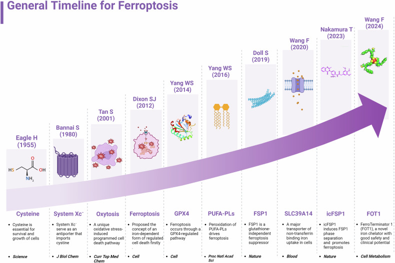
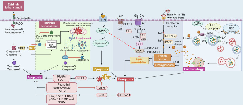
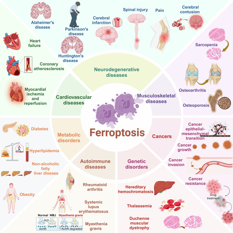
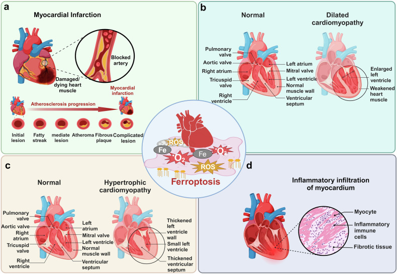
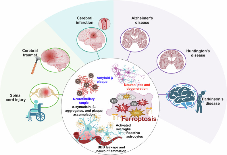
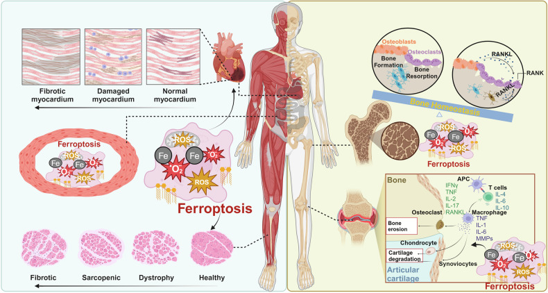
Iron, an essential mineral in the body, is involved in numerous physiological processes, making the maintenance of iron homeostasis crucial for overall health. Both iron overload and deficiency can cause various disorders and human diseases. Ferroptosis, a form of cell death dependent on iron, is characterized by the extensive peroxidation of lipids. Unlike other kinds of classical unprogrammed cell death, ferroptosis is primarily linked to disruptions in iron metabolism, lipid peroxidation, and antioxidant system imbalance. Ferroptosis is regulated through transcription, translation, and post-translational modifications, which affect cellular sensitivity to ferroptosis. Over the past decade or so, numerous diseases have been linked to ferroptosis as part of their etiology, including cancers, metabolic disorders, autoimmune diseases, central nervous system diseases, cardiovascular diseases, and musculoskeletal diseases. Ferroptosis-related proteins have become attractive targets for many major human diseases that are currently incurable, and some ferroptosis regulators have shown therapeutic effects in clinical trials although further validation of their clinical potential is needed. Therefore, in-depth analysis of ferroptosis and its potential molecular mechanisms in human diseases may offer additional strategies for clinical prevention and treatment. In this review, we discuss the physiological significance of iron homeostasis in the body, the potential contribution of ferroptosis to the etiology and development of human diseases, along with the evidence supporting targeting ferroptosis as a therapeutic approach. Importantly, we evaluate recent potential therapeutic targets and promising interventions, providing guidance for future targeted treatment therapies against human diseases.
Overview
The claims and statistics of this paper haven’t been checked yet.
Run the check — free, no account needed. It reads the open-access text and checks the sources it cites.
Integrity signals
Registry
Cites
Statistics
Figures
Verification
Claims the paper makes
The claims and statistics of this paper haven’t been checked yet.
Run the check — free, no account needed. It reads the open-access text and checks the sources it cites.
Claims other papers make about this one
Each is checked against this paper’s own text: does it say what the citing paper says it does?
The cited source says what the claim attributes to it.
In the citing paper
“Ferroptosis represents an iron-dependent RCD pathway characterized by lipid peroxidation and downregulation of the antioxidant defense of cells [39,40,41].”
From Role of Regulated Cell Death Pathways in Snakebite Envenomation: Mechanisms, Crosstalk, and Therapeutic Opportunities (2026) · cites this paper as [39]
Self-check
Percentages, sums and reported differences re-computed from the paper’s own numbers.
The self-check needs the paper’s full text.
Citations
Cites
1 retracted
RETRACTED: Ferroptosis and aerobic training in ageing
Kordi, N., Saydi, A., Karami, S., Bagherzadeh-Rahmani, B., Marzetti, E., Jung, F., & Stockwell, B. R. (2024) · Clinical Hemorheology and Microcirculation · doi:10.3233/ch-232076 · Cited by 13
RetractedSung, H., Ferlay, J., Siegel, R. L., Laversanne, M., Soerjomataram, I., Jemal, A., & Bray, F. (2021) · CA: A Cancer Journal for Clinicians · doi:10.3322/caac.21660 · Cited by 108,699
Ferroptosis: An Iron-Dependent Form of Nonapoptotic Cell Death
Dixon, S. J., Lemberg, K. M., Lamprecht, M. R., Skouta, R., Zaitsev, E. M., Gleason, C. E., Patel, D. N., Bauer, A. J., Cantley, A. M., Yang, W. S., et al. (2012) · Cell · doi:10.1016/j.cell.2012.03.042 · Cited by 18,691
Siegel, R. L., Miller, K. D., Fuchs, H. E., & Jemal, A. (2022) · CA: A Cancer Journal for Clinicians · doi:10.3322/caac.21708 · Cited by 17,196
Siegel, R. L., Miller, K. D., Fuchs, H. E., & Jemal, A. (2021) · CA: A Cancer Journal for Clinicians · doi:10.3322/caac.21654 · Cited by 16,522
Siegel, R. L., Miller, K. D., Wagle, N. S., & Jemal, A. (2023) · CA: A Cancer Journal for Clinicians · doi:10.3322/caac.21763 · Cited by 16,303
Positional cloning of the mouse obese gene and its human homologue
Zhang, Y., Proenca, R., Maffei, M., Barone, M., Leopold, L., & Friedman, J. M. (1994) · Nature · doi:10.1038/372425a0 · Cited by 13,419
Ferroptosis: mechanisms, biology and role in disease
Jiang, X., Stockwell, B. R., & Conrad, M. (2021) · Nature Reviews Molecular Cell Biology · doi:10.1038/s41580-020-00324-8 · Cited by 7,973
Regulation of Ferroptotic Cancer Cell Death by GPX4
Yang, W. S., SriRamaratnam, R., Welsch, M. E., Shimada, K., Skouta, R., Viswanathan, V. S., Cheah, J. H., Clemons, P. A., Shamji, A. F., Clish, C. B., et al. (2014) · Cell · doi:10.1016/j.cell.2013.12.010 · Cited by 7,713
Ferroptosis: A Regulated Cell Death Nexus Linking Metabolism, Redox Biology, and Disease
Stockwell, B. R., Friedmann Angeli, J. P., Bayir, H., Bush, A. I., Conrad, M., Dixon, S. J., Fulda, S., Gascón, S., Hatzios, S. K., Kagan, V. E., et al. (2017) · Cell · doi:10.1016/j.cell.2017.09.021 · Cited by 7,695
Research works printed but not linked: 1
- Heit, B. S. et al. Tonic extracellular glutamate and ischaemia. glutamate antiporter Syst. x(c.) (-) regulates anoxic depolarization hippocampus. J. Physiol. 601, 607–629 (2023).
Cited by
Sorted by citations
Items 401–500 of 724
Soleimani, A., Khalilian, S., Fathi, M., Tajik, Z., & Ghafouri-Fard, S. (2026) · Human Gene · doi:10.1016/j.humgen.2026.201541
Nurkolis, F. (2026) · Diabetology & Metabolic Syndrome · doi:10.1186/s13098-026-02131-1 · Cited by 1
Dysregulated iron metabolism and kidney stone risk: an epidemiological and experimental study
Wan, W., Yuan, D., Xun, Y., & Yu, X. (2026) · Renal Failure · doi:10.1080/0886022x.2026.2631316 · Cited by 1
Şahin, T. (2026) · Pharmedicine journal. · doi:10.62482/pmj.53 · Cited by 1
Abediasl, M., Siavash, M., Esfahani, S. S., Amini, Z., Amiri, K., Saket, S., Esmaeili, E., Rafizadeh, F., Saraei, R., & Fakhr, M. S. (2026) · Clinical & Translational Oncology · doi:10.1007/s12094-026-04225-8 · Cited by 1
Cendali, F. I., Lisk, C., Argabright, A., Dzieciątkowska, M., Rana, N. K., Swindle, D. C., Stephenson, D., McAfee, J., Westover, N., Lucero, M. J., et al. (2026) · HemaSphere · doi:10.1002/hem3.70318 · Cited by 1
Liu, A., Xing, L., Li, J., Yao, M., Song, J., Guo, W., Duan, P., & Li, H. (2026) · Journal of Alzheimer s Disease · doi:10.1177/13872877261420211 · Cited by 1
PROTACs in cancer therapy: targeted degradation of GPX4, PARP and epigenetic regulators
Periyasamy, S., Jarrett, T., Truong, J., & Skouta, R. (2026) · Journal of Enzyme Inhibition and Medicinal Chemistry · doi:10.1080/14756366.2026.2636394 · Cited by 2
Rao, Q., Gong, B., Liu, T., Lin, G., Tang, H., Liao, Y., Zhang, S., Zou, Y., Peng, Y., Li, W., et al. (2026) · Journal of Agricultural and Food Chemistry · doi:10.1021/acs.jafc.6c01532 · Cited by 1
Zhao, K., Ludwig-Radtke, L., Wang, Y., Rusch, T., Nimsky, C., Taudte, R. V., & Bartsch, J. W. (2026) · BioMetals · doi:10.1007/s10534-026-00809-7 · Cited by 1
Guo, K., Cao, H., Wu, J., Wang, W. H., Lu, G., Wei, R., Huang, T., Wang, Y., Gao, Y., & Zhang, K. (2026) · Journal of Ethnopharmacology · doi:10.1016/j.jep.2026.121590 · Cited by 1
Lee, J., & Roh, J.-L. (2026) · Cells · doi:10.3390/cells15070630 · Cited by 2
Hua, S., Tong, S., Hu, Y., He, C.-W., Bo, Y., Li, K., Weng, W., Chen, P., Xu, A., Wang, L., et al. (2026) · ACS Sensors · doi:10.1021/acssensors.5c04701 · Cited by 1
Targeting ferroptosis mediated cell death: a novel strategy for mitigating acute pancreatitis
Brahadeeswaran, S., Sreedhar, S., Ramesh, S., & Tamizhselvi, R. (2026) · Molecular Biology Reports · doi:10.1007/s11033-026-11772-4 · Cited by 1
An ultrasensitive cyanine dye-based fluorescent probe for the determination of iron(III) and oxalate
Shabelko, A. R., Halina, S. S., Zinko, L. S., Derevyanko, N., & Tananaiko, O. Y. (2026) · Spectrochimica Acta Part A Molecular and Biomolecular Spectroscopy · doi:10.1016/j.saa.2026.127881 · Cited by 1
Ferroptosis as a therapeutic vulnerability in ARID1A-deficient bladder cancer
Zhao, M., Wang, X., Duan, X., Wu, C., Guo, R., Wen, Y., Liang, J., Zhou, J., Lei, X., Fu, S., et al. (2026) · Communications Biology · doi:10.1038/s42003-026-09998-w · Cited by 2
Xing, Z., Yang, J., Zhang, H., Xie, Y., Yang, Q., Wu, M., Wang, W., Gao, S., & Hua, C. (2026) · Free Radical Biology and Medicine · doi:10.1016/j.freeradbiomed.2026.04.024 · Cited by 1
From Integrated Analysis to Clinical Insight: ncRNA-Mediated Ferroptosis in Glioblastoma
Seerapu, V. N., Amaresan, R., & Gopal, U. (2026) · Cancers · doi:10.3390/cancers18081238 · Cited by 1
Luo, X., Zhu, X., Chen, Y., Guo, Y., Xu, B., Mou, J., Wang, M., Gu, Q., Sun, Q., Lv, Y., et al. (2026) · Redox Biology · doi:10.1016/j.redox.2026.104138 · Cited by 1
Lipidized deferoxamine nanomedicines (DFOsomes) against iron overload
Li, Y. W., Duan, J., Jia, J., Zhu, F., Tan, E., Wang, Y., Zhai, Y., Wang, X., & Cheng, Y. (2026) · Journal of Controlled Release · doi:10.1016/j.jconrel.2026.114939 · Cited by 1
Self-citationA Rhodamine Appended Iron-Recognizing Probe for On-Demand Theranostics in Anemic Drosophila
Samal, D., Hembram, M., Mishra, L., Priyadarshini, A., Panda, S. P., Sahoo, S. C., Pramanik, S., Manna, S., Mundlapati, V. R., Dutta, B., et al. (2026) · Inorganic Chemistry · doi:10.1021/acs.inorgchem.6c00293 · Cited by 1
Qi, J., Liu, K., Wang, Y., Huang, Y., & Gan, M. (2026) · Frontiers in Oncology · doi:10.3389/fonc.2026.1821007 · Cited by 1
Poly-GR promotes ferroptosis-associated vulnerability in C9orf72-ALS
Lin, C., Hsieh, W.-C., & Wang, S. (2026) · Cell & Bioscience · doi:10.1186/s13578-026-01574-3 · Cited by 1
Ferroptosis: a promising therapeutic target for periodontitis
Li, Y., Sun, L., Xie, J., Li, L., Guo, L., & Lan, Y. (2026) · Frontiers in Immunology · doi:10.3389/fimmu.2026.1823915 · Cited by 1
Self-citationEmerging tumor therapies mediated by layered double hydroxides
Wu, J., Liu, Z., 裴寅清, Pei, Z., Li, L., & Cheng, L. (2026) · Coordination Chemistry Reviews · doi:10.1016/j.ccr.2026.218004 · Cited by 1
Xie, Y., Jiang, Q., Huang, H., Zhang, X., Zheng, X., Yang, Y., Wang, S., Zhao, J., & Li, Z. (2026) · Biomaterials Research · doi:10.34133/bmr.0369 · Cited by 1
Wang, M., ying, Q., Li, Q., Lou, X., Dai, S., & Liu, Z. (2026) · Cells · doi:10.3390/cells15100895 · Cited by 1
Cao, B., Tan, Y., Liu, P., & Pei, L. (2026) · Multiple Sclerosis and Related Disorders · doi:10.1016/j.msard.2026.107276 · Cited by 1
Novel insights of ferroptosis in atherosclerosis progression
Liang, P., & Huang, Y. (2026) · Frontiers in Cell and Developmental Biology · doi:10.3389/fcell.2026.1851553 · Cited by 2
Iron dysregulation in the central nervous system: implications for autism spectrum disorder
Bjørklund, G., Harutyunyan, H. S., Mkrtchyan, Z., Baghdasaryan, E., Avetisyan, T., Meguid, N. A., Hussein, F., & Yenkoyan, K. B. (2026) · BioMetals · doi:10.1007/s10534-026-00833-7 · Cited by 1
Liu, Y., Yin, L., Zhang, P., & Li, W. (2026) · Frontiers in Immunology · doi:10.3389/fimmu.2026.1825000 · Cited by 2
Karim, A., Shafarin, J., Suresh, S., Muhammad, J. S., Qaisar, R., Ahmad, F., & Hamad, M. A. (2026) · Cell and Tissue Research · doi:10.1007/s00441-026-04082-5 · Cited by 1
Ubiquitination-dependent regulation of ferroptosis in ischemic heart and brain
Zhang, Y.-Y., Long, X.-Y., Tian, J., Luo, X., & Peng, J. (2026) · Redox Biology · doi:10.1016/j.redox.2026.104249 · Cited by 1
Luo, H., Chen, W., Yang, Y., Li, P., Yu, Z., Wu, S., Wang, H., Du, B., & Yang, P. (2026) · ACS Applied Materials & Interfaces · doi:10.1021/acsami.6c05257 · Cited by 1
Zheng, H., Liang, Y., & Tian, Y. (2026) · Journal of Nanobiotechnology · doi:10.1186/s12951-026-04652-3 · Cited by 1
Kantapan, J., Innuan, P., Daranarong, D., Roytrakul, S., Jaikumkao, K., Rolin, G., Viennet-Steiner, C., Khaodee, W., Sangthong, P., Wantanajittikul, K., et al. (2026) · Regenerative Biomaterials · doi:10.1093/rb/rbag132 · Cited by 1
Li, J., Cao, Y., Wang, Y., & Shi, H. (2026) · Toxicology and Applied Pharmacology · doi:10.1016/j.taap.2026.117912 · Cited by 1
Chen, X., Xiong, Z., Xie, Y., Ruan, K., Huang, G., & Mao, J. (2026) · Pharmacological Research · doi:10.1016/j.phrs.2026.108308 · Cited by 1
Yuan, J., Li, F., Chen, J., Yu, S., Zhou, Y., Song, C., Fu, Z., & Liu, Y. (2026) · Molecular Biology Reports · doi:10.1007/s11033-026-12224-9 · Cited by 1
Zhang, Y., 黄娅林, Xie, G., Qin, C., & Chen, S. (2026) · Scientific Reports · doi:10.1038/s41598-026-60227-0 · Cited by 1
Ferroptosis in the tumor immune microenvironment: A double-edged sword in immunotherapy
Yang, R., Zhu, T., Jiang, J., Deng, M., & Miao, J. (2026) · Biochimica et Biophysica Acta (BBA) - Reviews on Cancer · doi:10.1016/j.bbcan.2026.189646 · Cited by 1
Ouyang, S., Chen, Y., Yan, S., Li, X., Liu, Y., Xiao, Y., Li, G., Sun, Z., & 胡晓芳 (2026) · Peritoneal Dialysis International · doi:10.1177/08968608261463605 · Cited by 1
Cheng, L., & Song, G. (2026) · Frontiers in Veterinary Science · doi:10.3389/fvets.2026.1874206 · Cited by 1
Iron and lipid dysregulation as key determinants of ferroptosis in cardiometabolic disease
Cho, S., Sung, H. K., Tang, J., Summers, S. A., & Sweeney, G. (2026) · Progress in Lipid Research · doi:10.1016/j.plipres.2026.101395 · Cited by 1
Wei, S., Zhou, S., Tu, J., Zhi, T., Wang, Y., Shen, Q.-H., Ni, C., Yao, M., & Ni, H. (2026) · Chinese Medicine · doi:10.1186/s13020-026-01466-1 · Cited by 1
Li, X. Z., & Hao, H. (2026) · European Journal of Pharmacology · doi:10.1016/j.ejphar.2026.179131 · Cited by 1
Kulyar, M. F., Pachaleva, J., Brazaite, S., Akhtar, M., Hasan, M. M., Kaboli, S., Lebedis, I., & Bernotienė, E. (2026) · Journal of Orthopaedic Translation · doi:10.1016/j.jot.2026.101178 · Cited by 2
Ferroptosis in liver diseases: molecular mechanisms, biomarker potential, and clinical translation
Tian, H., Yang, Y., Chen, L., Wei, R., Liu, W., Liu, Y., Wang, J., Zhuo, E., Gu, K., & Xing, J. (2026) · Frontiers in Cell and Developmental Biology · doi:10.3389/fcell.2026.1865349 · Cited by 1
Self-citationZhang, N., Yang, S., Amona, F. M., Yu, Y., Sun, H., Wang, Y., Liao, G., Gong, Z., & Zhang, Y. (2026) · Materials & Design · doi:10.1016/j.matdes.2026.116713 · Cited by 1
Precision medicine targeting ferroptosis in cerebrovascular diseases
Cao, L., Zhang, R., Zhang, Y., Yong, V. W., & Xue, M. (2026) · Trends in Molecular Medicine · doi:10.1016/j.molmed.2026.07.003 · Cited by 1
Ten controversies defining the boundaries of ferroptosis
Tang, D., & Kang, R. (2026) · Element · doi:10.20517/element.2026.07 · Cited by 1
Deng, L., Liu, K., Peng, J., & Luo, L. (2026) · Medicinal Research Reviews · doi:10.1002/med.70099 · Cited by 1
A Brake on Toxicity: Iron Suppresses Organic-Induced Superoxide Generation in Macrophages
Shen, Y., Edwards, K. C., Kapur, S., Pan, T., Hu, W., Zhou, S., Shiraiwa, M., & Fang, T. (2026) · Environmental Science & Technology · doi:10.1021/acs.est.6c06554 · Cited by 1
Sanghai, N., Pierce, K., Sharma, N. K., Leggett, S., Yadav, T. C., Masrori, P., Atukorallaya, D., Palaniyandi, S., Marcogliese, P. C., & Tranmer, G. K. (2026) · CNS Neuroscience & Therapeutics · doi:10.1002/cns.71078 · Cited by 1
The Glial Coupling in the Developing Brain: Astrocytes, Oligodendrocytes and Myelin Formation
Canepa, M., Santucci, C., Mariano, D., Mangini, S., Calandrino, A., Gagliardi, L., Petretto, A., Striano, P., & Ramenghi, L. A. (2026) · Metabolites · doi:10.3390/metabo16100774 · Cited by 1
Shen, X., Li, J., Ma, X., Li, H., Shao, K., Wei, S., Zhang, X., Li, M., Wu, X., Li, Y., et al. (2025) · ASAIO Journal · doi:10.1097/mat.0000000000002469 · Cited by 1
Self-citationBui, M. X., Linh, N. T., Le, P. T. V., & Nguyen, P. D. (2025) · The International Journal of Psychiatry in Medicine · doi:10.1177/00912174251352405 · Cited by 1
Cooperative targeting of NF-κB enhances ferroptosis-driven HCC therapy with Alisertib and Donafenib
Zhou, Q., & Wang, R. (2025) · Frontiers in Cell and Developmental Biology · doi:10.3389/fcell.2025.1637767 · Cited by 1
Zhu, H., Li, P., Xing, Z., Yang, B., Zheng, F., Yu, Z., Zhang, W., Yang, X., Luo, J., Tang, T., et al. (2025) · Phytochemistry Reviews · doi:10.1007/s11101-025-10171-0 · Cited by 1
GPR91 mediates low shear stress-induced endothelial ferroptosis via EGR1-dependent GPX4 repression
Su, M., Wu, Q., Zhang, Y., Wang, D., Yuan, C., Lu, S., Liang, L., Yu, B., & Wei, D. (2025) · Cellular Signalling · doi:10.1016/j.cellsig.2025.112127 · Cited by 1
Alangode, A., Rajasekhar, A., Sabu, J. J., Krishna, S., Anuja, D., Sreeja, A., Remesh, G., Kaladevi, A., & Nair, B. G. (2026) · International Journal of Molecular Sciences · doi:10.3390/ijms27156830
“Ferroptosis represents an iron-dependent RCD pathway characterized by lipid peroxidation and downregulation of the antioxidant defense of cells [39–41].”
Chen, S., Yu, Q., Yang, D., Ran, B., Pan, J., Song, Y., Cui, M., Mei, C., Wang, H., Li, H., et al. (2024) · Research Square · doi:10.21203/rs.3.rs-5419195/v1
Self-citationYue, Z., Liu, Q., Wang, Y., Xu, W., Wang, R., Zhang, W., Luo, P., Shahab Uddin, M. S., Huang, F., Deng, H., et al. (2025) · SSRN Electronic Journal · doi:10.2139/ssrn.5078070
Self-citationResearch Progress on the Effects of Iron on Human Body
王, 洪. (2025) · Advances in Clinical Medicine · doi:10.12677/acm.2025.153659
Li, J., Zhou, T., Feng, Z., Zhai, X., Li, X., Luo, T., Tong, H. H. Y., Li, K., & Huang, M. (2025) · doi:10.2196/preprints.77798
Zou, Y., Wei, X., Wu, G., Li, D., Cao, H., Chen, W., Zeng, L., & Chen, Z. (2025) · Chinese Journal of Analytical Chemistry · doi:10.1016/j.cjac.2025.100578
Nolt, M. J., Neely, E. B., Kheirabadi, S., Jaberi, A., Sheikhi, A., & Connor, J. R. (2025) · FASEB BioAdvances · doi:10.1096/fba.2025-00188
A Nir Fluorescent Probe for Imaging Labile Iron(Ii) Pools in Ferroptosis-Related DisordersPreprint
Yi, M., Wang, Y., Shen, R., Ma, T., Zhao, J., Han, F., Liu, J., Li, X., Zhang, B., & Fang, J. (2025) · SSRN Electronic Journal · doi:10.2139/ssrn.5360716
Mousavian, A. S., Shakeryan, S., Tabandeh, M. R., & Habibi, A. (2025) · Jentashapir Journal of Cellular and Molecular Biology · doi:10.5812/jjcmb-162429
Zhang, L., Wang, H., Liang, G., Chen, T. X., & Ge, T. (2025) · Journal of Molecular Histology · doi:10.1007/s10735-025-10502-7
Zhang, W., Yang, L. C., & Zhang, R. (2025) · PubMed Central · doi:10.11817/j.issn.1672-7347.2025.250206
Liu, P., Wang, Y., Shi, W., & Li, X. (2025) · SSRN Electronic Journal · doi:10.2139/ssrn.5388331
Tang, H., Wang, S., Liu, F., Xiong, X., Zhu, H., Lu, Y., Yang, J., Wu, J., Zhou, B., & Sun, X. (2025) · SSRN Electronic Journal · doi:10.2139/ssrn.5399334
Liu, Y., Wen, C. P., Pan, J., Cui, J., Lü, W., Sun, T. S., Ning, X., Lee, J. H., Li, W., Tu, H., et al. (2025) · European Journal of Epidemiology · doi:10.1007/s10654-025-01295-w
Zhou, M., Mo, D., Wang, Y., & Gao, Y. (2025) · medRxiv · doi:10.1101/2025.09.08.25335383
Bakhsh, A., Salama, S. A., Althobaiti, M. M., Alsufyani, S. E., Almalki, A. M., & Alanazi, S. (2025) · Journal of Trace Elements in Medicine and Biology · doi:10.1016/j.jtemb.2025.127758
Liu, W., Jiang, X., Ma, Y., Qing, H., Bao, Y., & Jiao, Z. (2025) · Medical Oncology · doi:10.1007/s12032-025-03053-0
Cao, Z., & Huang, J. (2025) · Pracatical anatomic pathology · doi:10.1007/978-3-031-97506-6_4
Lin, W., Xiong, J., Yan, Y., Bian, J., Zhang, Z., Xu, X., Diao, Y., Lan, X., Zhong, Y., Ji, H., et al. (2025) · bioRxiv (Cold Spring Harbor Laboratory) · doi:10.1101/2025.09.23.677952
Argon Alleviates Bone Cancer Pain by Mitigating Neuronal Ferroptosis via the TLR2-COX-2 Pathway
Kang, P., Cong, G., Meng, X., Zhang, Y., Sun, H., Wang, L., Zhang, C., Ma, J., Liu, T., & Ji, F. (2025) · Frontiers in Bioscience-Landmark · doi:10.31083/fbl43761
Iron Deficiency Anemia and Public Health: A New Narrative Related to India
Purandare, D., Deshmukh, C. D., Ramanathan, V., Tayade, V., Jadhav, S., & Polshettiwar, S. A. (2025) · Springer eBooks · doi:10.1007/978-3-031-32047-7_130-1
HMOX1 controls a heme-ferritin switch that protects cells from ferroptosis
de Souza, I., de Andrade, I. A. M., Freitas, F. P., Teixeira, A. B. D. S., Ramalho, M. C. C., Almeida, K., dos Santos, A. F., Schmitz, W., Fazeli, G., Rocha, C. R. R., et al. (2026) · Science Advances · doi:10.1126/sciadv.aeg3424
Weiskirchen, R. (2025) · Exploration of Digestive Diseases · doi:10.37349/edd.2025.100599
Computational Analysis of Iron-binding Proteins in the Mutational Landscape of Cancer
Chauhan, S., Kulharia, M., & Verma, S. K. (2025) · Biological Trace Element Research · doi:10.1007/s12011-025-04881-x
Harmonizing Iron Terminology and Nomenclature
Nashwan, A. J., Zakir, B., Hawas, Y., & Yassin, M. A. (2025) · Cureus · doi:10.7759/cureus.97362
Computational analysis and molecular dynamics insights into deleterious SNPs of the HFE gene
Islam, M. S., Tanha, T. H., Zarin, N., Haque, S., Shiddiky, M. J. A., Lam, A. K.-Y., & Gopalan, V. (2025) · Journal of Biomolecular Structure and Dynamics · doi:10.1080/07391102.2025.2593504
Bone antiresorptives as potential chelators for iron overload diseases
Konno, J. T. L., & Espósito, B. P. (2025) · BioMetals · doi:10.1007/s10534-025-00777-4
Zhou, D., Wang, X., Yu, Y., Yangyang, C., Yang, S. X., & Chen, J. (2025) · New Journal of Chemistry · doi:10.1039/d5nj03255e
Shen, Y., Li, D., Chen, Q., Wang, C., & Yu, L. (2025) · All Life · doi:10.1080/26895293.2025.2595807 · Cited by 1
Cucurbiturils in Oxygen Delivery and Their Potential in Anemia Management
Papiu, D., Nadaban, A., Palcu, A., Sasu, A., Mara, G., Albu, P., Boru, C., & Cotoraci, C. A. (2025) · Journal of Clinical Medicine · doi:10.3390/jcm14238571
Aldehyde dehydrogenase 2 mitigates acrolein-driven ferroptosis to preserve kidney function
Kuo, Y.-M., Chung, S., Chang, J.-T., Yang, H., Lee, C.-C., Lin, C., & Wang, H. (2025) · Environmental Pollution · doi:10.1016/j.envpol.2025.127555
Excess iron may accelerate amyloid beta accumulation in the brains of older mice
Song, S., & Shin, J. (2025) · Neurobiology of Aging · doi:10.1016/j.neurobiolaging.2025.12.003
宗少晖, Zeng, G., Peng, X., Zhao, W., Yang, F., Wei, Y., Chen, S., Wei, J. Y., Huang, J., Teng, Y., et al. (2025) · SSRN Electronic Journal · doi:10.2139/ssrn.5392070
Occupational and environmental iron exposure: a hidden risk factor in breast cancer etiology
Obeagu, E. I. (2025) · Annals of Medicine and Surgery · doi:10.1097/ms9.0000000000004305
Sharma, A., Pathangey, L. B., Chirackal, S. S., Shim, K. G., Fonseca, R., & Swaminathan, S. (2025) · European Journal Of Haematology · doi:10.1111/ejh.70084
Majidinia, L., Alabdali, A. Y. M., Panneerselvam, J., Ranganathan, V., Chinnappan, S., & Mani, R. R. (2025) · Tropical Journal of Natural Product Research · doi:10.26538/tjnpr/v9i12.13
Targeting Ferroptosis to Restore Salivary Gland Homeostasis in an Obesity Model
Park, G. C., Park, H., Bang, S.-Y., Kim, J. M., Shin, S.-C., Cheon, Y.-I., Kwon, H.-N., Cho, J. H., & Lee, B.-J. (2026) · International Journal of Molecular Sciences · doi:10.3390/ijms27010514
Sun, N., Xie, F., Wang, Z., Liu, Y.-W., Yang, H., Chen, L., Lu, J., He, Y., He, C.-Q., & Liu, S. (2026) · Frontiers in Neurology · doi:10.3389/fneur.2025.1708739
Self-citationAlghadir, A. H., Iqbal, Z. A., Alghadir, T., Alghadir, F., & Iqbal, A. (2026) · BMC Public Health · doi:10.1186/s12889-025-26162-x
Nutritional Strategies for Neuroprotection: Vitamins, Minerals, and Beyond
Kumari, M. (2026) · Springer eBooks · doi:10.1007/978-981-95-4169-0_4
Metadata
- Authors
- Ru, Q., Li, Y., Chen, L., Wu, Y., Min, J., & Wang, F.
- Affiliations
- Jianghan University (CN)
- Central South University (CN)
- First Affiliated Hospital Zhejiang University (CN)
- Zhejiang University (CN)
- Corresponding author
- Min, Junxia
- Wang, Fudi
- Venue
- Signal Transduction and Targeted Therapy
- Published
- 2024-10-14
- Volume
- 9
- Issue
- 1
- Pages
- 271-271
- ISSN
- 2059-3635 (ISSN-L)
- PMC
- PMC11486532
- OpenAlex
- W4403369705
- Cited by
- 724
- References
- 1,003
- Citations by year
- 2026: 407 · 2025: 310 · 2024: 7
- FWCI
- 53.50
- Type
- journal article
- Language
- en
- Open access
- gold
- doi.org
- Locations
- Signal Transduction and Targeted Therapy (publishedVersion, cc-by)
- PubMed Central (publishedVersion, cc-by)
- PubMed (publishedVersion)
- DOAJ (DOAJ: Directory of Open Access Journals) (submittedVersion)
- Funders
- National Natural Science Foundation of China (32330047, 82072506, 82071970, 31970689)
- Natural Science Foundation of Hubei Province (2024AFB971)
- Topics
- Life Sciences › Biochemistry, Genetics and Molecular Biology › Cancer Research › Ferroptosis and cancer prognosis
- Health Sciences › Medicine › Hematology › Iron Metabolism and Disorders
- Life Sciences › Neuroscience › Neurology › Neurological diseases and metabolism
- Keywords
- iron homeostasis, ferroptosis, iron overload, iron deficiency, lipid peroxidation, antioxidant imbalance, ferroptosis regulators, cancer, metabolic disorders, autoimmune disease, central nervous system disorders, cardiovascular disease, musculoskeletal disorders, ferroptosis-targeted therapy
- MeSH terms
- Ferroptosis, Autoimmune Diseases, Cardiovascular Diseases, Homeostasis, Humans, Iron, Metabolic Diseases, Neoplasms, Lipid Peroxidation
About these checks
Registry, retraction, Cites and statistics results are computed, not judged, with one exception: a printed reference that no index lists is read once by a model before it counts as a citation error. Claims are extracted by a model and kept only when their quote appears verbatim in the text; each cited source is then checked, with a model’s help, for whether it says what the claim relies on. Those verdicts can be wrong — read the claim’s quote and the cited source before relying on one.
Statistics and arithmetic are recomputed, not judged.
Confidence bands are shown only for fields calibrated on our annotated test set. None is calibrated yet, so no band is shown.
What counts as a claim ›How we measure confidence ›How we check this ›
Are you an author, or do you see a problem with this page? Request a review
