

The DOI matches its Crossref record.
A liquid biopsy to detect multidrug resistance and disease burden in multiple myeloma
Open accessAbstract
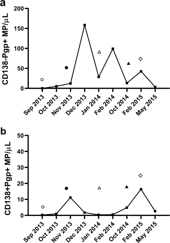
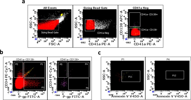
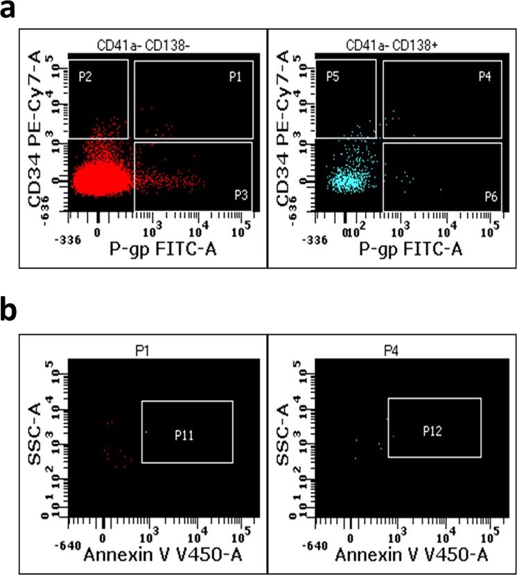
Multiple myeloma is an incurable cancer of bone marrow plasma cells, with a 5-year survival rate of 43%. Its incidence has increased by 126% since 1990. Treatment typically involves high-dose combination chemotherapy, but therapeutic response and patient survival are unpredictable and highly variable—attributed largely to the development of multidrug resistance (MDR). MDR is the simultaneous cross-resistance to a range of unrelated chemotherapeutic agents and is associated with poor prognosis and survival. Currently, no clinical procedures allow for a direct, continuous monitoring of MDR. We identified circulating large extracellular vesicles (specifically microparticles (MPs)) that can be used to monitor disease burden, disease progression and development of MDR in myeloma. These MPs differ phenotypically in the expression of four protein biomarkers: a plasma-cell marker (CD138), the MDR protein, P-glycoprotein (P-gp), the stem-cell marker (CD34); and phosphatidylserine (PS), an MP marker and mediator of cancer spread. Elevated levels of P-gp+and PS+MPs correlate with disease progression and treatment unresponsiveness. Furthermore, P-gp, PS and CD34 are predominantly expressed in CD138−MPs in advanced disease. In particular, a dual-positive (CD138−P-gp+CD34+) population is elevated in aggressive/unresponsive disease. Our test provides a personalised liquid biopsy with potential to address the unmet clinical need of monitoring MDR and treatment failure in myeloma.
Overview
The claims and statistics of this paper haven’t been checked yet.
Run the check — free, no account needed. It reads the open-access text and checks the sources it cites.
Integrity signals
Registry
Cites
Statistics
Figures
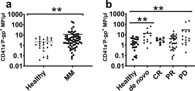
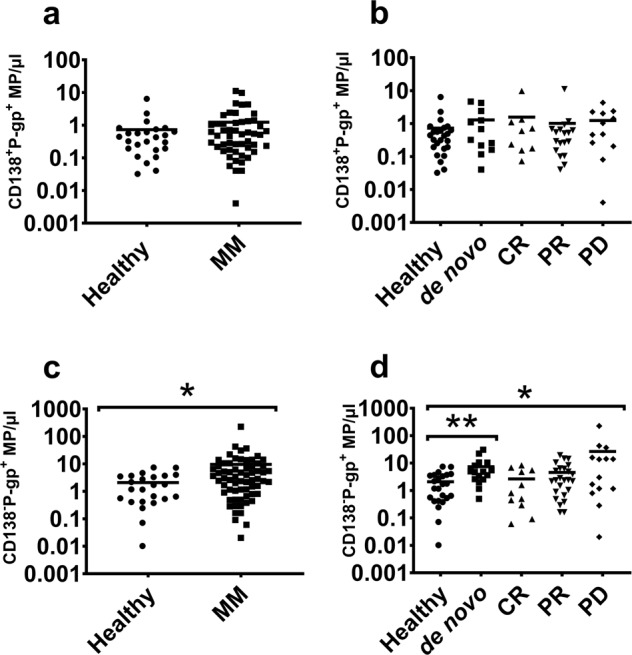
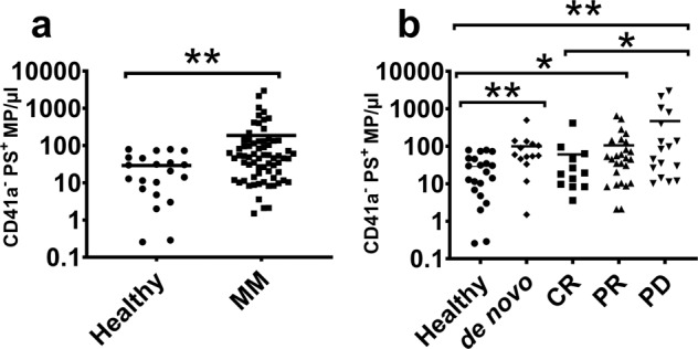
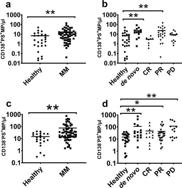
Verification
Claims the paper makes
The claims and statistics of this paper haven’t been checked yet.
Run the check — free, no account needed. It reads the open-access text and checks the sources it cites.
Claims other papers make about this one
Each is checked against this paper’s own text: does it say what the citing paper says it does?
The source backs part of the claim, not all of it — or backs it for a different population, condition or comparison.
In the citing paper
“In addition, P-gp expression rises in patients by up to 75% after therapy.”
From Mesenchymal stromal cells in bone marrow niche of patients with multiple myeloma: a double-edged sword (2025) · cites this paper as [242]
Experimental — accuracy not yet measuredThe cited source says what the claim attributes to it.
In the citing paper
“The overexpression of resistance proteins, such as P-glycoprotein (P-gp), which are members of the ATP class of drug transporters, is one of the most significant mechanisms in MDR tumors.”
From Mesenchymal stromal cells in bone marrow niche of patients with multiple myeloma: a double-edged sword (2025) · cites this paper as [242]
The cited source says what the claim attributes to it.
In the citing paper
“However, there is evidence that more recent forms of treatment, such as proteasome inhibitors and immunomodulatory medications, are similarly hampered since they are P-gp substrates.”
From Mesenchymal stromal cells in bone marrow niche of patients with multiple myeloma: a double-edged sword (2025) · cites this paper as [242]
Self-check
Percentages, sums and reported differences re-computed from the paper’s own numbers.
The self-check needs the paper’s full text.
Citations
Cites
No references could be resolved yet.
Research works printed but not linked: 53
- Kumar, S. K. et al. Multiple myeloma. Nat. Rev. Dis. Primers 3, 17046 (2017). · DOI
- Siegel, R. L., Miller, K. D. & Jemal, A. Cancer statistics, 2016. CA Cancer J. Clin. 66, 7–30 (2016). · DOI · Paper page
- Bray, F. et al. Global cancer statistics 2018: GLOBOCAN estimates of incidence and mortality worldwide for 36 cancers in 185 countries. CA Cancer J. Clin. 68, 394–424 (2018). · DOI · Paper page
- Keats, J. J. et al. Clonal competition with alternating dominance in multiple myeloma. Blood 120, 1067–1076 (2012). · DOI
- Bryant, C. et al. Long-term survival in multiple myeloma is associated with a distinct immunological profile, which includes proliferative cytotoxic T-cell clones and a favourable Treg/Th17 balance. Blood Cancer J. 3, e148 (2013). · DOI
- Decaux, O. et al. Prediction of survival in multiple myeloma based on gene expression profiles reveals cell cycle and chromosomal instability signatures in high-risk patients and hyperdiploid signatures in low-risk patients: a study of the Intergroupe Francophone du Myelome. J. Clin. Oncol. 26, 4798–4805 (2008). · DOI
- Ludwig, H. et al. European perspective on multiple myeloma treatment strategies in 2014. Oncologist 19, 829–844 (2014). · DOI
- Chaidos, A. et al. Clinical drug resistance linked to interconvertible phenotypic and functional states of tumor-propagating cells in multiple myeloma. Blood 121, 318–328 (2013). · DOI
- Gong, J. et al. Microparticles and their emerging role in cancer multidrug resistance. Cancer Treat. Rev. 38, 226–234 (2012). · DOI
- Begicevic, R. R. & Falasca, M. ABC transporters in cancer stem cells: beyond chemoresistance. Int. J. Mol. Sci. 18, 2362 (2017). · DOI
- Grogan, T. M. et al. P-glycoprotein expression in human plasma cell myeloma: correlation with prior chemotherapy. Blood 81, 490–495 (1993). · DOI
- O’Connor, R. et al. The interaction of bortezomib with multidrug transporters: implications for therapeutic applications in advanced multiple myeloma and other neoplasias. Cancer Chemother. Pharmacol. 71, 1357–1368 (2013). · DOI
- Hofmeister, C. C. et al. Phase I trial of lenalidomide and CCI-779 in patients with relapsed multiple myeloma: evidence for lenalidomide-CCI-779 interaction via P-glycoprotein. J. Clin. Oncol. 29, 3427–3434 (2011). · DOI
- Krishnan, S. R. et al. Multiple myeloma and persistence of drug resistance in the age of novel drugs (Review). Int. J. Oncol. 49, 33–50 (2016). · DOI
- Freyssinet, J. M. Cellular microparticles: what are they bad or good for? J. Thromb. Haemost. 1, 1655–1662 (2003). · DOI
- Caivano, A. et al. Extracellular vesicles in hematological malignancies: from biology to therapy. Int. J. Mol. Sci. 18, 1183 (2017). · DOI
- Minciacchi, V. R., Freeman, M. R. & Di Vizio, D. Extracellular vesicles in cancer: exosomes, microvesicles and the emerging role of large oncosomes. Semin. Cell Dev. Biol. 40, 41–51 (2015). · DOI
- Taylor, J. & Bebawy, M. Proteins regulating microvesicle biogenesis and multidrug resistance in cancer. Proteomics 19, e1800165 (2018). · DOI
- Giusti, I., D’Ascenzo, S. & Dolo, V. Microvesicles as potential ovarian cancer biomarkers. Biomed. Res. Int. 2013, 703048 (2013). · DOI
- van Doormaal, F. F. et al. Cell-derived microvesicles and cancer. Neth. J. Med. 67, 266–273 (2009).
- van Niel, G., D’Angelo, G. & Raposo, G. Shedding light on the cell biology of extracellular vesicles. Nat. Rev. Mol. Cell Biol. 19, 213–228 (2018). · DOI · Paper page
- Wang, C. C. et al. Circulating microparticles are prognostic biomarkers in advanced non-small cell lung cancer patients. Oncotarget 8, 75952–75967 (2017). · DOI
- Krishnan, S. R. et al. Isolation of human CD138(+) microparticles from the plasma of patients with multiple myeloma. Neoplasia 18, 25–32 (2016). · DOI
- Julich-Haertel, H. et al. Cancer-associated circulating large extracellular vesicles in cholangiocarcinoma and hepatocellular carcinoma. J. Hepatol. 67, 282–292 (2017). · DOI
- De Rubis, G., Rajeev Krishnan, S. & Bebawy, M. Liquid biopsies in cancer diagnosis, monitoring, and prognosis. Trends Pharmacol. Sci. 40, 172–186 (2019). · DOI
- Lu, J. F. et al. Microparticles mediate MRP1 intercellular transfer and the re-templating of intrinsic resistance pathways. Pharmacol. Res. 76, 77–83 (2013). · DOI
- Jaiswal, R. et al. Microparticle conferred microRNA profiles-implications in the transfer and dominance of cancer traits. Mol. Cancer 11, 37 (2012). · DOI
- Jaiswal, R. et al. Breast cancer-derived microparticles display tissue selectivity in the transfer of resistance proteins to cells. PLoS ONE 8, e61515 (2013). · DOI
- Jaiswal, R. et al. Microparticle-associated nucleic acids mediate trait dominance in cancer. Faseb J. 26, 420–429 (2012). · DOI
- Bebawy, M. et al. Membrane microparticles mediate transfer of P-glycoprotein to drug sensitive cancer cells. Leukemia 23, 1643–1649 (2009). · DOI
- Durie, B. G. et al. International uniform response criteria for multiple myeloma. Leukemia 20, 1467–1473 (2006). · DOI · Paper page
- Kumar, S. et al. International Myeloma Working Group consensus criteria for response and minimal residual disease assessment in multiple myeloma. Lancet Oncol. 17, e328–e346 (2016). · DOI · Paper page
- Yuana, Y., Bertina, R. M. & Osanto, S. Pre-analytical and analytical issues in the analysis of blood microparticles. Thromb. Haemost. 105, 396–408 (2011). · DOI
- Krishnan, S. R. et al. Isolation of human CD138+ microparticles from the plasma of patients with multiple myeloma. Neoplasia 18, 25–32 (2016). · DOI
- Sharma, B. & Kanwar, S. S. Phosphatidylserine: a cancer cell targeting biomarker. Semin. Cancer Biol. 52(Pt 1), 17–25 (2018). · DOI
- Riedl, S. et al. In search of a novel target—phosphatidylserine exposed by non-apoptotic tumor cells and metastases of malignancies with poor treatment efficacy. Biochim. Biophys. Acta 1808, 2638–2645 (2011). · DOI
- Wei, X. et al. Surface phosphatidylserine is responsible for the internalization on microvesicles derived from hypoxia-induced human bone marrow mesenchymal stem cells into human endothelial cells. PLoS ONE 11, e0147360 (2016). · DOI
- Matsui, W. et al. Characterization of clonogenic multiple myeloma cells. Blood 103, 2332–2336 (2004). · DOI
- Matsui, W. et al. Clonogenic multiple myeloma progenitors, stem cell properties, and drug resistance. Cancer Res. 68, 190–197 (2008). · DOI
- Kawano, Y. et al. Multiple myeloma cells expressing low levels of CD138 have an immature phenotype and reduced sensitivity to lenalidomide. Int. J. Oncol. 41, 876–884 (2012). · DOI
- Civin, C. I. et al. Antigenic analysis of hematopoiesis. III. A hematopoietic progenitor cell surface antigen defined by a monoclonal antibody raised against KG-1a cells. J. Immunol. 133, 157–165 (1984). · DOI
- Kuranda, K. et al. A subpopulation of malignant CD34+ CD138+ B7-H1+ plasma cells is present in multiple myeloma patients. Exp. Hematol. 38, 124–131.e4 (2010). · DOI
- O’Connell, F. P., Pinkus, J. L. & Pinkus, G. S. CD138 (syndecan-1), a plasma cell marker immunohistochemical profile in hematopoietic and nonhematopoietic neoplasms. Am. J. Clin. Pathol. 121, 254–263 (2004). · DOI
- Pilarski, L. M. & Belch, A. R. Intrinsic expression of the multidrug transporter, P-glycoprotein 170, in multiple myeloma: implications for treatment. Leuk. Lymphoma 17, 367–374 (1995). · DOI
- Pilarski, L. M. & Belch, A. R. Circulating monoclonal B cells expressing P glycoprotein may be a reservoir of multidrug-resistant disease in multiple myeloma. Blood 83, 724–736 (1994). · DOI
- Szczepek, A. J. et al. CD34+ cells in the blood of patients with multiple myeloma express CD19 and IgH mRNA and have patient-specific IgH VDJ gene rearrangements. Blood 89, 1824–1833 (1997). · DOI
- Roseblade, A. et al. Cell-derived microparticles: new targets in the therapeutic management of disease. J. Pharm. Pharm. Sci. 16, 238–253 (2013). · DOI
- Morel, O. et al. Cellular mechanisms underlying the formation of circulating microparticles. Arterioscler. Thromb. Vasc. Biol. 31, 15–26 (2011). · DOI
- Jimenez, J. J. et al. Endothelial cells release phenotypically and quantitatively distinct microparticles in activation and apoptosis. Thromb. Res. 109, 175–180 (2003). · DOI
- Sharma, P. & Allison, J. P. The future of immune checkpoint therapy. Science 348, 56–61 (2015). · DOI · Paper page
- Gray, M. J. et al. Phosphatidylserine-targeting antibodies augment the anti-tumorigenic activity of anti-PD-1 therapy by enhancing immune activation and downregulating pro-oncogenic factors induced by T-cell checkpoint inhibition in murine triple-negative breast cancers. Breast Cancer Res. 18, 50 (2016). · DOI
- Yu, M. et al. Phosphatidylserine on microparticles and associated cells contributes to the hypercoagulable state in diabetic kidney disease. Nephrol. Dial. Transplant. 33, 2115–2127 (2018). · DOI
- Palumbo, A. & Palladino, C. Venous and arterial thrombotic risks with thalidomide: evidence and practical guidance. Ther. Adv. Drug Saf. 3, 255–266 (2012). · DOI
Cited by
Sorted by citations
Kamrani, S., Naseramini, R., Khani, P., Razavi, Z. S., Afkhami, H., Atashzar, M. R., Nasri, F., Alavimanesh, S., Saeidi, F., & Ronaghi, H. (2025) · Cancer Cell International · doi:10.1186/s12935-025-03741-x · Cited by 27
Metadata
- Authors
- Rajeev Krishnan, S., De Rubis, G., Suen, H., Joshua, D., Lam Kwan, Y., & Bebawy, M.
- Affiliations
- Royal Prince Alfred Hospital (AU)
- University of Technology Sydney (AU)
- Corresponding author
- Bebawy, Mary
- Venue
- Blood Cancer Journal
- Published
- 2020-03-13
- Volume
- 10
- Issue
- 3
- Pages
- 37-37
- ISSN
- 2044-5385 (ISSN-L)
- PMC
- PMC7070076
- OpenAlex
- W3011733886
- Cited by
- 34
- References
- 0
- Citations by year
- 2026: 1 · 2025: 7 · 2024: 2 · 2023: 3 · 2022: 7 · 2021: 10 · 2020: 4
- FWCI
- 3.79
- Type
- journal article
- Language
- en
- Open access
- gold
- doi.org
- Locations
- Blood Cancer Journal (publishedVersion, cc-by)
- PubMed Central (publishedVersion, cc-by)
- PubMed (publishedVersion)
- RUNE (Research UNE) (submittedVersion, cc-by)
- UTS ePRESS (University of Technology Sydney) (submittedVersion, other-oa)
- Funders
- NSW Health Pathology
- Topics
- Health Sciences › Medicine › Hematology › Multiple Myeloma Research and Treatments
- Life Sciences › Biochemistry, Genetics and Molecular Biology › Molecular Biology › Extracellular vesicles in disease
- Health Sciences › Medicine › Hematology › Chronic Lymphocytic Leukemia Research
- Keywords
- multiple myeloma, multidrug resistance, liquid biopsy, circulating microparticles, extracellular vesicles, P-glycoprotein, phosphatidylserine, syndecan-1, CD34, disease burden monitoring, treatment response, disease progression
- MeSH terms
- Liquid Biopsy, Humans, Multiple Myeloma, Drug Resistance, Multiple
About these checks
Registry, retraction, Cites and statistics results are computed, not judged, with one exception: a printed reference that no index lists is read once by a model before it counts as a citation error. Claims are extracted by a model and kept only when their quote appears verbatim in the text; each cited source is then checked, with a model’s help, for whether it says what the claim relies on. Those verdicts can be wrong — read the claim’s quote and the cited source before relying on one.
Statistics and arithmetic are recomputed, not judged.
Confidence bands are shown only for fields calibrated on our annotated test set. None is calibrated yet, so no band is shown.
What counts as a claim ›How we measure confidence ›How we check this ›
Are you an author, or do you see a problem with this page? Request a review
