

The DOI matches its Crossref record; 220 of the 220 references it cites were found in a registry or an index.
Molecular mechanosensors in osteocytes
Open accessAbstract
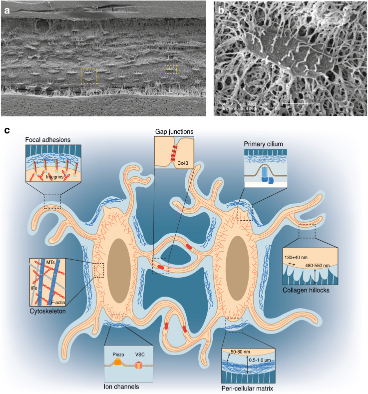
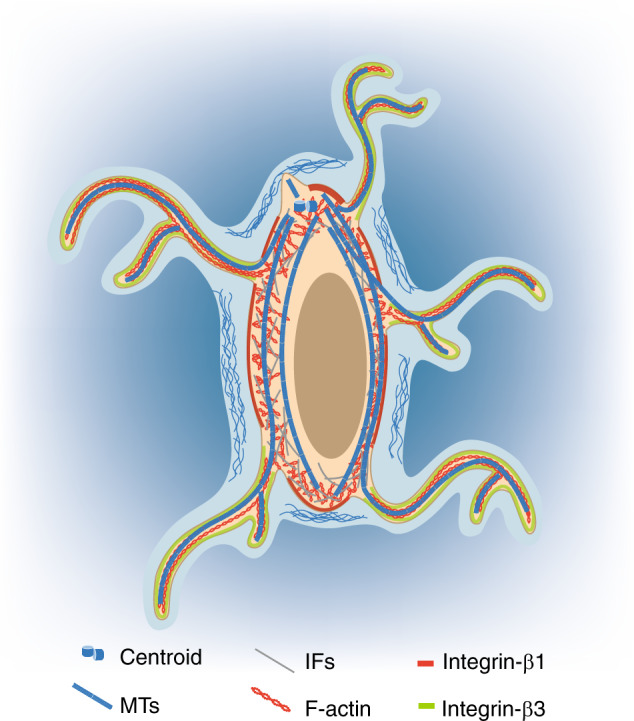
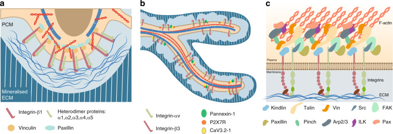
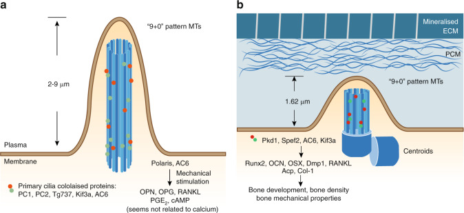
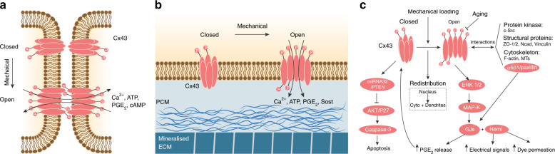
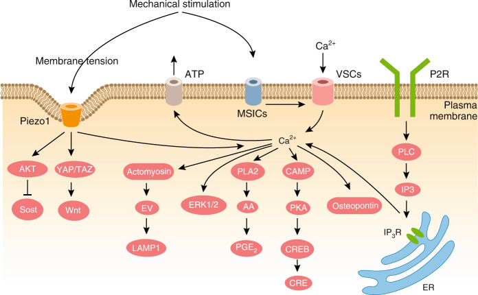
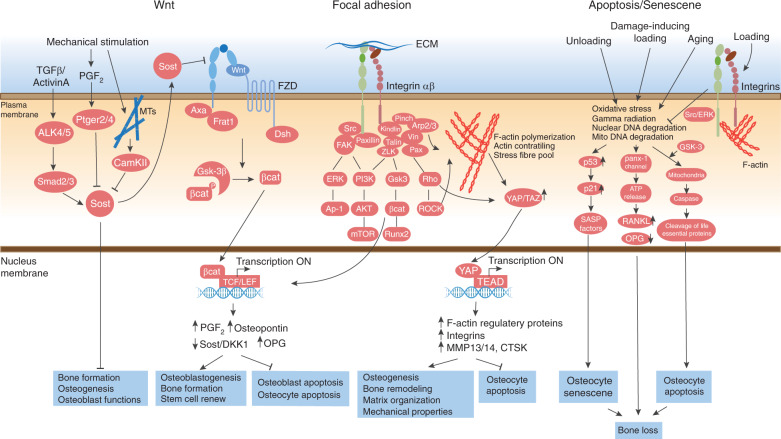
Osteocytes, the most abundant and long-lived cells in bone, are the master regulators of bone remodeling. In addition to their functions in endocrine regulation and calcium and phosphate metabolism, osteocytes are the major responsive cells in force adaptation due to mechanical stimulation. Mechanically induced bone formation and adaptation, disuse-induced bone loss and skeletal fragility are mediated by osteocytes, which sense local mechanical cues and respond to these cues in both direct and indirect ways. The mechanotransduction process in osteocytes is a complex but exquisite regulatory process between cells and their environment, between neighboring cells, and between different functional mechanosensors in individual cells. Over the past two decades, great efforts have focused on finding various mechanosensors in osteocytes that transmit extracellular mechanical signals into osteocytes and regulate responsive gene expression. The osteocyte cytoskeleton, dendritic processes, Integrin-based focal adhesions, connexin-based intercellular junctions, primary cilium, ion channels, and extracellular matrix are the major mechanosensors in osteocytes reported so far with evidence from both in vitro and in vitro studies. This review aims to give a systematic introduction to osteocyte mechanobiology, provide details of osteocyte mechanosensors, and discuss the roles of osteocyte mechanosensitive signaling pathways in the regulation of bone homeostasis.
Overview
The claims and statistics of this paper haven’t been checked yet.
Run the check — free, no account needed. It reads the open-access text and checks the sources it cites.
Integrity signals
Registry
Cites
Statistics
Figures
Verification
Claims the paper makes
The claims and statistics of this paper haven’t been checked yet.
Run the check — free, no account needed. It reads the open-access text and checks the sources it cites.
Claims other papers make about this one
Each is checked against this paper’s own text: does it say what the citing paper says it does?
The source backs part of the claim, not all of it — or backs it for a different population, condition or comparison.
In the citing paper
“Muscle-derived mechanical loading activates mechanosensors in osteocytes and osteoblast-lineage cells, including integrins and Piezo1, thereby inducing Ca2+-dependent signaling and Wnt/β-catenin-associated osteogenic pathways that promote osteoblast activity [47,48]”
From Interorgan Communications in Skeletal Pathophysiology: From Molecular Pathways to Multidisciplinary Therapies (2026) · cites this paper as [47]
Experimental — accuracy not yet measuredIn the citing paper
“The strain-induced flow of BEF in the lacuno-canalicular network exerts a shear stress (fluid flow shear stress, FFSS) on osteocyte bodies, which undergo dendrogenesis to activate several classes of mechano-sensors regulating specific gene expression patterns.28”
From Bone remodeling: an operational process ensuring survival and bone mechanical competence (2022) · cites this paper as [28]
Self-check
Percentages, sums and reported differences re-computed from the paper’s own numbers.
The self-check needs the paper’s full text.
Citations
Cites
Role of YAP/TAZ in mechanotransduction
Dupont, S., Morsut, L., Aragona, M., Enzo, E., Giulitti, S., Cordenonsi, M., Zanconato, F., Le Digabel, J., Forcato, M., Bicciato, S., et al. (2011) · Nature · doi:10.1038/nature10137 · Cited by 5,979
Stöckl, S., Göttl, C., Grifka, J. K., & Grässel, S. (2013) · Cellular Physiology and Biochemistry · doi:10.1159/000350089 · Cited by 4,670
Manolagas, S. C. (2000) · Endocrine Reviews · doi:10.1210/edrv.21.2.0395 · Cited by 2,639
Environmental sensing through focal adhesions
Geiger, B., Spatz, J. P., & Bershadsky, A. D. (2009) · Nature Reviews Molecular Cell Biology · doi:10.1038/nrm2593 · Cited by 2,543
Bonewald, L. F. (2011) · Journal of Bone and Mineral Research · doi:10.1002/jbmr.320 · Cited by 2,287
Bone “mass” and the “mechanostat”: A proposal
Frost, H. M. (1987) · The Anatomical Record · doi:10.1002/ar.1092190104 · Cited by 2,132
Biology of Bone Tissue: Structure, Function, and Factors That Influence Bone Cells
Florencio-Silva, R., Sasso, G. R. D. S., Sasso-Cerri, E., Simões, M. J., & Cerri, P. S. (2015) · BioMed Research International · doi:10.1155/2015/421746 · Cited by 2,044
Das Gesetz der Transformation der Knochen
Wolff, J. (1893) · DMW - Deutsche Medizinische Wochenschrift · doi:10.1055/s-0028-1144106 · Cited by 2,037
The mechanisms of Hedgehog signalling and its roles in development and disease
Briscoe, J., & Thérond, P. P. (2013) · Nature Reviews Molecular Cell Biology · doi:10.1038/nrm3598 · Cited by 1,751
Barczyk, M., Carracedo, S., & Gullberg, D. (2010) · Cell and Tissue Research · doi:10.1007/s00441-009-0834-6 · Cited by 1,512
Research works printed but not linked: 1
- Michael Sheetz, H. Y. The Cell as a Machine 1 edn, 0–434 (Cambridge university press, 2019).
Cited by
Sorted by citations
Items 501–526 of 526
Lv, X., Ran, X., Wei, C., Liu, X., Wang, J., & Huo, L. (2026) · Frontiers in Cell and Developmental Biology · doi:10.3389/fcell.2026.1938041
Tian, B., Chen, X., & Kang, X. (2026) · Molecular Medicine Reports · doi:10.3892/mmr.2026.14009
Wen, P., Bai, Y., & Yang, Z. (2026) · Pharmacological Research · doi:10.1016/j.phrs.2026.108441
Ramakrishnan, P., Thajuddin, N., Mohamed, S. B., Vinayagam, S., Gnanasekaran, L., Shanmugapriya, D., Rajamohan, R., Jalaludeen, A. M., & Sundaram, T. (2026) · Cell Biology International · doi:10.1002/cbin.70208
Bone Aging and Glycative Stress: Convergent and Divergent Mechanisms Driving Skeletal Deterioration
Peñarrubia, S., Martín‐Guerrero, E., Gortázar, A. R., & Ardura, J. A. (2026) · Cells · doi:10.3390/cells15181712
hMSCs form Osteocyte-like Cell Networks within Strain-Stiffening Bottlebrush Polymer HydrogelsPreprint
Ohnsorg, M. L., Friend, N. E., Givens, S. E., Saeb, D., Mash, K. M., & Anseth, K. S. (2026) · bioRxiv (Cold Spring Harbor Laboratory) · doi:10.64898/2026.09.22.753603
Zhu, Y. (2026) · International Journal of Public Health and Medical Research · doi:10.62051/ijphmr.v6n8.02
Recent progress in the biomarkers of peripheral artery disease
Chen, B.-W., Chen, W., Yu, X.-H., & Wang, G. (2026) · Clinica Chimica Acta · doi:10.1016/j.cca.2026.122864
Microgravity-Induced Osteocyte Dysfunction: An Osteocyte Mechanobiology Unit Framework
Roque, A. M., Acosta, O. B. L., Lozano, M. J. R., Suarez, H. N., & Bacabac, R. G. (2026) · Current Osteoporosis Reports · doi:10.1007/s11914-026-00989-8
Identificazione di nuove molecole coinvolte nell'omeostasi degli osteoblasti
Puri, C. (n.d.) · Archivio Istituzionale della Ricerca - Università degli Studi dell Aquila
"Pre-clinical development of siRNA Therapy for Autosomal Dominant Osteopetrosis (ADO)"
Patrizii, P. (n.d.) · Archivio Istituzionale della Ricerca - Università degli Studi dell Aquila
Laughrey, L. E., Castillo, N. A. C., Xie, Y., Scott, J., Moore, D., Johnson, M. L., Dallas, S. L., Johnson, M. L., & Thiagarajan, G. (2026) · JBMR Plus · doi:10.1093/jbmrpl/ziag162
Guest, S. L., & Dawson, S. C. (2026) · Current Biology · doi:10.1016/j.cub.2026.09.032
Shahid, E., Najum, H., Raza, M. T., & Majid, A. (2026) · RSC Advances · doi:10.1039/d6ra08442g
Fetuin-A Expression and Differentiation of MG-63 Cells Under Mechanical Stimulation
Korpela, V. (2026) · UTUPub (University of Turku)
Obszańska, A., Tesyna, J., Gendasz, A., & Marchlik, K. (2026) · Quality in Sport · doi:10.12775/qs.2026.77.76176
Bae, E., Goswami, A., Yaung, J., Kim, S. Y., Baik, S. S., Yu, K. A., Park, C., Huh, J., Kim, J., Hong, C., et al. (2026) · Clinical Oral Implants Research · doi:10.1111/clr.70191
Research Progress in Smart Hydrogel-Based Colloidal Systems for Osteoporosis Treatment
Wang, Z., Zhang, J., Zhang, B., & Jiang, Y. (2026) · International Journal of Nanomedicine · doi:10.2147/ijn.s644186
Multidimensional biomimetic strategies for artificial bone scaffolds in bone defect repair
Zhang, D., Lu, J., & He, W. (2026) · Bio-Medical Materials and Engineering · doi:10.1177/09592989261499320
Брониславовна, Л. Ю., Валентиновна, В. М., & Викторович, М. А. (n.d.) · CyberLeninK (CyberLeninka)
Molecular and systemic mechanisms of tumor-associated bone metabolic disorders
Chen, S., Jiang, Y., Chen, Y., 张春英, Deng, S., Liu, Y., Xu, X., Zhang, T., Liao, J., Zeng, T., et al. (2026) · Journal of bone oncology · doi:10.1016/j.jbo.2026.100811
Akhtar, S., & Al-Suhaimi, E. A. (2026) · Microscopy Research and Technique · doi:10.1002/jemt.70175
Zhu, Q., Liu, X., & Zhao, Y. (2026) · Medical Hypotheses · doi:10.1016/j.mehy.2026.112222
Guo, T., Zhou, Z.-M., Li, J.-T., Deng, J.-H., Chen, J.-Y., Zhou, H.-F., Yu, B., Wang, Z.-G., Zhou, W., & Yu, F. (2026) · Materials Advances · doi:10.1039/d6ma01152g
Franchi, M., Franchi, L., Mangani, S., Spanopoulos, K., Piperigkou, Z., & Karamanos, N. K. (2026) · Frontiers in Bioscience-Landmark · doi:10.31083/fbl53120
Li, W., Meng, S., & Zhu, K. (2026) · Frontiers in Cell and Developmental Biology · doi:10.3389/fcell.2026.2014138
Metadata
- Authors
- Qin, L., Liu, W., Cao, H., & Xiao, G.
- Affiliations
- Southern University of Science and Technology (CN)
- Corresponding author
- Cao, Huiling
- Xiao, Guozhi
- Venue
- Bone Research
- Published
- 2020-06-08
- Volume
- 8
- Issue
- 1
- Pages
- 23-23
- ISSN
- 2095-4700 (ISSN-L)
- 2095-6231
- PMC
- PMC7280204
- OpenAlex
- W3033561017
- Cited by
- 484
- References
- 219
- Citations by year
- 2026: 87 · 2025: 91 · 2024: 87 · 2023: 81 · 2022: 69 · 2021: 51 · 2020: 16
- FWCI
- 6.26
- Type
- journal article
- Language
- en
- Open access
- gold
- doi.org
- Locations
- Bone Research (publishedVersion, cc-by)
- PubMed Central (publishedVersion, cc-by)
- PubMed (publishedVersion)
- Funders
- National Natural Science Foundation of China (2017B030301018, 81991513, 81972100, 8163066, 2019YFA0906001, 81870532)
- China Postdoctoral Science Foundation
- Southern University of Science and Technology
- National Key Research and Development Program of China (2019YFA0906001, 2019YFA0906004)
- Topics
- Life Sciences › Biochemistry, Genetics and Molecular Biology › Cell Biology › Cellular Mechanics and Interactions
- Life Sciences › Biochemistry, Genetics and Molecular Biology › Molecular Biology › Bone Metabolism and Diseases
- Health Sciences › Medicine › Orthopedics and Sports Medicine › Bone health and osteoporosis research
- Keywords
- osteocytes, mechanosensors, mechanotransduction, mechanobiology, bone remodeling, bone homeostasis, force adaptation, primary cilia, mechanosensitive ion channels, extracellular matrix, bone fragility
About these checks
Registry, retraction, Cites and statistics results are computed, not judged, with one exception: a printed reference that no index lists is read once by a model before it counts as a citation error. Claims are extracted by a model and kept only when their quote appears verbatim in the text; each cited source is then checked, with a model’s help, for whether it says what the claim relies on. Those verdicts can be wrong — read the claim’s quote and the cited source before relying on one.
Statistics and arithmetic are recomputed, not judged.
Confidence bands are shown only for fields calibrated on our annotated test set. None is calibrated yet, so no band is shown.
What counts as a claim ›How we measure confidence ›How we check this ›
Are you an author, or do you see a problem with this page? Request a review
