

The DOI matches its Crossref record; 61 of the 62 references it cites were found in a registry or an index.
Adoption of artificial intelligence in breast imaging: evaluation, ethical constraints and limitations
Open accessAbstract
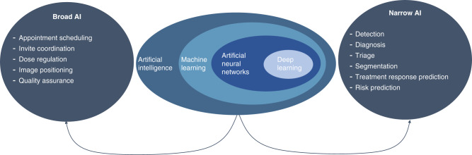
Retrospective studies have shown artificial intelligence (AI) algorithms can match as well as enhance radiologist’s performance in breast screening. These tools can facilitate tasks not feasible by humans such as the automatic triage of patients and prediction of treatment outcomes. Breast imaging faces growing pressure with the exponential growth in imaging requests and a predicted reduced workforce to provide reports. Solutions to alleviate these pressures are being sought with an increasing interest in the adoption of AI to improve workflow efficiency as well as patient outcomes. Vast quantities of data are needed to test and monitor AI algorithms before and after their incorporation into healthcare systems. Availability of data is currently limited, although strategies are being devised to harness the data that already exists within healthcare institutions. Challenges that underpin the realisation of AI into everyday breast imaging cannot be underestimated and the provision of guidance from national agencies to tackle these challenges, taking into account views from a societal, industrial and healthcare prospective is essential. This review provides background on the evaluation and use of AI in breast imaging in addition to exploring key ethical, technical, legal and regulatory challenges that have been identified so far.
Overview
The claims and statistics of this paper haven’t been checked yet.
Run the check — free, no account needed. It reads the open-access text and checks the sources it cites.
Integrity signals
Registry
Cites
Statistics
Figures
Verification
Claims the paper makes
The claims and statistics of this paper haven’t been checked yet.
Run the check — free, no account needed. It reads the open-access text and checks the sources it cites.
Claims other papers make about this one
Each is checked against this paper’s own text: does it say what the citing paper says it does?
The source backs part of the claim, not all of it — or backs it for a different population, condition or comparison.
In the citing paper
“The presence of AI experts, perhaps a multidisciplinary team, particularly with clinical scientists, data science and subject matter experts with AI skills, an innovation manager, AI experts to provide training, and local champions within departments involved in the end-to-end process, was considered an important element for adoption [32,57,59,62,71].”
From Barriers to and facilitators of artificial intelligence adoption in health care: scoping review (2024) · cites this paper as [71]
Experimental — accuracy not yet measuredThe cited source says what the claim attributes to it.
In the citing paper
“Another facilitator of trust is the engagement of patients in the development of AI.”
From Barriers to and facilitators of artificial intelligence adoption in health care: scoping review (2024) · cites this paper as [71]
The cited source says what the claim attributes to it.
In the citing paper
“Hickman et al [71], in particular, noted the lack of technological (infrastructure) maturity to allow for the integration of AI.”
From Barriers to and facilitators of artificial intelligence adoption in health care: scoping review (2024) · cites this paper as [71]
Self-check
Percentages, sums and reported differences re-computed from the paper’s own numbers.
The self-check needs the paper’s full text.
Citations
Cites
LeCun, Y., Bengio, Y., & Hinton, G. (2015) · Nature · doi:10.1038/nature14539 · Cited by 84,126
The FAIR Guiding Principles for scientific data management and stewardship
Wilkinson, M. D., Dumontier, M., Aalbersberg, I. J., Appleton, G., Axton, M., Baak, A., Blomberg, N., Boiten, J.-W., da Silva Santos, L. B., Bourne, P. E., et al. (2016) · Scientific Data · doi:10.1038/sdata.2016.18 · Cited by 18,423
Artificial intelligence in radiology
Hosny, A., Parmar, C., Quackenbush, J., Schwartz, L. H., & Aerts, H. J. W. L. (2018) · Nature Reviews Cancer · doi:10.1038/s41568-018-0016-5 · Cited by 3,803
International evaluation of an AI system for breast cancer screening
McKinney, S. M., Sieniek, M., Godbole, V., Godwin, J., Antropova, N., Ashrafian, H., Back, T., Chesus, M., Corrado, G. S., Darzi, A., et al. (2020) · Nature · doi:10.1038/s41586-019-1799-6 · Cited by 3,287
Self-citationLiu, X., Faes, L., Kale, A. U., Wagner, S. K., Fu, D. J., Bruynseels, A., Mahendiran, T., Moraes, G., Shamdas, M., Kern, C., et al. (2019) · The Lancet Digital Health · doi:10.1016/s2589-7500(19)30123-2 · Cited by 1,883
Checklist for Artificial Intelligence in Medical Imaging (CLAIM): a guide for authors and reviewers
Mongan, J., Moy, L., & Kahn, C. E. (2020) · Radiology: Artificial Intelligence · doi:10.1148/ryai.2020200029 · Cited by 1,443
Liu, X., Cruz Rivera, S., Moher, D., Calvert, M. J., Denniston, A. K., The SPIRIT-AI and CONSORT-AI Working Group, SPIRIT-AI and CONSORT-AI Steering Group, Chan, A.-W., Darzi, A., Holmes, C., et al. (2020) · Nature Medicine · doi:10.1038/s41591-020-1034-x · Cited by 1,421
Moreira, I. C., Amaral, I., Domingues, I., Cardoso, A. N. L., Cardoso, M. J., & Cardoso, J. S. (2011) · Academic Radiology · doi:10.1016/j.acra.2011.09.014 · Cited by 1,256
The Mammographic Image Analysis Society digital mammogram database
John Suckling, A., Parker, J., Astley, S., Hutt, I., Boggis, C. R. M., Ricketts, I. W., Stamatakis, E. A., Cerneaz, N., Kok, S., Taylor, P., et al. (1994) · Cited by 1,155
Nagendran, M., Chen, Y., Lovejoy, C. A., Gordon, A. C., Komorowski, M., Harvey, H., Topol, E. J., Ioannidis, J. P. A., Collins, G. S., & Maruthappu, M. (2020) · BMJ · doi:10.1136/bmj.m689 · Cited by 1,152
Preparing Medical Imaging Data for Machine Learning
Willemink, M. J., Koszek, W. A., Hardell, C., Wu, J., Fleischmann, D., Harvey, H., Folio, L. R., Summers, R. M., Rubin, D. L., & Lungren, M. P. (2020) · Radiology · doi:10.1148/radiol.2020192224 · Cited by 1,020
Cruz Rivera, S., Liu, X., Chan, A.-W., Denniston, A. K., Calvert, M. J., The SPIRIT-AI and CONSORT-AI Working Group, SPIRIT-AI and CONSORT-AI Steering Group, Darzi, A., Holmes, C., Yau, C., et al. (2020) · Nature Medicine · doi:10.1038/s41591-020-1037-7 · Cited by 933
Park, S. H., & Han, K. (2018) · Radiology · doi:10.1148/radiol.2017171920 · Cited by 894
General Data Protection Regulation (GDPR)
Deaves, M. (2017) · Cited by 815
Reporting of artificial intelligence prediction models
Collins, G. S., & Moons, K. G. M. (2019) · The Lancet · doi:10.1016/s0140-6736(19)30037-6 · Cited by 701
Diagnostic Accuracy of Digital Screening Mammography With and Without Computer-Aided Detection
Lehman, C. D., Wellman, R. D., Buist, D. S. M., Kerlikowske, K., Tosteson, A. N. A., & Miglioretti, D. L. (2015) · JAMA Internal Medicine · doi:10.1001/jamainternmed.2015.5231 · Cited by 678
Ethical Machine Learning in Healthcare
Chen, I. Y., Pierson, E., Rose, S., Joshi, S., Ferryman, K., & Ghassemi, M. (2021) · Annual Review of Biomedical Data Science · doi:10.1146/annurev-biodatasci-092820-114757 · Cited by 645
Schaffter, T., Buist, D. S. M., Lee, C. I., Nikulin, Y., Ribli, D., Guan, Y., Lotter, W., Jie, Z., Du, H., Wang, S., et al. (2020) · JAMA Network Open · doi:10.1001/jamanetworkopen.2020.0265 · Cited by 409
Geis, J. R., Brady, A. P., Wu, C. C., Spencer, J., Ranschaert, E., Jaremko, J. L., Langer, S. G., Borondy Kitts, A., Birch, J., Shields, W. F., et al. (2019) · Radiology · doi:10.1148/radiol.2019191586 · Cited by 388
Dembrower, K., Wåhlin, E., Liu, Y., Salim, M., Smith, K., Lindholm, P., Eklund, M., & Strand, F. (2020) · The Lancet Digital Health · doi:10.1016/s2589-7500(20)30185-0 · Cited by 311
Salim, M., Wåhlin, E., Dembrower, K., Azavedo, E., Foukakis, T., Liu, Y., Smith, K., Eklund, M., & Strand, F. (2020) · JAMA Oncology · doi:10.1001/jamaoncol.2020.3321 · Cited by 301
Single Reading with Computer-Aided Detection for Screening Mammography
Gilbert, F. J., Astley, S., Gillan, M. G., Agbaje, O. F., Wallis, M., James, J. J., Boggis, C. R. M., & Duffy, S. W. (2008) · New England Journal of Medicine · doi:10.1056/nejmoa0803545 · Cited by 296
Self-citationAnelli, F., Leoni, G., Monaco, R., Nume, C., Rossi, R. C., Marinoni, G., Spata, G., Giorgi, D. D., Peccarisi, L., Miani, A., et al. (2020) · BMJ · doi:10.1136/bmj.m1254 · Cited by 287
Artificial intelligence in breast imaging
Le, E., Wang, Y., Huang, Y., Hickman, S. E., & Gilbert, F. J. (2019) · Clinical Radiology · doi:10.1016/j.crad.2019.02.006 · Cited by 283
Self-citationThe royal college of radiologists
Steiner, R. E. (1980) · Clinical Radiology · doi:10.1016/s0009-9260(80)80176-0 · Cited by 278
Sounderajah, V., Ashrafian, H., Aggarwal, R., De Fauw, J., Denniston, A. K., Greaves, F., Karthikesalingam, A., King, D., Liu, X., Markar, S. R., et al. (2020) · Nature Medicine · doi:10.1038/s41591-020-0941-1 · Cited by 270
Recht, M. P., Dewey, M., Dreyer, K., Langlotz, C., Niessen, W., Prainsack, B., & Smith, J. J. (2020) · European Radiology · doi:10.1007/s00330-020-06672-5 · Cited by 268
Halligan, S., Altman, D. G., & Mallett, S. (2015) · European Radiology · doi:10.1007/s00330-014-3487-0 · Cited by 255
Conant, E. F., Toledano, A. Y., Periaswamy, S., Fotin, S. V., Go, J., Boatsman, J. E., & Hoffmeister, J. W. (2019) · Radiology Artificial Intelligence · doi:10.1148/ryai.2019180096 · Cited by 243
Continuous Learning AI in Radiology: Implementation Principles and Early Applications
Pianykh, O. S., Langs, G., Dewey, M., Enzmann, D. R., Herold, C. J., Schoenberg, S. O., & Brink, J. A. (2020) · Radiology · doi:10.1148/radiol.2020200038 · Cited by 237
Mammographic density. Measurement of mammographic density
Yaffe, M. J. (2008) · Breast Cancer Research · doi:10.1186/bcr2102 · Cited by 235
Rodríguez‐Ruiz, A., Lång, K., Gubern‐Mérida, A., Teuwen, J., Broeders, M. J. M., Gennaro, G., Clauser, P., Helbich, T. H., Chevalier, M. L., Mertelmeier, T., et al. (2019) · European Radiology · doi:10.1007/s00330-019-06186-9 · Cited by 213
Latent bias and the implementation of artificial intelligence in medicine
DeCamp, M., & Lindvall, C. (2020) · Journal of the American Medical Informatics Association · doi:10.1093/jamia/ocaa094 · Cited by 198
Breast cancer diagnosis in DCE-MRI using mixture ensemble of convolutional neural networks
Rasti, R., Teshnehlab, M., & Phung, S. L. (2017) · Pattern Recognition · doi:10.1016/j.patcog.2017.08.004 · Cited by 186
Zhou, J., Luo, L., Dou, Q., Chen, H., Chen, C., Li, G., Jiang, Z., & Heng, P. (2019) · Journal of Magnetic Resonance Imaging · doi:10.1002/jmri.26721 · Cited by 158
Rexhepaj, E., Brennan, D. J., Holloway, P., Kay, E. W., McCann, A. H., Landberg, G., Duffy, M. J., Jirström, K., & Gallagher, W. M. (2008) · Breast Cancer Research · doi:10.1186/bcr2187 · Cited by 137
Qu, Y., Zhu, H., Cao, K., Li, X., Ye, M., & Sun, Y. (2020) · Thoracic Cancer · doi:10.1111/1759-7714.13309 · Cited by 96
REEVES, G. K., Pirie, K. L., Green, J., Bull, D., Beral, V., & Collaborators, F. T. M. W. S. (2011) · International Journal of Cancer · doi:10.1002/ijc.26460 · Cited by 88
Dembrower, K., Lindholm, P., & Strand, F. (2019) · Journal of Imaging Informatics in Medicine · doi:10.1007/s10278-019-00278-0 · Cited by 88
Fully automated detection of breast cancer in screening MRI using convolutional neural networks
Dalmış, M. U., Vreemann, S., Kooi, T., Mann, R. M., Karssemeijer, N., & Gubern‐Mérida, A. (2018) · Journal of medical imaging · doi:10.1117/1.jmi.5.1.014502 · Cited by 81
De-identification of Medical Images with Retention of Scientific Research Value
Moore, S., Maffitt, D. R., Smith, K. E., Kirby, J., Clark, K. W., Freymann, J., Vendt, B. A., Tarbox, L., & Prior, F. (2015) · Radiographics · doi:10.1148/rg.2015140244 · Cited by 80
Curated Breast Imaging Subset of Digital Database for Screening Mammography (CBIS-DDSM)
Sawyer-Lee, R., Gimenez, F., Hoogi, A., & Rubin, D. L. (2016) · The Cancer Imaging Archive · doi:10.7937/k9/tcia.2016.7o02s9cy · Cited by 78
Cohort Profile: the Million Women Study
Green, J., REEVES, G. K., Floud, S., Barnes, I. M., Cairns, B. J., Gathani, T., Pirie, K. L., Sweetland, S., Yang, T. O., Beral, V., et al. (2018) · International Journal of Epidemiology · doi:10.1093/ije/dyy065 · Cited by 77
Gray, J. A. (1989) · BMJ · doi:10.1136/bmj.298.6665.48-c · Cited by 73
Ha, R. S., Chang, P., Mutasa, S., Karcich, J., Goodman, S., Blum, E. S., Kalinsky, K. M., Liu, M. Z., & Jambawalikar, S. (2018) · Journal of Magnetic Resonance Imaging · doi:10.1002/jmri.26244 · Cited by 73
Ravichandran, K., Braman, N. M., Janowczyk, A. R., & Madabhushi, A. (2018) · SPIE Medical Imaging: Computer-Aided Diagnosis · doi:10.1117/12.2294056 · Cited by 63
Impact of adding breast density to breast cancer risk models: A systematic review
Vilmun, B. M., Vejborg, I., Lynge, E., Lillholm, M., Nielsen, M., Nielsen, M. B., & Carlsen, J. F. (2020) · European Journal of Radiology · doi:10.1016/j.ejrad.2020.109019 · Cited by 59
The ethical adoption of artificial intelligence in radiology
Mudgal, K. S., & Das, N. (2020) · BJR|Open · doi:10.1259/bjro.20190020 · Cited by 52
Ming, C., Viassolo, V., Probst-Hensch, N. M., Dinov, I. D., Chappuis, P. O., & Katapodi, M. C. (2020) · British Journal of Cancer · doi:10.1038/s41416-020-0937-0 · Cited by 48
Salim, M., Dembrower, K., Eklund, M., Lindholm, P., & Strand, F. (2020) · Radiology · doi:10.1148/radiol.2020192212 · Cited by 42
Multi-center breast DCE-MRI data and segmentations from patients in the I-SPY 1/ACRIN 6657 trials
Newitt, D., & Hylton, N. (2016) · The Cancer Imaging Archive · doi:10.7937/k9/tcia.2016.hdhpgjlk · Cited by 39
Huynh, B. Q., Antropova, N., & Giger, M. L. (2017) · Proceedings of SPIE, the International Society for Optical Engineering/Proceedings of SPIE · doi:10.1117/12.2255316 · Cited by 38
The Cancer Genome Atlas Breast Invasive Carcinoma Collection (TCGA-BRCA)
Lingle, W. L., Erickson, B. J., Zuley, M. L., Jarosz, R., Bonaccio, E., Filippini, J., Net, J. M., Levi, L., Morris, E. A., Figler, G. G., et al. (2016) · The Cancer Imaging Archive · doi:10.7937/k9/tcia.2016.ab2nazrp · Cited by 37
Nagendran, M., Chen, Y., Lovejoy, C. A., et al. (2020) · Cited by 31
Artificial intelligence in clinical imaging: a health system approach
Gilbert, F. J., Smye, S., & Schönlieb, C. (2019) · Clinical Radiology · doi:10.1016/j.crad.2019.09.122 · Cited by 24
Self-citationDench, E. K., Bond‐Smith, D., Darcey, E., Lee, G., Aung, Y. K., Chan, A. H. L., Cuzick, J. M., Ding, Z. Y., Evans, C. F., Harvey, J. A., et al. (2019) · BMJ Open · doi:10.1136/bmjopen-2019-031041 · Cited by 20
Braman, N. M., Adoui, M. E., Vulchi, M., Turk, P., Etesami, M., Fu, P., Bera, K., Drisis, S., Varadan, V., Plecha, D. M., et al. (2020) · Repository of the University of Namur · doi:10.48550/arxiv.2001.08570 · Cited by 15
Ethical Machine Learning in HealthPreprint
Chen, I. Y., Pierson, E., Rose, S., Joshi, S. D., Ferryman, K., & Ghassemi, M. (2020) · arXiv (Cornell University) · Cited by 10
Google health-data scandal spooks researchers
Ledford, H. K. (2019) · Nature · doi:10.1038/d41586-019-03574-5 · Cited by 9
Cross, L., Carson, L. E., Jewell, A., Heslin, M., Osborn, D., Downs, J. M., & Stewart, R. J. (2020) · International Journal for Population Data Science · doi:10.23889/ijpds.v5i1.1355 · Cited by 6
NHS data: Maximising its impact on the health and wealth of the United Kingdom
Ghafur, S., Fontana, G., Halligan, J., O'Shaughnessy, J., & Darzi, A. W. (2020) · Spiral (Imperial College London) · doi:10.25561/76409 · Cited by 5
Cited by
Sorted by citations
Redefining Radiology: A Review of Artificial Intelligence Integration in Medical Imaging
Najjar, R. (2023) · Diagnostics · doi:10.3390/diagnostics13172760 · Cited by 1,025
Dembrower, K., Crippa, A., Colón, E., Eklund, M., & Strand, F. (2023) · The Lancet Digital Health · doi:10.1016/s2589-7500(23)00153-x · Cited by 397
Barriers to and facilitators of artificial intelligence adoption in health care: scoping review
Hassan, M., Kushniruk, A., & Borycki, E. (2024) · JMIR Human Factors · doi:10.2196/48633 · Cited by 435
Artificial Intelligence in Breast Cancer Diagnosis and Personalized Medicine
Ahn, J. S., Shin, S., Yang, S., Park, E. K., Kim, K. H., Cho, S. I., Ock, C., & Kim, S. (2023) · Journal of Breast Cancer · doi:10.4048/jbc.2023.26.e45 · Cited by 281
Saheb, T., Saheb, T., & Carpenter, D. O. (2021) · Computers in Biology and Medicine · doi:10.1016/j.compbiomed.2021.104660 · Cited by 148
Metadata
- Authors
- Hickman, S. E., Baxter, G. C., & Gilbert, F. J.
- Affiliations
- University of Cambridge (GB)
- Corresponding author
- Gilbert, Fiona J.
- Venue
- British Journal of Cancer
- Published
- 2021-03-26
- Volume
- 125
- Issue
- 1
- Pages
- 15-22
- ISSN
- 0007-0920 (ISSN-L)
- 1532-1827
- PMC
- PMC8257639
- OpenAlex
- W3136635219
- Cited by
- 110
- References
- 62
- Citations by year
- 2026: 12 · 2025: 23 · 2024: 21 · 2023: 27 · 2022: 21 · 2021: 6
- FWCI
- 1.34
- Type
- journal article
- Language
- en
- Open access
- hybrid
- doi.org
- Locations
- British Journal of Cancer (publishedVersion, cc-by)
- PubMed Central (publishedVersion, cc-by)
- PubMed (publishedVersion)
- Apollo (University of Cambridge) (submittedVersion, cc-by)
- Apollo (University of Cambridge) (submittedVersion)
- Apollo
- Funders
- Cancer Research UK (C543/A26884)
- Topics
- Physical Sciences › Computer Science › Artificial Intelligence › AI in cancer detection
- Health Sciences › Medicine › Health Informatics › Artificial Intelligence in Healthcare and Education
- Health Sciences › Medicine › Radiology, Nuclear Medicine and Imaging › Digital Radiography and Breast Imaging
- Keywords
- artificial intelligence, breast imaging, breast cancer screening, artificial intelligence adoption, AI evaluation, ethical considerations, legal and regulatory challenges, data availability, radiologist workload, triage, treatment outcome prediction, workflow efficiency
- MeSH terms
- Artificial Intelligence, Breast, Female, Humans, Prospective Studies, Radiographic Image Interpretation, Computer-Assisted, Retrospective Studies, Practice Guidelines as Topic
About these checks
Registry, retraction, Cites and statistics results are computed, not judged, with one exception: a printed reference that no index lists is read once by a model before it counts as a citation error. Claims are extracted by a model and kept only when their quote appears verbatim in the text; each cited source is then checked, with a model’s help, for whether it says what the claim relies on. Those verdicts can be wrong — read the claim’s quote and the cited source before relying on one.
Statistics and arithmetic are recomputed, not judged.
Confidence bands are shown only for fields calibrated on our annotated test set. None is calibrated yet, so no band is shown.
What counts as a claim ›How we measure confidence ›How we check this ›
Are you an author, or do you see a problem with this page? Request a review
