

The DOI matches its Crossref record; 47 of the 47 references it cites were found in a registry or an index.
Exosomal miR-21-5p derived from gastric cancer promotes peritoneal metastasis via mesothelial-to-mesenchymal transition
Open accessAbstract
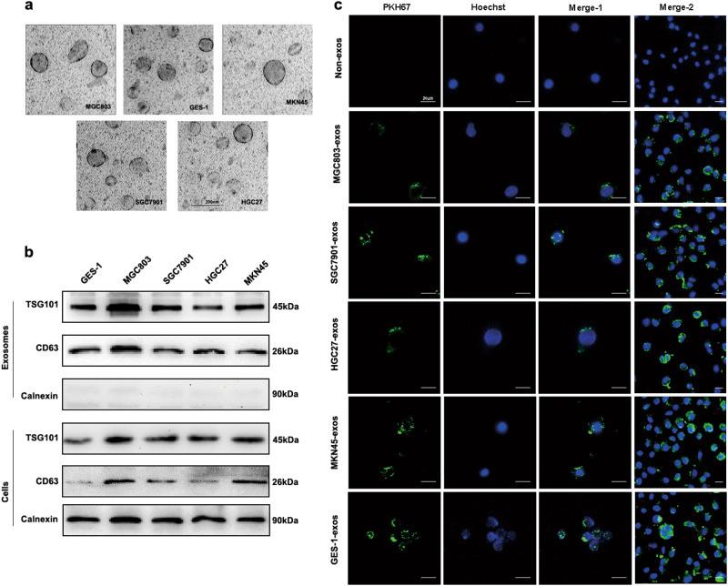
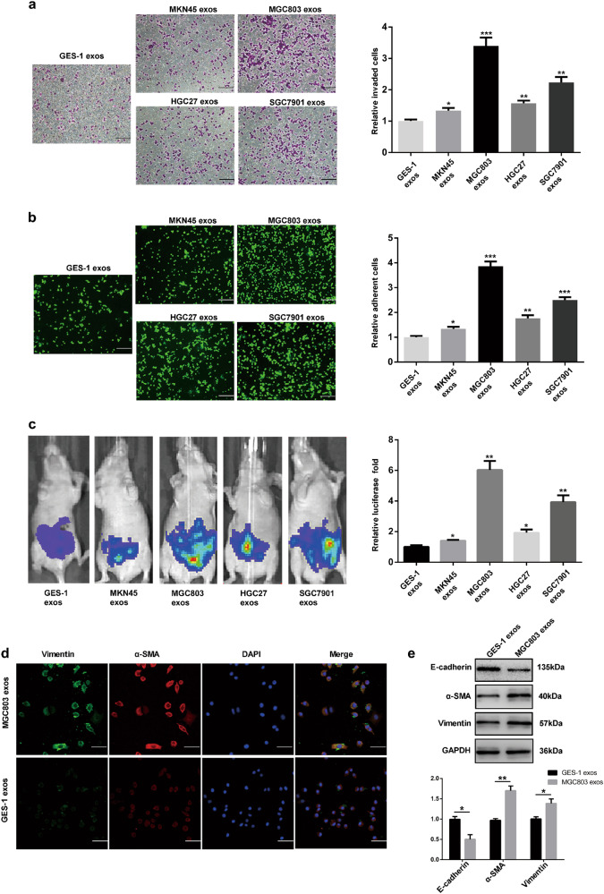
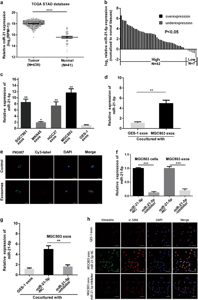
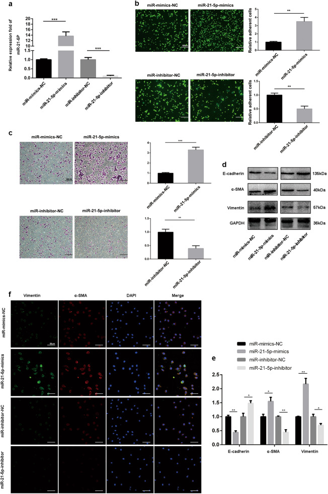
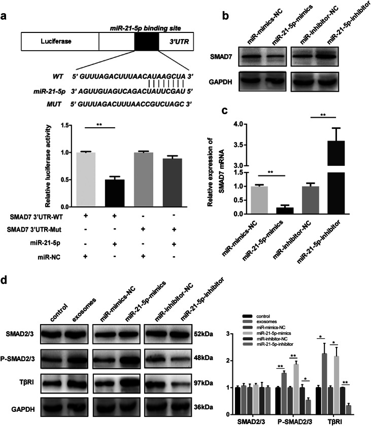
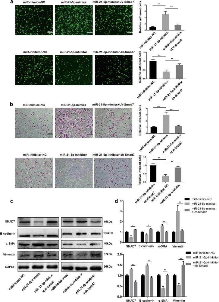
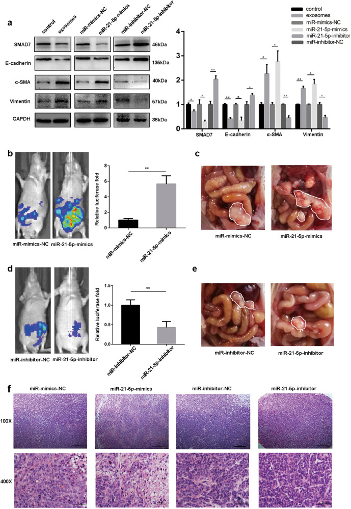
Peritoneal metastasis is a primary metastatic route for gastric cancers, and the mechanisms underlying this process are still unclear. Peritoneal mesothelial cells (PMCs) undergo mesothelial-to-mesenchymal transition (MMT) to provide a favorable environment for metastatic cancer cells. In this study, we investigated how the exosomal miR-21-5p induces MMT and promotes peritoneal metastasis. Gastric cancer (GC)-derived exosomes were identified by transmission electron microscopy and western blot analysis, then the uptake of exosomes was confirmed by PKH-67 staining. The expression of miR-21-5p and SMAD7 were measured by quantitative real-time polymerase chain reaction (qRT-PCR) and western blot, and the interactions between miR-21-5p and its target genes SMAD7 were confirmed by Luciferase reporter assays. The MMT of PMCs was determined by invasion assays, adhesion assays, immunofluorescent assay, and western blot. Meanwhile, mouse model of tumor peritoneal dissemination model was performed to investigate the role of exosomal miR-21-5p in peritoneal metastasis in vivo. We found that PMCs could internalize GC-derived exosomal miR-21-5p and led to increased levels of miR-21-5p in PMCs. Through various types of in vitro and in vivo assays, we confirmed that exosomal miR-21-5p was able to induce MMT of PMCs and promote tumor peritoneal metastasis. Moreover, our study revealed that this process was promoted by exosomal miR-21-5p through activating TGF-β/Smad pathway via targeting SMAD7. Altogether, our data suggest that exosomal miR-21-5p induces MMT of PMCs and promote cancer peritoneal dissemination by targeting SMAD7. The exosomal miR-21-5p may be a novel therapeutic target for GC peritoneal metastasis.
Overview
The claims and statistics of this paper haven’t been checked yet.
Run the check — free, no account needed. It reads the open-access text and checks the sources it cites.
Integrity signals
Registry
Cites
Statistics
Figures
Verification
Claims the paper makes
The claims and statistics of this paper haven’t been checked yet.
Run the check — free, no account needed. It reads the open-access text and checks the sources it cites.
Claims other papers make about this one
Each is checked against this paper’s own text: does it say what the citing paper says it does?
The cited source says what the claim attributes to it.
In the citing paper
“microRNA's presence in exosomes have been already reported in different cells and cancer types (44–50).”
From Potential Therapeutic Effects of Exosomes Packed With a miR-21-Sponge Construct in a Rat Model of Glioblastoma (2019) · cites this paper as [47]
Self-check
Percentages, sums and reported differences re-computed from the paper’s own numbers.
The self-check needs the paper’s full text.
Citations
Cites
1 retracted
Lv, Z., Wang, H., Dong, Q., Kong, B., Li, J., Yang, Z., Qu, H., Cao, W., & Xu, H. (2013) · Molecular and Cellular Biochemistry · doi:10.1007/s11010-013-1583-0 · Cited by 21
RetractedValadi, H., Ekström, K., Bossios, A., Sjöstrand, M., Lee, J. J., & Lötvall, J. O. (2007) · Nature Cell Biology · doi:10.1038/ncb1596 · Cited by 12,834
The distribution of secondary growths in cancer of the breast.
Paget, S. (1889) · The Lancet · doi:10.1016/s0140-6736(00)49915-0 · Cited by 5,277
Extracellular Vesicles in Cancer: Cell-to-Cell Mediators of Metastasis
Becker, A., Thakur, B. K., Weiss, J. M., Kim, H. S., Peinado, H., & Lyden, D. (2016) · Cancer Cell · doi:10.1016/j.ccell.2016.10.009 · Cited by 2,003
Asangani, I. A., Rasheed, S. A. K., Nikolova, D. A., Leupold, J. H., Colburn, N. H., Post, S., & Allgayer, H. (2008) · Oncogene · doi:10.1038/sj.onc.1210856 · Cited by 1,882
Identification of Smad7, a TGFβ-inducible antagonist of TGF-β signalling
Nakao, A., Afrakhte, M., Morn, A., Nakayama, T., Christian, J. L., Heuchel, R. L., Itoh, S., Kawabata, M., Heldin, N., Heldin, C., et al. (1997) · Nature · doi:10.1038/39369 · Cited by 1,796
Smad7 Binds to Smurf2 to Form an E3 Ubiquitin Ligase that Targets the TGFβ Receptor for Degradation
Kavsak, P., Rasmussen, R. K., Causing, C. G., Bonni, S., Zhu, H., Thomsen, G. H., & Wrana, J. L. (2000) · Molecular Cell · doi:10.1016/s1097-2765(00)00134-9 · Cited by 1,205
ExoCarta 2012: database of exosomal proteins, RNA and lipids
Mathivanan, S., Fahner, C. J., Reid, G. E., & Simpson, R. J. (2011) · Nucleic Acids Research · doi:10.1093/nar/gkr828 · Cited by 1,096
Exfoliation of membrane ecto-enzymes in the form of micro-vesicles
Trams, E. G., Lauter, C. J., Salem, N., & Heine, U. (1981) · Biochimica et Biophysica Acta (BBA) - Biomembranes · doi:10.1016/0005-2736(81)90512-5 · Cited by 1,094
Fang, T., Lv, H., Lv, G., Li, T., Wang, C., Han, Q., Yu, L., Su, B., Guo, L., Huang, S., et al. (2018) · Nature Communications · doi:10.1038/s41467-017-02583-0 · Cited by 995
Cited by
Sorted by citations
Items 201–222 of 222
Wang, C., Wang, J., Shen, X., Li, M., Yue, Y., Cheng, X., Lü, W., Wang, X., & Xie, X. (2021) · Figshare · doi:10.6084/m9.figshare.14228108
Zhang, Y., Pan, Y., Liu, Y., Li, X., Tang, L. V., Duan, M., Li, J., & Zhang, G. (2021) · Figshare · doi:10.6084/m9.figshare.15102856
Gjorgjieva, T., Chaloemtoem, A., Shahin, T., Bayaraa, O., Dieng, M. M., Alshaikh, M., Abdalbaqi, M., Monte, J. D., Begum, G., Leonor, C., et al. (2023) · Figshare · doi:10.6084/m9.figshare.23499429
Prospects of Exosomes in the Diagnosis and Treatment of Gastric Cancer
赵, 姣. (2024) · Advances in Clinical Medicine · doi:10.12677/acm.2024.1492493
Meta-analysis and Database Validation of Exosomal microRNAs and Prognosis in Gastric Cancer Patients
Liang, T., Ding, C., Yang, Z., & Da, M. (2025) · Current Medicinal Chemistry · doi:10.2174/0109298673356200250612183707
Li, K., Gong, Q., Xiang, X.-D., Guo, G., Liu, J., Zhao, L., Li, J., Chen, N., Li, H., Zhang, L.-J., et al. (2023) · Figshare · doi:10.6084/m9.figshare.23505084
Li, K., Gong, Q., Xiang, X.-D., Guo, G., Liu, J., Zhao, L., Li, J., Chen, N., Li, H., Zhang, L.-J., et al. (2023) · Figshare · doi:10.6084/m9.figshare.23505093
Zhang, Y., Pan, Y., Liu, Y., Li, X., Tang, L., Duan, M., Li, J., & Zhang, G. (2021) · Figshare · doi:10.6084/m9.figshare.15102853
Luan, W., Ding, Y., Xi, H., Ruan, H., Lu, F., Ma, S., & Wang, J. (2021) · Figshare · doi:10.6084/m9.figshare.14252235
Li, K., Gong, Q., Xiang, X.-D., Guo, G., Liu, J., Zhao, L., Li, J., Chen, N., Li, H., Zhang, L.-J., et al. (2023) · Figshare · doi:10.6084/m9.figshare.23505090
Gjorgjieva, T., Chaloemtoem, A., Shahin, T., Bayaraa, O., Dieng, M. M., Alshaikh, M., Abdalbaqi, M., Monte, J. D., Begum, G., Leonor, C., et al. (2023) · Figshare · doi:10.6084/m9.figshare.23499426
Li, K., Gong, Q., Xiang, X.-D., Guo, G., Liu, J., Zhao, L., Li, J., Chen, N., Li, H., Zhang, L.-J., et al. (2023) · Figshare · doi:10.6084/m9.figshare.23505087
Li, K., Gong, Q., Xiang, X.-D., Guo, G., Liu, J., Zhao, L., Li, J., Chen, N., Li, H., Zhang, L.-J., et al. (2023) · Figshare · doi:10.6084/m9.figshare.23505081
Li, P., Xue, Y., & Gu, X. (2025) · Cancer Cell International · doi:10.1186/s12935-025-04136-8
Huang, C., Jiang, Z., Shen, X., Ding, Q., Wang, H., Cheng, Z., Liu, G., Zhao, X., & You, X. (2026) · iScience · doi:10.1016/j.isci.2026.115908
Li, Y., Lv, M., Xiong, W., Wei, J., Li, J., Guo, N., Zhang, S., Sui, C., & Jin, L. (2026) · Reproduction · doi:10.1093/reprod/xaag065
Zeng, X., Wu, Y., Zhong, W., & Yang, L. (2026) · Critical Reviews in Oncology/Hematology · doi:10.1016/j.critrevonc.2026.105438
Self-citationAdvances in molecular mechanism of gastric cancer peritoneal metastasis
Yan, H.-Q., Lai, Q., Huang, M., Wang, T., Wu, L., & Qin, Y. (2026) · World Journal of Gastrointestinal Oncology · doi:10.4251/wjgo.v18.i8.121799
Wang, Y., Gao, L., Zhou, Y., Jiang, L., Xue, Z., Zhu, W., Ge, X., Xu, W., Jiang, J., & Zang, X. (2026) · Frontiers in Cell and Developmental Biology · doi:10.3389/fcell.2026.1883199
AlRamadneh, T. N., Abdulsahib, W. K., Abdal-Wahab, S. T., Malathi, H., Mangaraj, S., Samikan, K., Mukherjee, G., Sinha, A., & Aminov, Z. (2026) · Cancer Treatment and Research Communications · doi:10.1016/j.ctarc.2026.101402
Yao, S., Chai, H., Tang, L., Li, X., Liu, F., Jin, H., & Tuo, B. (2026) · Frontiers in Immunology · doi:10.3389/fimmu.2026.1915650
Niches in cancer: from primary tumors to metastatic disease
Chao, Z., Wang, X., Wang, Z., Wu, K., & Li, L. (2026) · Journal of Hematology & Oncology · doi:10.1186/s13045-026-01858-8
Metadata
- Authors
- Li, Q., Li, B., Li, Q., Wei, S., He, Z., Huang, X., Wang, L., Xia, Y., Xu, Z., Li, Z., Wang, W., Yang, L., Zhang, D., & Xu, Z.
- Affiliations
- Jiangsu Province Hospital (CN)
- Corresponding author
- Zhang, Diancai
- ORCID
- Venue
- Cell Death & Disease
- Published
- 2018-08-28
- Volume
- 9
- Issue
- 9
- Pages
- 854-854
- ISSN
- 2041-4889 (ISSN-L)
- PMC
- PMC6113299
- OpenAlex
- W2889190369
- Cited by
- 219
- References
- 47
- Citations by year
- 2026: 11 · 2025: 21 · 2024: 31 · 2023: 34 · 2022: 45 · 2021: 46 · 2020: 17 · 2019: 14
- FWCI
- 7.03
- Type
- journal article
- Language
- en
- Open access
- gold
- doi.org
- Locations
- Cell Death and Disease (publishedVersion, cc-by)
- Europe PMC (PubMed Central) (publishedVersion, other-oa)
- PubMed Central (publishedVersion, cc-by)
- PubMed (publishedVersion)
- DOAJ (DOAJ: Directory of Open Access Journals) (submittedVersion)
- Funders
- National Natural Science Foundation of China (JX10231801, 30901443)
- Priority Academic Program Development of Jiangsu Higher Education Institutions (PAPD, JX10231801, JX 10231801)
- National Institutes of Health
- Topics
- Life Sciences › Biochemistry, Genetics and Molecular Biology › Molecular Biology › Extracellular vesicles in disease
- Life Sciences › Biochemistry, Genetics and Molecular Biology › Cancer Research › MicroRNA in disease regulation
- Health Sciences › Medicine › Oncology › Cancer Cells and Metastasis
- Keywords
- gastric cancer, peritoneal carcinomatosis, exosomes, miR-21-5p, mesothelial-to-mesenchymal transition, peritoneal mesothelial cells, Smad7, TGF-β/Smad signaling, extracellular vesicles, tumor microenvironment
- MeSH terms
- Animals, Cell Movement, Epithelium, Female, Humans, Male, Mice, Nude, Middle Aged, Neoplasm Metastasis, Peritoneal Neoplasms, Peritoneum, Stomach Neoplasms, Signal Transduction, Gene Expression Regulation, Neoplastic, Transforming Growth Factor beta, MicroRNAs, Cell Line, Tumor, Cell Proliferation, Mice, Smad7 Protein, Exosomes, Epithelial-Mesenchymal Transition
About these checks
Registry, retraction, Cites and statistics results are computed, not judged, with one exception: a printed reference that no index lists is read once by a model before it counts as a citation error. Claims are extracted by a model and kept only when their quote appears verbatim in the text; each cited source is then checked, with a model’s help, for whether it says what the claim relies on. Those verdicts can be wrong — read the claim’s quote and the cited source before relying on one.
Statistics and arithmetic are recomputed, not judged.
Confidence bands are shown only for fields calibrated on our annotated test set. None is calibrated yet, so no band is shown.
What counts as a claim ›How we measure confidence ›How we check this ›
Are you an author, or do you see a problem with this page? Request a review
