

Oxidative stress-mediated mitochondrial dysfunction facilitates mesenchymal stem cell senescence in ankylosing spondylitis
Open accessThe DOI matches its Crossref record; 66 of the 66 references it cites were found in a registry or an index.
Abstract
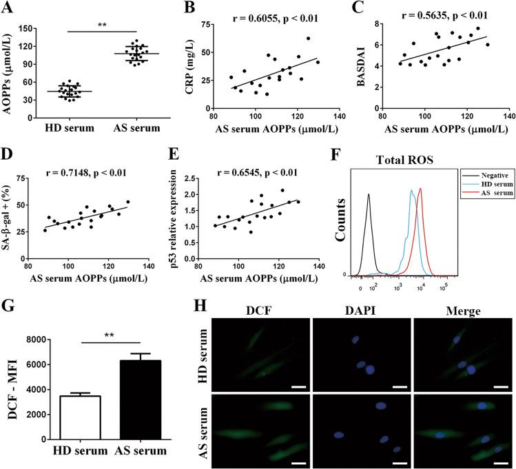
Ankylosing spondylitis (AS) is a chronic inflammatory disease possessing a morbid serum microenvironment with enhanced oxidative stress. Long-term exposure to an oxidative environment usually results in cellular senescence alone with cellular dysfunction. Mesenchymal stem cells (MSCs) are a kind of stem cell possessing strong capabilities for immunoregulation, and senescent MSCs may increase inflammation and participate in AS pathogenesis. The objective of this study was to explore whether and how the oxidative serum environment of AS induces MSC senescence. Here, we found that AS serum facilitated senescence of MSCs in vitro, and articular tissues from AS patients exhibited higher expression levels of the cell cycle arrest-related proteins p53, p21 and p16. Importantly, the levels of advanced oxidative protein products (AOPPs), markers of oxidative stress, were increased in AS serum and positively correlated with the extent of MSC senescence induced by AS serum. Furthermore, MSCs cultured with AS serum showed decreased mitochondrial membrane potential and ATP production together with a reduced oxygen consumption rate. Finally, we discovered that AS serum-induced mitochondrial dysfunction resulted in elevated reactive oxygen species (ROS) in MSCs, and ROS inhibition successfully rescued MSCs from senescence. In conclusion, our data demonstrated that the oxidative serum environment of AS facilitated MSC senescence through inducing mitochondrial dysfunction and excessive ROS production. These results may help elucidate the pathogenesis of AS and provide potential targets for AS treatment.
Overview
The claims and statistics of this paper haven’t been checked yet.
Free: it reads the open-access text and checks the first 5 claims that cite a source we can check. Create a free account to check every claim.
Integrity signals
Registry
Cites
Statistics
Figures




Verification
Claims the paper makes
The claims and statistics of this paper haven’t been checked yet.
Free: it reads the open-access text and checks the first 5 claims that cite a source we can check. Create a free account to check every claim.
Claims other papers make about this one
Each is checked against this paper’s own text: does it say what the citing paper says it does?
The source backs part of the claim, not all of it — or backs it for a different population, condition or comparison.
In the citing paper
“The production of ROS and oxidized proteins is increased in mesenchymal stem cells incubated with sera from patients with ankylosing spondylarthritis compared with cells incubated with healthy sera. This finding suggests that circulating factors mediate mitochondrial oxidative stress in the pathophysiology of the disease64”
From The role of mitochondria in rheumatic diseases (2022) · cites this paper as [64]
Experimental — accuracy not yet measured
Self-check
Percentages, sums and reported differences re-computed from the paper’s own numbers.
The self-check needs the paper’s full text.
Citations
Cites
1 retracted
Wu, Y., Mingliang, R., Yang, R., Liang, X., Ma, Y., Tang, Y., Huang, L. C., Ye, J., Chen, K., Wang, P., et al. (2011) · Arthritis Research & Therapy · doi:10.1186/ar3257 · Cited by 88
Self-citationRetractedEvaluation of Diagnostic Criteria for Ankylosing Spondylitis
Linden, S. V. D., Valkenburg, H. A., & Cats, A. (1984) · Arthritis & Rheumatism · doi:10.1002/art.1780270401 · Cited by 5,697
Oxidative stress, inflammation, and cancer: How are they linked?
Reuter, S., Gupta, S. C., Chaturvedi, M. M., & Aggarwal, B. B. (2010) · Free Radical Biology and Medicine · doi:10.1016/j.freeradbiomed.2010.09.006 · Cited by 5,441
Reactive Oxygen Species in Inflammation and Tissue Injury
Mittal, M., Siddiqui, M. R., Tran, K., Reddy, S. P., & Malik, A. B. (2014) · Antioxidants & Redox Signaling · doi:10.1089/ars.2012.5149 · Cited by 4,897
Mitochondrial free radical generation, oxidative stress, and aging
Cadenas, E., & Davies, K. J. A. (2000) · Free Radical Biology and Medicine · doi:10.1016/s0891-5849(00)00317-8 · Cited by 3,010
A DNA damage checkpoint response in telomere-initiated senescence
Fagagna, F. D. D., Reaper, P. M., Clay-Farrace, L., Fiegler, H., Carr, P., von Zglinicki, T., Saretzki, G., Carter, N. P., & Jackson, S. P. (2003) · Nature · doi:10.1038/nature02118 · Cited by 2,917
Cellular senescence: from physiology to pathology
Muñoz-Espín, D., & Serrano, M. (2014) · Nature Reviews Molecular Cell Biology · doi:10.1038/nrm3823 · Cited by 2,818
The essence of senescence: Figure 1.
Kuilman, T., Michaloglou, C., Mooi, W. J., & Peeper, D. S. (2010) · Genes & Development · doi:10.1101/gad.1971610 · Cited by 2,038
Braun, J., & Sieper, J. (2007) · The Lancet · doi:10.1016/s0140-6736(07)60635-7 · Cited by 1,806
Senescence in Health and Disease
He, S., & Sharpless, N. E. (2017) · Cell · doi:10.1016/j.cell.2017.05.015 · Cited by 1,766
Cited by
Sirt3-mediated mitophagy regulates AGEs-induced BMSCs senescence and senile osteoporosis
Guo, Y., Jia, X., Cui, Y., Song, Y., Wang, S., Geng, Y., Li, R., Gao, W., & Fu, D. (2021) · Redox Biology · doi:10.1016/j.redox.2021.101915 · Cited by 296
Self-citationThe role of mitochondria in rheumatic diseases
Becker, Y. L. C., Duvvuri, B., Fortin, P. R., Lood, C., & Boilard, E. (2022) · Nature Reviews Rheumatology · doi:10.1038/s41584-022-00834-z · Cited by 81
Metadata
- Authors
- Ye, G., Xie, Z., Zeng, H., Wang, P., Li, J., Zheng, G., Wang, S., Cao, Q., Li, M., Liu, W., Cen, S., Li, Z., Wu, Y., Ye, Z., & Shen, H.
- Affiliations
- Sun Yat-sen University (CN)
- Corresponding author
- Wu, Yanfeng
- Ye, Zhizhong
- Shen, Huiyong
- ORCID
- Venue
- Cell Death & Disease
- Published
- 2020-09-17
- Volume
- 11
- Issue
- 9
- Pages
- 775-775
- ISSN
- 2041-4889 (ISSN-L)
- PMC
- PMC7498590
- OpenAlex
- W3087134616
- Cited by
- 115
- References
- 66
- Citations by year
- 2026: 10 · 2025: 21 · 2024: 32 · 2023: 23 · 2022: 17 · 2021: 12
- FWCI
- 7.81
- Type
- journal article
- Language
- en
- Open access
- gold
- doi.org
- Licence
- cc-by
- creativecommons.org/licenses/by/4.0 (tdm)
- creativecommons.org/licenses/by/4.0 (vor)
- Locations
- Cell Death and Disease (publishedVersion, cc-by)
- PubMed (publishedVersion)
- Funders
- National Natural Science Foundation of China (81971518, 81700884)
- Fundamental Research Funds for the Central Universities (19ykpy01)
- Topics
- Life Sciences › Biochemistry, Genetics and Molecular Biology › Cell Biology › Telomeres, Telomerase, and Senescence
- Life Sciences › Biochemistry, Genetics and Molecular Biology › Cell Biology › Mesenchymal stem cell research
- Health Sciences › Medicine › Rheumatology › Spondyloarthritis Studies and Treatments
- Keywords
- ankylosing spondylitis, mesenchymal stem cells, cellular senescence, oxidative stress, mitochondrial dysfunction, reactive oxygen species, advanced oxidation protein products, p53, p21, p16, mitochondrial membrane potential, ATP production, articular tissues
- MeSH terms
- Adenosine Triphosphate, Adult, Cell Cycle, Female, Humans, Inflammation, Male, Mitochondria, Organophosphorus Compounds, Oxygen, Oxygen Consumption, Spondylitis, Ankylosing, Stem Cells, Ubiquinone, Signal Transduction, Cytokines, Cellular Senescence, Reactive Oxygen Species, Oxidative Stress, Cell Proliferation, Membrane Potential, Mitochondrial, Young Adult, Cell Cycle Checkpoints, Mesenchymal Stem Cells
About these checks
Registry, retraction, Cites and statistics results are computed, not judged, with one exception: a printed reference that no index lists is read once by a model before it counts as a citation error. Claims are extracted by a model and kept only when their quote appears verbatim in the text; each cited source is then checked, with a model’s help, for whether it says what the claim relies on. Those verdicts can be wrong — read the claim’s quote and the cited source before relying on one.
Statistics and arithmetic are recomputed, not judged.
Confidence bands are shown only for fields calibrated on our annotated test set. None is calibrated yet, so no band is shown.
What counts as a claim ›How we measure confidence ›How we check this ›
Are you an author, or do you see a problem with this page? Request a review
How to cite
Cite CoChat for the checks on this page, and the paper for its own findings.
- This page
- CoChat. (2026). Citation check: Oxidative stress-mediated mitochondrial dysfunction facilitates mesenchymal stem cell senescence in ankylosing spondylitis. https://app.cochat.ai/papers/10.1038/s41419-020-02993-x
- The paper
- Ye, G., Xie, Z., Zeng, H., Wang, P., Li, J., Zheng, G., Wang, S., Cao, Q., Li, M., Liu, W., Cen, S., Li, Z., Wu, Y., Ye, Z., & Shen, H. (2020). Oxidative stress-mediated mitochondrial dysfunction facilitates mesenchymal stem cell senescence in ankylosing spondylitis. Cell Death & Disease. https://doi.org/10.1038/s41419-020-02993-x
Data sources
Bibliographic data: Semantic Scholar, OpenAlex, Crossref.
Contains information from the Semantic Scholar Academic Graph Datasets, made available under the ODC Attribution License (ODC-BY 1.0).
Semantic Scholar’s data is described in: Kinney, R., et al. (2023). The Semantic Scholar Open Data Platform. arXiv:2301.10140.
