

The DOI matches its Crossref record; 35 of the 35 references it cites were found in a registry or an index.
ROS induces NETosis by oxidizing DNA and initiating DNA repair
Open accessAbstract
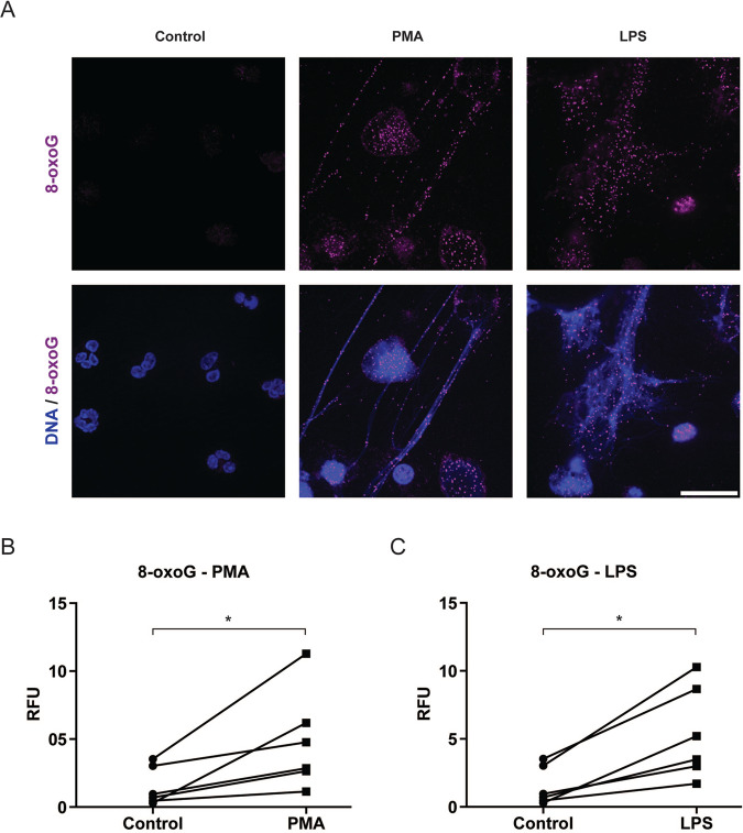
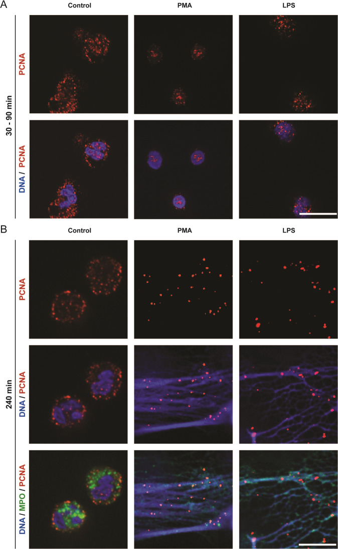
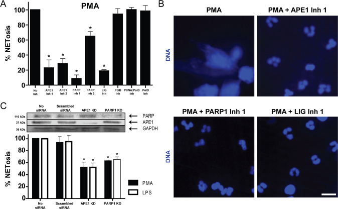
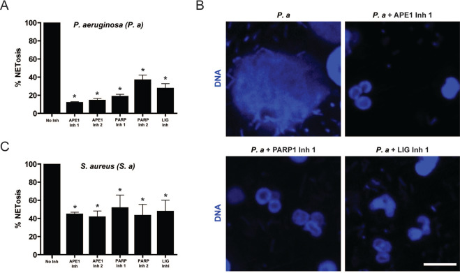
Reactive oxygen species (ROS) are essential for neutrophil extracellular trap (NET) formation or NETosis. Nevertheless, how ROS induces NETosis is unknown. Neutrophil activation induces excess ROS production and a meaningless genome-wide transcription to facilitate chromatin decondensation. Here we show that the induction of NADPH oxidase-dependent NETosis leads to extensive DNA damage, and the subsequent translocation of proliferating cell nuclear antigen (PCNA), a key DNA repair protein, stored in the cytoplasm into the nucleus. During the activation of NETosis (e.g., by phorbol myristate acetate, Escherichia coli LPS, Staphylococcus aureus (RN4220), or Pseudomonas aeruginosa ), preventing the DNA-repair-complex assembly leading to nick formation that decondenses chromatin causes the suppression of NETosis (e.g., by inhibitors to, or knockdown of, Apurinic endonuclease APE1, poly ADP ribose polymerase PARP, and DNA ligase). The remaining repair steps involving polymerase activity and PCNA interactions with DNA polymerases β/δ do not suppress agonist-induced NETosis. Therefore, excess ROS produced during neutrophil activation induces NETosis by inducing extensive DNA damage (e.g., oxidising guanine to 8-oxoguanine), and the subsequent DNA repair pathway, leading to chromatin decondensation.
Overview
The claims and statistics of this paper haven’t been checked yet.
Run the check — free, no account needed. It reads the open-access text and checks the sources it cites.
Integrity signals
Registry
Cites
Statistics
Figures
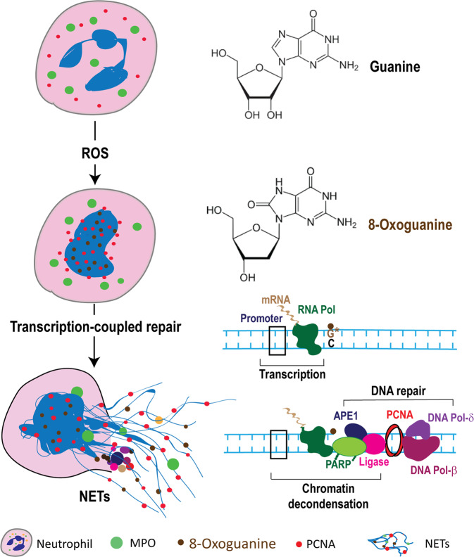
Verification
Claims the paper makes
The claims and statistics of this paper haven’t been checked yet.
Run the check — free, no account needed. It reads the open-access text and checks the sources it cites.
Claims other papers make about this one
Each is checked against this paper’s own text: does it say what the citing paper says it does?
In the citing paper
“Its induction depends on ROS, the main source being NOx [61].”
From The Role of Reactive Species on Innate Immunity (2022) · cites this paper as [61]
Self-check
Percentages, sums and reported differences re-computed from the paper’s own numbers.
The self-check needs the paper’s full text.
Citations
Cites
Neutrophil Extracellular Traps Kill Bacteria
Brinkmann, V., Reichard, U., Goosmann, C., Fauler, B., Uhlemann, Y., Weiss, D. S., Weinrauch, Y., & Zychlinsky, A. (2004) · Science · doi:10.1126/science.1092385 · Cited by 10,268
Novel cell death program leads to neutrophil extracellular traps
Fuchs, T. A., Abed, U., Goosmann, C., Hurwitz, R., Schulze, I., Wahn, V., Weinrauch, Y., Brinkmann, V., & Zychlinsky, A. (2007) · The Journal of Cell Biology · doi:10.1083/jcb.200606027 · Cited by 3,197
Krokan, H. E., & Bjoras, M. (2013) · Cold Spring Harbor Perspectives in Biology · doi:10.1101/cshperspect.a012583 · Cited by 1,322
Neutrophil Extracellular Traps: Double-Edged Swords of Innate Immunity
Kaplan, M. J., & Radic, M. Z. (2012) · The Journal of Immunology · doi:10.4049/jimmunol.1201719 · Cited by 1,214
Structural basis for recognition and repair of the endogenous mutagen 8-oxoguanine in DNA
Bruner, S. D., Norman, D. P. G., & Verdine, G. L. (2000) · Nature · doi:10.1038/35002510 · Cited by 972
Neutrophil extracellular trap cell death requires both autophagy and superoxide generation
Remijsen, Q., Berghe, T. V., Wirawan, E., Asselbergh, B., Parthoens, E., De Rycke, R., Noppen, S., Delforge, M., Willems, J., & Vandenabeele, P. (2011) · Cell Research · doi:10.1038/cr.2010.150 · Cited by 818
Proliferating cell nuclear antigen (PCNA): a key factor in DNA replication and cell cycle regulation
Strzałka, W., & Ziemienowicz, A. (2010) · Annals of Botany · doi:10.1093/aob/mcq243 · Cited by 804
Douda, D. N., Khan, M. A., Grasemann, H., & Palaniyar, N. (2015) · Proceedings of the National Academy of Sciences · doi:10.1073/pnas.1414055112 · Cited by 712
Self-citationGupta, A. K., Joshi, M. B., Philippova, M., Erné, P., Hasler, P., Hahn, S., & Resink, T. J. (2010) · FEBS Letters · doi:10.1016/j.febslet.2010.06.006 · Cited by 547
Gehrke, N., Mertens, C., Zillinger, T., Wenzel, J., Bald, T., Zahn, S., Tüting, T., Hartmann, G., & Barchet, W. (2013) · Immunity · doi:10.1016/j.immuni.2013.08.004 · Cited by 485
Takei, H., Araki, A., Watanabe, H. K., Ichinose, A., & Sendo, F. (1996) · Journal of Leukocyte Biology · doi:10.1002/jlb.59.2.229 · Cited by 451
Byrd, A. S., O’Brien, X. M., Johnson, C. M., Lavigne, L. M., & Reichner, J. S. (2013) · The Journal of Immunology · doi:10.4049/jimmunol.1202671 · Cited by 321
Base Excision Repair and Lesion-Dependent Subpathways for Repair of Oxidative DNA Damage
Svilar, D., Goellner, E. M., Almeida, K. H., & Sobol, R. W. (2010) · Antioxidants and Redox Signaling · doi:10.1089/ars.2010.3466 · Cited by 277
Cell-Cycle Proteins Control Production of Neutrophil Extracellular Traps
Amulic, B., Knackstedt, S. L., Abed, U. A., Deigendesch, N., Harbort, C. J., Caffrey, B. E., Brinkmann, V., Heppner, F. L., Hinds, P. W., & Zychlinsky, A. (2017) · Developmental Cell · doi:10.1016/j.devcel.2017.10.013 · Cited by 275
Mammalian base excision repair by DNA polymerases δ and ε
Stucki, M., Pascucci, B., Parlanti, E., Fortini, P., Wilson, S. H., Hübscher, U., & Eugenia Dogliotti, Y. (1998) · Oncogene · doi:10.1038/sj.onc.1202001 · Cited by 192
Khan, M. A., Farahvash, A., Douda, D. N., Licht, J.-C., Grasemann, H., Sweezey, N., & Palaniyar, N. (2017) · Scientific Reports · doi:10.1038/s41598-017-03257-z · Cited by 178
Self-citationTranscriptional firing helps to drive NETosis
Khan, M. A., & Palaniyar, N. (2017) · Scientific Reports · doi:10.1038/srep41749 · Cited by 175
Self-citationWitko‐Sarsat, V., Mocek, J., Bouayad, D., Tamassia, N., Ribeil, J., Candalh, C., Davezac, N., Reuter, N., Mouthon, L., Hermine, O., et al. (2010) · The Journal of Experimental Medicine · doi:10.1084/jem.20092241 · Cited by 173
Thakur, S., Sarkar, B., Cholia, R. P., Gautam, N., Dhiman, M., & Mantha, A. K. (2014) · Experimental & Molecular Medicine · doi:10.1038/emm.2014.42 · Cited by 167
Akt is essential to induce NADPH-dependent NETosis and to switch the neutrophil death to apoptosis
Douda, D. N., Yip, L., Khan, M. A., Grasemann, H., & Palaniyar, N. (2014) · Blood · doi:10.1182/blood-2013-09-526707 · Cited by 150
Self-citationInfluences of Chloride and Hypochlorite on Neutrophil Extracellular Trap Formation
Akong, K., Chow, O. A., von Köckritz‐Blickwede, M., & Nizet, V. (2012) · PLoS ONE · doi:10.1371/journal.pone.0042984 · Cited by 135
The Role of 8-Oxoguanine DNA Glycosylase-1 in Inflammation
Ba, X., Aguilera-Aguirre, L., Rashid, Q. T. A., Bácsi, A., Radak, Z. O., Sur, S., Hosoki, K., Hegde, M. L., & Boldogh, I. R. (2014) · International Journal of Molecular Sciences · doi:10.3390/ijms150916975 · Cited by 128
Evasion of Neutrophil Extracellular Traps by Respiratory Pathogens
Storisteanu, D. M. L., Pocock, J., Cowburn, A. S., Juss, J. K., Nadesalingam, A., Nizet, V., & Chilvers, E. R. (2017) · American Journal of Respiratory Cell and Molecular Biology · doi:10.1165/rcmb.2016-0193ps · Cited by 112
Hegde, M. L., Theriot, C. A., Das, A., Hegde, P. M., Guo, Z., Gary, R. K., Hazra, T. K., Shen, B., & MITRA, S. P. (2008) · Journal of Biological Chemistry · doi:10.1074/jbc.m802712200 · Cited by 109
Nucleosome Disruption by DNA Ligase III-XRCC1 Promotes Efficient Base Excision Repair
Odell, I. D., Barbour, J.-E. R., Murphy, D. L., Della-Maria, J., Sweasy, J. B., Tomkinson, A. E., Wallace, S. S., & Pederson, D. S. (2011) · Molecular and Cellular Biology · doi:10.1128/mcb.05715-11 · Cited by 92
Role of Nox2 in elimination of microorganisms
Rada, B., Hably, C., Meczner, A., Tímár, C. I., Lakatos, G., Enyedi, P., & Ligeti, E. (2008) · Seminars in Immunopathology · doi:10.1007/s00281-008-0126-3 · Cited by 90
The BER necessities: the repair of DNA damage in human-adapted bacterial pathogens
van der Veen, S., & Tang, C. M. (2015) · Nature Reviews Microbiology · doi:10.1038/nrmicro3391 · Cited by 86
Hanssen‐Bauer, A., Solvang‐Garten, K., Sundheim, O., Peña-Dı́az, J., Andersen, S. B., Slupphaug, G., Krokan, H. E., Wilson, D. M., Akbari, M., & Otterlei, M. (2011) · Environmental and Molecular Mutagenesis · doi:10.1002/em.20663 · Cited by 69
DNA polymerase β-dependent long patch base excision repair in living cells
Asagoshi, K., Liu, Y., Masaoka, A., Lan, L., Prasad, R., Horton, J. K., Brown, A. R., Wang, X.-H., Bdour, H. M., Sobol, R. W., et al. (2010) · DNA Repair · doi:10.1016/j.dnarep.2009.11.002 · Cited by 51
Modulation of DNA base excision repair during neuronal differentiation
Sýkora, P., Yang, J., Ferrarelli, L. K., Tian, J., Tadokoro, T., Kulkarni, A., Weissman, L., Keijzers, G., Wilson, D. M., Mattson, M. P., et al. (2013) · Neurobiology of Aging · doi:10.1016/j.neurobiolaging.2012.12.016 · Cited by 51
Small-Molecule Inhibitors of APE1 DNA Repair Function: An Overview
Al-Safi, R. I., Odde, S., Shabaik, Y., & Neamati, N. (2011) · Current Molecular Pharmacology · doi:10.2174/1874467211205010014 · Cited by 48
Efficiency and fidelity of human DNA polymerases λ and β during gap-filling DNA synthesis
Brown, J. A., Pack, L. R., Sanman, L. E., & Suo, Z. (2010) · DNA repair · doi:10.1016/j.dnarep.2010.09.005 · Cited by 46
Polymerase Stalling during Replication, Transcription and Translation
Edenberg, E. R., Downey, M., & Toczyski, D. P. (2014) · Current Biology · doi:10.1016/j.cub.2014.03.060 · Cited by 42
Salati, S., Bianchi, E., Zini, R., Tenedini, E., Quaglino, D., Manfredini, R., & Ferrari, S. (2007) · Haematologica · doi:10.3324/haematol.11472 · Cited by 22
Larmonier, C. B., Shehab, K. W., McFadden, R.-M. T., Ghishan, F. K., & Kiela, P. R. (2014) · The FASEB Journal · doi:10.1096/fasebj.28.1_supplement.902.5 · Cited by 5
Cited by
Sorted by citations
Mitochondria in oxidative stress, inflammation and aging: from mechanisms to therapeutic advances
Xu, X., Pang, Y., & Fan, X. (2025) · Signal Transduction and Targeted Therapy · doi:10.1038/s41392-025-02253-4 · Cited by 1,016
Neutrophil extracellular traps: from physiology to pathology
Hidalgo, A., Libby, P., Soehnlein, O., Aramburu, I. V., Papayannopoulos, V., & Silvestre-Roig, C. (2021) · Cardiovascular Research · doi:10.1093/cvr/cvab329 · Cited by 426
Neutrophil intrinsic and extrinsic regulation of NETosis in health and disease
Poli, V., & Zanoni, I. (2022) · Trends in Microbiology · doi:10.1016/j.tim.2022.10.002 · Cited by 223
Recent Advances in Diabetic Kidney Diseases: From Kidney Injury to Kidney Fibrosis
Hung, P., Hsu, Y., Chen, T., & Lin, C. (2021) · International Journal of Molecular Sciences · doi:10.3390/ijms222111857 · Cited by 154
The Role of Reactive Species on Innate Immunity
Andrés, C., Pérez de la Lastra, J., Juan, C., Plou, F., & Pérez-Lebeña, E. (2022) · Vaccines · doi:10.3390/vaccines10101735 · Cited by 142
Metadata
- Authors
- Azzouz, D., Khan, M. A., & Palaniyar, N.
- Affiliations
- University of Toronto (CA)
- Hospital for Sick Children (CA)
- Corresponding author
- Palaniyar, Nades
- ORCID
- Palaniyar, Nades
- Venue
- Cell Death Discovery
- Published
- 2021-05-18
- Volume
- 7
- Issue
- 1
- Pages
- 113-113
- ISSN
- 2058-7716 (ISSN-L)
- PMC
- PMC8128883
- OpenAlex
- W3163218484
- Cited by
- 202
- References
- 35
- Citations by year
- 2026: 37 · 2025: 61 · 2024: 50 · 2023: 22 · 2022: 28 · 2021: 4
- FWCI
- 9.89
- Type
- journal article
- Language
- en
- Open access
- gold
- doi.org
- Locations
- Cell Death Discovery (publishedVersion, cc-by)
- PubMed Central (publishedVersion, cc-by)
- PubMed (publishedVersion)
- DOAJ (DOAJ: Directory of Open Access Journals) (submittedVersion, cc-by-sa)
- Funders
- Cystic Fibrosis Canada (3180)
- Gouvernement du Canada | Natural Sciences and Engineering Research Council of Canada (RGPIN436250-13)
- Gouvernement du Canada | Canadian Institutes of Health Research (MOP-111012)
- Hospital for Sick Children
- Topics
- Life Sciences › Immunology and Microbiology › Immunology › Neutrophil, Myeloperoxidase and Oxidative Mechanisms
- Life Sciences › Biochemistry, Genetics and Molecular Biology › Molecular Biology › Redox biology and oxidative stress
- Health Sciences › Medicine › Hematology › Blood disorders and treatments
- Keywords
- neutrophil extracellular traps, NETosis, reactive oxygen species, NADPH oxidase, DNA damage, DNA repair, chromatin decondensation, 8-oxoguanine, APE1, poly(ADP-ribose) polymerase, DNA ligase, neutrophil activation, DNA polymerase beta, DNA polymerase delta
About these checks
Registry, retraction, Cites and statistics results are computed, not judged, with one exception: a printed reference that no index lists is read once by a model before it counts as a citation error. Claims are extracted by a model and kept only when their quote appears verbatim in the text; each cited source is then checked, with a model’s help, for whether it says what the claim relies on. Those verdicts can be wrong — read the claim’s quote and the cited source before relying on one.
Statistics and arithmetic are recomputed, not judged.
Confidence bands are shown only for fields calibrated on our annotated test set. None is calibrated yet, so no band is shown.
What counts as a claim ›How we measure confidence ›How we check this ›
Are you an author, or do you see a problem with this page? Request a review
