

The DOI matches its Crossref record; 66 of the 66 references it cites were found in a registry or an index.
Biogenic manganese oxide nanoparticle formation by a multimeric multicopper oxidase Mnx
Open accessAbstract
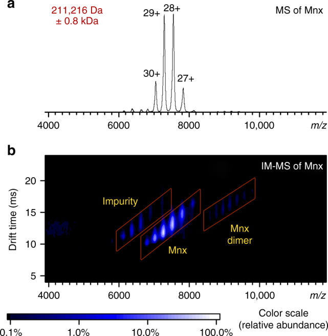
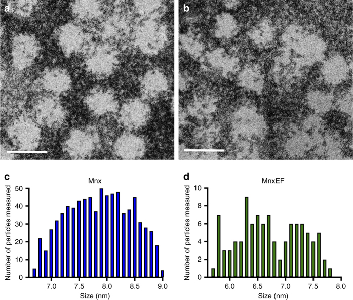
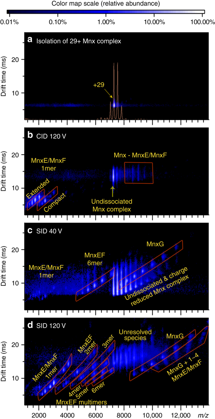
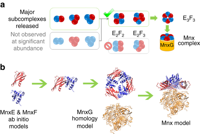
Bacteria that produce Mn oxides are extraordinarily skilled engineers of nanomaterials that contribute significantly to global biogeochemical cycles. Their enzyme-based reaction mechanisms may be genetically tailored for environmental remediation applications or bioenergy production. However, significant challenges exist for structural characterization of the enzymes responsible for biomineralization. The active Mn oxidase in Bacillus sp. PL-12, Mnx, is a complex composed of a multicopper oxidase (MCO), MnxG, and two accessory proteins, MnxE and MnxF. MnxG shares sequence similarity with other, structurally characterized MCOs. MnxE and MnxF have no similarity to any characterized proteins. The ~200 kDa complex has been recalcitrant to crystallization, so its structure is unknown. Here, we show that native mass spectrometry defines the subunit topology and copper binding of Mnx, while high-resolution electron microscopy visualizes the protein and nascent Mn oxide minerals. These data provide critical structural information for understanding Mn biomineralization by such unexplored enzymes.
Overview
The claims and statistics of this paper haven’t been checked yet.
Run the check — free, no account needed. It reads the open-access text and checks the sources it cites.
Integrity signals
Registry
Cites
Statistics
Figures
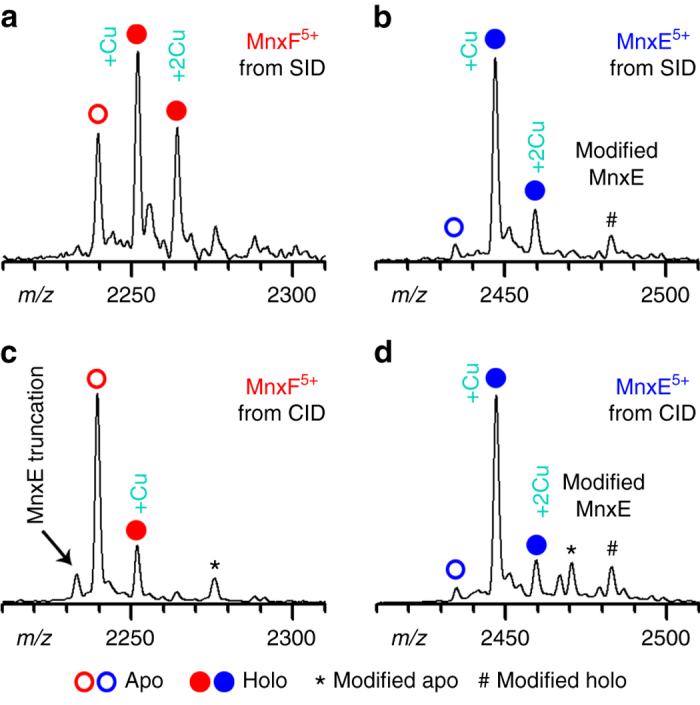
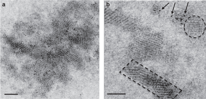
Verification
Claims the paper makes
The claims and statistics of this paper haven’t been checked yet.
Run the check — free, no account needed. It reads the open-access text and checks the sources it cites.
Claims other papers make about this one
Each is checked against this paper’s own text: does it say what the citing paper says it does?
In the citing paper
“Still, heterocomplexes that have multiple subunits with very similar masses are difficult to characterise just from the intact mass, especially when there is high uncertainty in mass measurement.”
From Higher-order structural characterisation of native proteins and complexes by top-down mass spectrometry (2020) · cites this paper as [50]
Self-check
Percentages, sums and reported differences re-computed from the paper’s own numbers.
The self-check needs the paper’s full text.
Citations
Cites
NIH Image to ImageJ: 25 years of image analysis
Schneider, C. A., Rasband, W. S., & Eliceiri, K. W. (2012) · Nature Methods · doi:10.1038/nmeth.2089 · Cited by 64,638
The I-TASSER Suite: protein structure and function prediction
Yang, J., Yan, R., Roy, A., Xu, D., Poisson, J., & Zhang, Y. (2015) · Nature Methods · doi:10.1038/nmeth.3213 · Cited by 5,596
Multicopper oxidases and oxygenases
Solomon, E. I., Sundaram, U. M., & Machonkin, T. E. (1996) · Chemical Reviews · doi:10.1021/cr950046o · Cited by 3,664
Fungal laccases – occurrence and properties
Baldrián, P. (2006) · FEMS Microbiology Reviews · doi:10.1111/j.1574-4976.2005.00010.x · Cited by 2,251
ZDOCK server: interactive docking prediction of protein–protein complexes and symmetric multimers
Pierce, B. G., Wiehe, K. J., Hwang, H., Kim, B., Vreven, T., & Weng, Z. (2014) · Bioinformatics · doi:10.1093/bioinformatics/btu097 · Cited by 1,767
Imlay, J. A. (2013) · Nature Reviews Microbiology · doi:10.1038/nrmicro3032 · Cited by 1,749
BIOGENIC MANGANESE OXIDES: Properties and Mechanisms of Formation
Tebo, B. M., Bargar, J., Clement, B., Dick, G. J., Murray, K. J., Parker, D. L., Verity, R., & Webb, S. M. (2004) · Annual Review of Earth and Planetary Sciences · doi:10.1146/annurev.earth.32.101802.120213 · Cited by 1,414
Self-citationDirection-Specific Interactions Control Crystal Growth by Oriented Attachment
Li, D., Nielsen, M. H., Lee, J. R. I., Frandsen, C., Banfield, J. F., & Yoreo, J. J. D. (2012) · Science · doi:10.1126/science.1219643 · Cited by 1,133
Xu, D. H., & Zhang, Y. (2012) · Proteins Structure Function and Bioinformatics · doi:10.1002/prot.24065 · Cited by 876
Mineralization in ferritin: an efficient means of iron storage
Chasteen, N. D., & Harrison, P. M. (1999) · Journal of Structural Biology · doi:10.1006/jsbi.1999.4118 · Cited by 815
Research works printed but not linked: 1
- Zhou M., et al. Unveiling the heterogeneity of an uncharacterized manganese oxidizing multicopper oxidase using high resolution native mass spectrometry and surface induced dissociation. Proc. 64th Am. Soc. Mass Spectrom. Conf. 279055 (2016).
Cited by
Sorted by citations
Zhou, H., & Fu, C. (2020) · Reviews in Environmental Science and Bio/Technology · doi:10.1007/s11157-020-09541-1 · Cited by 160
Protein–Small Molecule Interactions in Native Mass Spectrometry
Bennett, J. L., Nguyen, G. T. H., & Donald, W. A. (2021) · Chemical Reviews · doi:10.1021/acs.chemrev.1c00293 · Cited by 161
Liu, J., Chen, Q., Yang, Y., Wei, H., Laipan, M., Zhu, R., He, H., & Hochella, M. F. (2022) · Earth-Science Reviews · doi:10.1016/j.earscirev.2022.104105 · Cited by 153
Zhou, M., Lantz, C., Brown, K. A., Ge, Y., Paša-Tolić, L., Loo, J. A., & Lermyte, F. (2020) · Chemical Science · doi:10.1039/d0sc04392c · Cited by 146
Self-citationSoldatova, A. V., Balakrishnan, G., Oyerinde, O. F., Romano, C. A., Tebo, B. M., & Spiro, T. G. (2019) · Environmental Science & Technology · doi:10.1021/acs.est.8b05806 · Cited by 131
Self-citation
Metadata
- Authors
- Romano, C. A., Zhou, M., Song, Y., Wysocki, V. H., Dohnalkova, A. C., Kovarik, L., Paša-Tolić, L., & Tebo, B. M.
- Affiliations
- Oregon Health & Science University (US)
- Pacific Northwest National Laboratory (US)
- The Ohio State University (US)
- Corresponding author
- Romano, Christine A.
- Zhou, Mowei
- Tebo, Bradley M.
- Venue
- Nature Communications
- Published
- 2017-09-29
- Volume
- 8
- Issue
- 1
- Pages
- 746-746
- ISSN
- 2041-1723 (ISSN-L)
- PMC
- PMC5622069
- OpenAlex
- W2760615945
- Cited by
- 102
- References
- 66
- Citations by year
- 2026: 11 · 2025: 14 · 2024: 10 · 2023: 12 · 2022: 11 · 2021: 14 · 2020: 11 · 2019: 13 · 2018: 6
- FWCI
- 3.14
- Type
- journal article
- Language
- en
- Open access
- gold
- doi.org
- Locations
- Nature Communications (publishedVersion, cc-by)
- PubMed Central (publishedVersion, cc-by)
- PubMed (publishedVersion)
- OSTI OAI (U.S. Department of Energy Office of Scientific and Technical Information) (submittedVersion)
- RePEc: Research Papers in Economics (submittedVersion)
- DOAJ (DOAJ: Directory of Open Access Journals) (submittedVersion)
- DOAJ (DOAJ: Directory of Open Access Journals) (submittedVersion, cc-by-sa)
- Funders
- National Science Foundation (1410688, 1202859)
- U.S. Department of Energy
- Bentham-Moxon Trust
- National Institutes of Health
- Office of Science
- National Institute of General Medical Sciences (R01GM113658)
- Division of Chemistry (CHE-1410688)
- Biological and Environmental Research
- Pacific Northwest National Laboratory
- Topics
- Physical Sciences › Chemistry › Inorganic Chemistry › Metal-Catalyzed Oxygenation Mechanisms
- Life Sciences › Biochemistry, Genetics and Molecular Biology › Structural Biology › Enzyme Structure and Function
- Physical Sciences › Earth and Planetary Sciences › Geochemistry and Petrology › Geochemistry and Elemental Analysis
- Keywords
- biogenic manganese oxides, multicopper oxidase, manganese nanoparticles, copper binding, native mass spectrometry, high-resolution electron microscopy
- MeSH terms
- Bacillus, Bacterial Proteins, Copper, Manganese, Oxides, Oxidoreductases, Mass Spectrometry, Manganese Compounds, Microscopy, Electron, Transmission, Nanoparticles
About these checks
Registry, retraction, Cites and statistics results are computed, not judged, with one exception: a printed reference that no index lists is read once by a model before it counts as a citation error. Claims are extracted by a model and kept only when their quote appears verbatim in the text; each cited source is then checked, with a model’s help, for whether it says what the claim relies on. Those verdicts can be wrong — read the claim’s quote and the cited source before relying on one.
Statistics and arithmetic are recomputed, not judged.
Confidence bands are shown only for fields calibrated on our annotated test set. None is calibrated yet, so no band is shown.
What counts as a claim ›How we measure confidence ›How we check this ›
Are you an author, or do you see a problem with this page? Request a review
