

The DOI matches its Crossref record; 67 of the 67 references it cites were found in a registry or an index.
Plasma cell survival in the absence of B cell memory
Open accessAbstract
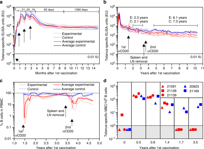
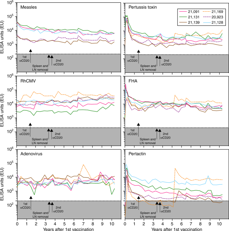
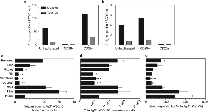
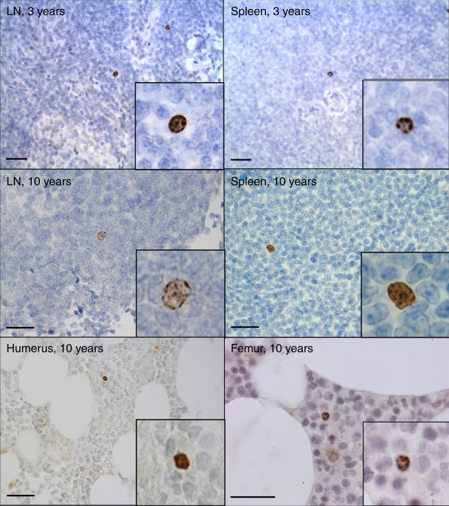
Pre-existing serum antibodies play an important role in vaccine-mediated protection against infection but the underlying mechanisms of immune memory are unclear. Clinical studies indicate that antigen-specific antibody responses can be maintained for many years, leading to theories that reactivation/differentiation of memory B cells into plasma cells is required to sustain long-term antibody production. Here, we present a decade-long study in which we demonstrate site-specific survival of bone marrow-derived plasma cells and durable antibody responses to multiple virus and vaccine antigens in rhesus macaques for years after sustained memory B cell depletion. Moreover, BrdU + cells with plasma cell morphology can be detected for 10 years after vaccination/BrdU administration, indicating that plasma cells may persist for a prolonged period of time in the absence of cell division. On the basis of these results, long-lived plasma cells represent a key cell population responsible for long-term antibody production and serological memory.
Overview
The claims and statistics of this paper haven’t been checked yet.
Run the check — free, no account needed. It reads the open-access text and checks the sources it cites.
Integrity signals
Registry
Cites
Statistics
Figures
Verification
Claims the paper makes
The claims and statistics of this paper haven’t been checked yet.
Run the check — free, no account needed. It reads the open-access text and checks the sources it cites.
Claims other papers make about this one
Each is checked against this paper’s own text: does it say what the citing paper says it does?
In the citing paper
“This pattern of rapid early decay is due to the short half-life of serum antibodies and to short-lived antibody-secreting cells, which decay with a half-life of weeks to leave a population of long-lived plasma cells with half-lives of months to years21.”
From Prospects for durable immune control of SARS-CoV-2 and prevention of reinfection (2021) · cites this paper as [21]
Self-check
Percentages, sums and reported differences re-computed from the paper’s own numbers.
The self-check needs the paper’s full text.
Citations
Cites
(2010) · Sage eBooks · doi:10.4135/9781412961288.n40 · Cited by 2,028
Maintenance of Serological Memory by Polyclonal Activation of Human Memory B Cells
Bernasconi, N. L., Traggiai, E., & Lanzavecchia, A. (2002) · Science · doi:10.1126/science.1076071 · Cited by 1,304
Duration of Humoral Immunity to Common Viral and Vaccine Antigens
Amanna, I. J., Carlson, N. E., & Slifka, M. K. (2007) · New England Journal of Medicine · doi:10.1056/nejmoa066092 · Cited by 1,286
Self-citationHumoral Immunity Due to Long-Lived Plasma Cells
Slifka, M. K., Antia, R., Whitmire, J. K., & Ahmed, R. (1998) · Immunity · doi:10.1016/s1074-7613(00)80541-5 · Cited by 1,249
Self-citationImpact of caloric restriction on health and survival in rhesus monkeys from the NIA study
Mattison, J. A., Roth, G. S., Beasley, T. M., Tilmont, E. M., Handy, A. M., Herbert, R. L., Longo, D. L., Allison, D. B., Young, J. E., Bryant, M., et al. (2012) · Nature · doi:10.1038/nature11432 · Cited by 1,145
Lifetime of plasma cells in the bone marrow
Manz, R. A., Thiel, A., & Radbruch, A. (1997) · Nature · doi:10.1038/40540 · Cited by 866
Long-Lived Plasma Cells Are Contained within the CD19−CD38hiCD138+ Subset in Human Bone Marrow
Halliley, J. L., Tipton, C. M., Liesveld, J. L., Rosenberg, A. F., Darce, J. R., Gregoretti, I. V., Popova, L., Kaminiski, D. A., Fucile, C., Albizua, I., et al. (2015) · Immunity · doi:10.1016/j.immuni.2015.06.016 · Cited by 545
Self-citationMetabolic properties of IgG subclasses in man
Morell, A., Terry, W. D., & Waldmann, T. A. (1970) · Journal of Clinical Investigation · doi:10.1172/jci106279 · Cited by 523
Eosinophils are required for the maintenance of plasma cells in the bone marrow
Chu, V. T., Fröhlich, A., Steinhauser, G., Scheel, T., Roch, T., Fillatreau, S., Lee, J. J., Löhning, M., & Berek, C. (2011) · Nature Immunology · doi:10.1038/ni.1981 · Cited by 519
Serologic changes following B lymphocyte depletion therapy for rheumatoid arthritis
Cambridge, G., Leandro, M. J., Edwards, J., Ehrenstein, M. R., Salden, M., Bodman‐Smith, M. D., & WEBSTER, A. D. B. (2003) · Arthritis & Rheumatism · doi:10.1002/art.11181 · Cited by 470
Research works printed but not linked: 2
- Wolters, K. L. & Dehmel, H. Abschliessende untersuchungen uber die Tetanus Prophylaxe durch active Immunisierung. Zeitschrift fur Hyeitschrift 124, 326–332 (1942).
- Wassilak, S. G., Roper, M. H., Kretsinger, K. & Orenstein, W. A. in Vaccines (eds. Plotkin, S. A., Orenstein, W. A. & Offit, P. A.) (Saunders Elsevier, Amsterdam, The Netherlands, 2008).
Cited by
Sorted by citations
Items 101–200 of 211
Humoral autoimmunity after solid organ transplantation: Germinal ideas may not be natural
Siu, J. H. Y., Motallebzadeh, R., & Pettigrew, G. J. (2020) · Cellular Immunology · doi:10.1016/j.cellimm.2020.104131 · Cited by 13
Do plasma cells contribute to the determination of their lifespan?
Tarlinton, D. M. (2020) · Immunology and Cell Biology · doi:10.1111/imcb.12346 · Cited by 12
Ponnan, S. M., Swaminathan, S., Tiruvengadam, K., Vidyavijayan, K. K., Cheedarla, N., Nesakumar, M., Kathirvel, S., Goyal, R., Singla, N., Mukherjee, J., et al. (2018) · PLoS ONE · doi:10.1371/journal.pone.0203037 · Cited by 12
Impact of vaccine type on HIV-1 vaccine elicited antibody durability and B cell gene signature
Palli, R., Seaton, K. E., Piepenbrink, M. S., Hural, J., Goepfert, P., Laher, F., Buchbinder, S., Churchyard, G., Gray, G., Robinson, H. L., et al. (2020) · Scientific Reports · doi:10.1038/s41598-020-69007-w · Cited by 11
Александровна, А. Н., Алексеевна, К. Н., Davydova, D. I., Aleksanin, S. S., & Rybnikov, V. Y. (2022) · Medical alphabet · doi:10.33667/2078-5631-2022-6-11-16 · Cited by 10
Chimeric HLA antibody receptor T cell therapy for humoral transplant rejection
Arana, C., García‐Busquets, A., Nicoli, M., Betriu, S., Gille, I., Heemskerk, M. H. M., Heidt, S., Palou, E., Rovira, J., & Diekmann, F. (2024) · Nephrology Dialysis Transplantation · doi:10.1093/ndt/gfae160 · Cited by 10
Pampusch, M. S., Sevcik, E. N., Quinn, Z. E., Davey, B. C., Berg, J. M., Gorrell-Brown, I., Abdelaal, H. M., Rakasz, E. G., Rendahl, A., & Skinner, P. J. (2023) · Frontiers in Immunology · doi:10.3389/fimmu.2023.1101446 · Cited by 10
Rational Design and In Vivo Characterization of Vaccine Adjuvants
Schmidt, S., Pedersen, G. K., & Christensen, D. (2018) · ILAR Journal · doi:10.1093/ilar/ily018 · Cited by 10
A cell-based in vitro assay for testing of immunological integrity of Tetanus toxoid vaccine antigen
Tichá, O., Klemm, D., Moos, L., & Bekeredjian‐Ding, I. (2021) · npj Vaccines · doi:10.1038/s41541-021-00344-1 · Cited by 10
Alexandre, M., Prague, M., McLean, C. M., Bockstal, V., Douoguih, M., Thiébaut, R., Consortia, F. T. E. 1. A. E. 2., Effelterre, T. P. V., Solforosi, L., Bockstal, V., et al. (2023) · npj Vaccines · doi:10.1038/s41541-023-00767-y · Cited by 11
Immunological memory to vaccines
Crotty, S. (2026) · Immunity · doi:10.1016/j.immuni.2026.02.019 · Cited by 9
Majority of human circulating IgG plasmablasts stop blasting in a cell-free pro-survival culture
Nguyen, D. C., Saney, C. L., Hentenaar, I., Cabrera-Mora, M., Capric, V., Woodruff, M. C., Andrews, J., Lonial, S., Sanz, I., & Lee, F. E.-H. (2024) · Scientific Reports · doi:10.1038/s41598-024-53977-2 · Cited by 10
The cellular biology of plasma cells: Unmet challenges and opportunities
Bonaud, A., Khamyath, M., & Espéli, M. (2023) · Immunology Letters · doi:10.1016/j.imlet.2023.01.005 · Cited by 10
Hirano, H., & Asada, H. (2024) · Journal of Medical Case Reports · doi:10.1186/s13256-024-04889-2 · Cited by 8
Cross-Neutralizing Breadth and Longevity Against SARS-CoV-2 Variants After Infections
Kurahashi, Y., Sutandhio, S., Furukawa, K., Tjan, L. H., Iwata, S., Sano, S., Tohma, Y., Ohkita, H., Nakamura, S., Nishimura, M., et al. (2022) · Frontiers in Immunology · doi:10.3389/fimmu.2022.773652 · Cited by 9
Dari, A., Boulton, M., Neyens, M., Gars, M. L., Valenzuela, B., Shukarev, G., Cárdenas, V., Ruiz‐Guiñazú, J., SADOFF, J. C., Hoetelmans, R. M. W., et al. (2022) · Clinical Pharmacology & Therapeutics · doi:10.1002/cpt.2796 · Cited by 7
Perturbations of immune landscape in COVID‐19 associated mucormycosis
Desai, N., Pradhan, V., Chougule, D., Tiwari, S., Mandke, C., Yadav, R. M., Athvale, A., Kawle, J., Pai, V., Pawaskar, S., et al. (2022) · Mycoses · doi:10.1111/myc.13546 · Cited by 8
Manoylov, I. K., Boneva, G., Doytchinova, I. A., Mihaylova, N., & Tchorbanov, A. I. (2019) · Clinical & Experimental Immunology · doi:10.1111/cei.13305 · Cited by 8
Network Organization of Antibody Interactions in Sequence and Structure Space: the RADARS Model
Prechl, J. (2020) · Antibodies · doi:10.3390/antib9020013 · Cited by 8
Cheng, S., Lien, C.-E., Hsieh, S., Cheng, C., Liu, W.-D., Lo, C.-L., Ko, W., Chen, Y., Huang, C., Chang, H., et al. (2022) · Vaccines · doi:10.3390/vaccines11010018 · Cited by 7
Mechanistic models of humoral kinetics following COVID-19 vaccination
Stocks, D., Thomas, A. C., Finn, A. H. R., Danon, L., & Brooks‐Pollock, E. (2025) · Journal of The Royal Society Interface · doi:10.1098/rsif.2024.0445 · Cited by 5
Self-citationRalchev, N., Markovski, A. M., Yankova, I. A., Manoylov, I. K., Doytchinova, I. A., Mihaylova, N., Shinkov, A. D., & Tchorbanov, A. I. (2022) · International Journal of Molecular Sciences · doi:10.3390/ijms232315083 · Cited by 7
Dari, A., Jacqmin, P., Iwaki, Y., Neyens, M., Gars, M. L., SADOFF, J. C., Hardt, K., Ruiz‐Guiñazú, J., & Ruixo, J. J. P. (2023) · CPT Pharmacometrics & Systems Pharmacology · doi:10.1002/psp4.13025 · Cited by 6
Khatri, I., Diks, A. M., van den Akker, E. B., Oosten, L. E. M., Zwaginga, J. J., Reinders, M. J. T., van Dongen, J. J. M., & Berkowska, M. A. (2021) · Vaccines · doi:10.3390/vaccines9111352 · Cited by 7
Long, A. H., Aftandilian, C. C., Barmettler, S., & Alexander, S. W. (2025) · Pediatric Blood & Cancer · doi:10.1002/pbc.31779 · Cited by 7
Liao, C., Chen, Y., Ye, C., Lin, S., Bai, L., Zhang, Z., Sun, W., Hu, Z., Cai, S., & Dong, L. (2026) · Autoimmunity Reviews · doi:10.1016/j.autrev.2026.104042 · Cited by 6
A biomimetic multi-component subunit vaccine via ratiometric loading of hierarchical hydrogels
Du, F., Yuk, S. A., Qian, Y., Mbaye, E. H. A., Vincent, M. P., Bobbala, S. K. R., Abbott, T. M., Kim, H., Li, Y., Li, H., et al. (2025) · Nature Communications · doi:10.1038/s41467-025-60416-x · Cited by 5
Jiang, R., Fichtner, M. L., Hoehn, K. B., Stathopoulos, P., Nowak, R. J., Kleinstein, S. H., & O’Connor, K. C. (2019) · bioRxiv (Cold Spring Harbor Laboratory) · doi:10.1101/840389 · Cited by 6
Kim, W. (2024) · Immune Network · doi:10.4110/in.2024.24.e28 · Cited by 5
Increased ocular plasma cells induce damaging α-synuclein+ microglia in autoimmune uveitis
Li, M., Feng, M., Liu, T., Duan, S., Man, X., Yuan, X., Wang, L., Sun, Y., Wei, X., Fu, Q., et al. (2025) · Mucosal Immunology · doi:10.1016/j.mucimm.2025.02.007 · Cited by 6
Lane, A., Quach, H. Q., Ovsyannikova, I. G., Kennedy, R. B., Ross, T. M., & Einav, T. (2025) · Vaccine · doi:10.1016/j.vaccine.2025.127677 · Cited by 6
Haas, M., & Fest, T. (2023) · Immunology Letters · doi:10.1016/j.imlet.2023.05.006 · Cited by 5
Long-term stability of antibody responses elicited by Dengue virus envelope DIII-based DNA vaccines
Slon-Campos, J., Poggianella, M., & Burrone, Ó. R. (2018) · Journal of General Virology · doi:10.1099/jgv.0.001094 · Cited by 5
Tissue resident T cell memory or how the magnificent seven are chilling in the bone
Zens, K. D., & Münz, C. (2019) · European Journal of Immunology · doi:10.1002/eji.201948208 · Cited by 5
Du, L., Hou, L., Yu, X., Cheng, H., Chen, J., Zheng, Q., & Hou, J. (2021) · Frontiers in Immunology · doi:10.3389/fimmu.2021.697292 · Cited by 5
Joyner, C. J., Ley, A. M., Nguyen, D. C., Ali, M., Corrado, A., Tipton, C. M., Scharer, C. D., Tian, M., Woodruff, M. C., Hom, J., et al. (2021) · bioRxiv (Cold Spring Harbor Laboratory) · doi:10.1101/2021.05.22.445269 · Cited by 5
Kreuser, S. A., Honaker, Y., Cheng, R. Y.-H., Dahl, N., Soligalla, R. D., Lopez, C., Rawlings, D. J., & James, R. G. (2021) · bioRxiv (Cold Spring Harbor Laboratory) · doi:10.1101/2021.09.30.462595 · Cited by 5
Human lymphoid tissue sampling for vaccinology
Kwiatkowska, K. M., Mkindi, C., & Nielsen, C. M. (2022) · Frontiers in Immunology · doi:10.3389/fimmu.2022.1045529 · Cited by 6
Kolachala, V. L., Wei, C., Venkateswaran, S., Hill, A., Warren, V., Espinoza, H., Sanz, I., & Gupta, N. A. (2024) · Autoimmunity · doi:10.1080/08916934.2024.2356089 · Cited by 4
Wilding, K. M., Molina-Parı́s, C., Kubicek-Sutherland, J. Z., McMahon, B. H., Perelson, A. S., & Ribeiro, R. M. (2025) · Frontiers in Immunology · doi:10.3389/fimmu.2025.1596518 · Cited by 5
Azar, J. H., Evans, J. P., Sikorski, M., Chakravarthy, K. B., McKenney, S., Carmody, I., Zeng, C., Teodorescu, R., Song, N., Hamon, J. L., et al. (2023) · JCI Insight · doi:10.1172/jci.insight.163434 · Cited by 4
Generation, expansion, gene delivery, and single-cell profiling in rhesus macaque plasma B cells
Cheng, R. Y.-H., Helmers, A. E., Kreuser, S. A., Dahl, N., Honaker, Y., Lopez, C., Rawlings, D. J., & James, R. G. (2024) · Cell Reports Methods · doi:10.1016/j.crmeth.2024.100878 · Cited by 4
Lineage tracing reveals fate bias and transcriptional memory in human B cells
Swift, M. A., Horns, F., & Quake, S. R. (2023) · Life Science Alliance · doi:10.26508/lsa.202201792 · Cited by 5
Zhen, Z.-D., Zhang, L., Li, Q., Zhu, Y., Wang, X., Fu, X., Ai, J., Wang, R., Xie, Z., & Ma, S.-X. (2023) · Infection Genetics and Evolution · doi:10.1016/j.meegid.2023.105477 · Cited by 5
Jeffery-Smith, A., Burton, A. R., Lens, S., Rees-Spear, C., Patel, M., Gopal, R., Muir, L., Aiano, F., Doores, K. J., Chow, J. Y., et al. (2021) · bioRxiv (Cold Spring Harbor Laboratory) · doi:10.1101/2021.05.30.446322 · Cited by 4
Antigen avidity drives the durability of the vaccine immune responsePreprint
Weidner, N. G., Padilla, M. V., Pruitt, L., Cottrell, C. A., Gonzales, K. A., Kalyuzhniy, O., Kubitz, M., Alavi, N. B., Phelps, N., Weglarz, M., et al. (2025) · bioRxiv (Cold Spring Harbor Laboratory) · doi:10.1101/2025.04.18.649567 · Cited by 4
Distinct autoreactive CD19– plasma cell subsets accumulate in lupus-prone mice
Dang, V. D., Szelinski, F., Mohr, E., Le, T. A., Ritter, J., Wiedemann, A., Ferreira‐Gomes, M., Guerra, G. M., Durek, P., Heinrich, F., et al. (2025) · Nature Communications · doi:10.1038/s41467-025-65906-6 · Cited by 3
Antigen avidity potentiates the durability of the vaccine immune response
Weidner, N. G., Padilla, M. V., Pruitt, L., Cottrell, C. A., Gonzales, K. A., Kalyuzhniy, O., Lu, D., Alavi, N. B., Phelps, N., Aniqua, M., et al. (2026) · Science Translational Medicine · doi:10.1126/scitranslmed.aee5273 · Cited by 8
Lessons learned from successful implementation of tetanus and diphtheria vaccination programs
Slifka, M. K., Thomas, A., Gao, L., Amanna, I. J., & Orenstein, W. A. (2025) · Clinical Microbiology Reviews · doi:10.1128/cmr.00031-25 · Cited by 5
Self-citationLong Live the Plasma Cell: The Basis of Sustained Humoral Immunity
Koutsakos, M., & Ellebedy, A. H. (2022) · The Journal of Immunology · doi:10.4049/jimmunol.2200121 · Cited by 3
Dari, A., Solforosi, L., Roozendaal, R., Hoetelmans, R. M. W., Ruixo, J. J. P., & Boulton, M. (2023) · Journal of Pharmacology and Experimental Therapeutics · doi:10.1124/jpet.123.001591 · Cited by 3
Eleftheriadis, T., Pissas, G., Liakopoulos, V., & Stefanidis, I. (2021) · Journal of Immunotherapy · doi:10.1097/cji.0000000000000365 · Cited by 3
Safety and Immunogenicity of SARS-CoV-2 S-2P Protein Vaccine MVC-COV1901 in People Living with HIVPreprint
Cheng, S., Lien, C.-E., Hsieh, S., Cheng, C., Liu, W.-D., Lo, C.-L., Ko, W., Chen, Y., Huang, C., Chang, H., et al. (2021) · medRxiv · doi:10.1101/2021.12.08.21267439 · Cited by 3
Amir, M. A., Huang, H., Tang, R., Zhang, Y., Xiao, Y., Yuan, Q., & Xiao, X. (2025) · Clinical Kidney Journal · doi:10.1093/ckj/sfaf248 · Cited by 2
Why are long-lived plasma cells long-lived?
Reinke, J. G., Schorr, C. R., & Lee, K. P. (2026) · Frontiers in Immunology · doi:10.3389/fimmu.2026.1785563 · Cited by 2
B-cell editing: An emerging alternative of precision oncotherapy
Choudhury, P. R., Chakravarti, M., Banerjee, S., Baral, R., & Bose, A. (2024) · Advances in immunology · doi:10.1016/bs.ai.2024.10.002 · Cited by 2
Notghi, A. A., Delahunty, J., Tattersall, R. S., & Stone, B. (2025) · The Lancet Infectious Diseases · doi:10.1016/s1473-3099(25)00223-3 · Cited by 2
A Panoramic View of the Immune Response to Trypanosoma cruzi Infection
Acevedo, G. R., Girard, M. C., & Gómez, K. A. (2019) · Birkhäuser advances in infectious diseases · doi:10.1007/978-3-030-00054-7_3 · Cited by 2
Landi, D., Nicoletti, C. G., Mauro, G. D., Cola, G., Grimaldi, A., Mataluni, G., & Marfia, G. A. (2021) · Multiple Sclerosis and Related Disorders · doi:10.1016/j.msard.2021.103334 · Cited by 2
Boussag-Abib, L., & Laraba‐Djebari, F. (2021) · Immunobiology · doi:10.1016/j.imbio.2021.152108 · Cited by 2
Cheng, R. Y.-H., Kreuser, S. A., Dahl, N., Honaker, Y., Soligalla, R. D., Lopez, C., Rawlings, D. J., & James, R. G. (2023) · bioRxiv (Cold Spring Harbor Laboratory) · doi:10.1101/2023.10.29.564645 · Cited by 2
Giannopoulou, M., Tarassi, K., Tsouka, G., Christodoulidou, C., Stefanidis, I., & Eleftheriadis, T. (2022) · Experimental and Clinical Transplantation · doi:10.6002/ect.2022.0146 · Cited by 1
van den Broek, D. A. J., Karahan, G. E., Meziyerh, S., de Vaal, Y. J. H., Bakker, K. H., Haasnoot, G. W., Rotmans, J. I., Teng, Y. K. O., van Kooten, C., Heidt, S., et al. (2025) · HLA · doi:10.1111/tan.70285 · Cited by 1
On the carrying capacity of the bone marrow survival niche in mice
Tonouchi, K., Yeh, C., Cain, D. W., Moseman, E. A., Haynes, B. F., Wagh, K., Wiehe, K. J., Kurosaki, T., & Kelsoe, G. H. (2025) · Frontiers in Immunology · doi:10.3389/fimmu.2025.1706810 · Cited by 1
Cell intrinsic versus cell extrinsic control of plasma cell longevity
Allman, D., & Bhandoola, A. (2025) · Trends in Immunology · doi:10.1016/j.it.2025.10.013 · Cited by 1
Quantification of the IgG antibody response half-life for hybrid immunity to SARS-CoV-2
Bongiovanni, F., Conway, E. J., Smith, L., Cumming, J., Mazhari, R., Kiernan‐Walker, N., Eriksson, E. M., McVernon, J., & Müeller, I. (2025) · BMC Medicine · doi:10.1186/s12916-025-04490-4 · Cited by 1
Therapeutic antibody delivery: vector tools to boost efficacy and affordability
Chiyyeadu, A., Khan, B., Ehrhardt, K., Büning, H., Morgan, M. A., & Schambach, A. (2026) · Frontiers in Immunology · doi:10.3389/fimmu.2025.1714390 · Cited by 1
Zhou, C., Zuo, Y., Ma, J., Liang, Z., & Zang, C. (2026) · Frontiers in Cellular and Infection Microbiology · doi:10.3389/fcimb.2026.1766140 · Cited by 1
Huang, M., Dong, F., Liu, Q., & Lin, S. (2026) · Frontiers in Immunology · doi:10.3389/fimmu.2026.1809668 · Cited by 1
B-cell biology, tolerance, and autoantibodies
Barnas, J. L., Meffre, E., & Anolik, J. H. (2021) · Elsevier eBooks · doi:10.1016/b978-0-12-820583-9.00036-1 · Cited by 1
Increased B Cell Understanding Puts Improved Vaccine Platforms Just Over the Horizon
Notario, G. R., & Kwak, K. (2022) · Immune Network · doi:10.4110/in.2022.22.e47 · Cited by 1
Significance of Thyroglobulin Autoantibodies in Patients With Thyroid Cancer Treated With Lenvatinib
Sgrò, D., Rossi, P., Piaggi, P., Brancatella, A., Lorusso, L., Bottici, V., Molinaro, E., Latrofa, F., Elisei, R., & Agate, L. (2023) · Journal of the Endocrine Society · doi:10.1210/jendso/bvad084 · Cited by 1
Ganusov, V. V. (2024) · ImmunoHorizons · doi:10.4049/immunohorizons.2300057 · Cited by 1
Hu, X., McAlister, A., Kinder, J. M., Johnson, A., White, K. A., Caruso, C. B., Wang, C., Basco, R., Gattis, C., Friera, A. M., et al. (2025) · Cytotherapy · doi:10.1016/j.jcyt.2025.06.013 · Cited by 1
Prager, K. C., Buhnerkempe, M., Greig, D. J., Orr, A. J., Jensen, E. D., Gomez, F. M., Galloway, R. L., Wu, Q., Gulland, F. M. D., & Lloyd‐Smith, J. O. (2019) · bioRxiv (Cold Spring Harbor Laboratory) · doi:10.1101/819532 · Cited by 1
Editorial: Advances in Plasma Cells in Health and Disease
Lee, F. E.-H., Wrammert, J., & Cenci, S. (2020) · Frontiers in Immunology · doi:10.3389/fimmu.2020.606737 · Cited by 1
Zhang, J., He, R., Liu, B., Zheng, X., Wu, Q., Wen, B., Wang, Q., Liu, Z., Chang, F., Hu, Y., et al. (2022) · bioRxiv (Cold Spring Harbor Laboratory) · doi:10.1101/2022.08.09.503302 · Cited by 1
Fichtner, M. L. (2023) · doi:10.26481/dis.20231024mf · Cited by 1
Glass, D. R., Dornisch, E. M., Yin, H., Ludmann, S. A., Samudre, A., Kuhl, S., Malone, J., Chander, A., Kaul, N., Phalen, C. G., et al. (2026) · bioRxiv (Cold Spring Harbor Laboratory) · doi:10.64898/2026.04.13.717827 · Cited by 1
Longitudinal dynamics of human B-cell response at single-cell level in response to Tdap vaccinationPreprint
Khatri, I., Diks, A. M., van den Akker, E. B., Oosten, L. E. M., Zwaginga, J. J., Reinders, M. J. T., van Dongen, J. J. M., & Berkowska, M. A. (2021) · bioRxiv (Cold Spring Harbor Laboratory) · doi:10.1101/2021.10.08.463649
Azar, J. H., Evans, J. P., Sikorski, M., Chakravarthy, K., McKenney, S., Carmody, I., Zeng, C., Teodorescu, R. N., Song, N. J., Hamon, J. L., et al. (2022) · medRxiv · doi:10.1101/2022.03.17.22272389
What are the prospects for durable immune control?
Lelièvre, J., & Bauer, J. (2022) · Infectious Diseases Now · doi:10.1016/j.idnow.2022.09.010
The Bone Marrow as a Hotbed for Plasma Cell Activation
Poulaki, A., Giannouli, S., & Voulgarelis, M. (2022) · Springer eBooks · doi:10.1007/978-3-031-10131-1_4
Majority of human circulating plasmablasts stop blasting: A probable misnomerPreprint
Nguyen, D. C., Saney, C. L., Hentenaar, I., Cabrera-Mora, M., Woodruff, M. C., Andrews, J., Lonial, S., Sanz, I., & Lee, F. E.-H. (2023) · bioRxiv (Cold Spring Harbor Laboratory) · doi:10.1101/2023.09.10.557057
Modelling antibody dynamics in humans after different Ad26.COV2.S vaccination schemes
Dari, A., Ruixo, J. J. P., Gars, M. L., Struyf, F., & Jacqmin, P. (2024) · British Journal of Clinical Pharmacology · doi:10.1111/bcp.16251
Stover, E. L., Rock, M. L., Olson, J. D., Schaaf, G. W., Oguin, T. H., Cline, J. M., & Macintyre, A. N. (2025) · Radiation Research · doi:10.1667/rade-23-00203.1
Ioannou, K., Karofylakis, E., Alkhammash, S., Baxendale, H., Eisen, S., Döffinger, R., Chandra, A., Manson, A. L., Thaventhiran, J. E. D., Gkrania‐Klotsas, E., et al. (2025) · medRxiv · doi:10.1101/2025.03.13.25323766
Kubo, R., Yoshikawa, R., Fujii, Y., Kawasaki, T., Takazono, T., Izumikawa, K., Ariyoshi, K., Mukae, H., & Yasuda, J. (2025) · Tropical Medicine and Health · doi:10.1186/s41182-025-00807-4
Zhao, J., Shao, J., Sui, X., Wei, M., Ma, X., Xu, Z., & Zhu, T. (2025) · Vaccines · doi:10.3390/vaccines13121226
Müller, M. K., Volzke, J., Subin, B., Müller, S. C., Sombetzki, M., Reisinger, E. C., & Müller‐Hilke, B. (2022) · Figshare · doi:10.6084/m9.figshare.18666522
Evaluating the contraceptive potential of active immunization against gonadotropin-releasing hormone
Urbanski, H. F., Slifka, M. K., Appleman, M. L., Thomas, A., Kohama, S. G., Erikson, D. W., & Lomniczi, A. (2026) · Reproduction · doi:10.1093/reprod/xaaf024
Self-citationCarvajal, R., Aroca-Alsina, M., Esperalba, J., Tur, C., Borràs-Bermejo, B., Cobo‐Calvo, Á., Ariño, H., Arrambide, G., Bollo, L., Bravo, G., et al. (2026) · Neurology Open Access · doi:10.1212/wn9.0000000000000061
Patel, J., Yang, Z., Ping, S., Ahmed, A., Simon, T. W., Waggoner, J. J., & Scherer, E. M. (2026) · Journal of Virology · doi:10.1128/jvi.02150-25
Characterizing the antibody response at the single cell level with droplet microfluidics
Castrillon, C. E. (2018) · theses.fr (ABES) · doi:10.70675/12610ff4z549bz4f5bz96c0z6895763079fa
Lefsihane, K. (2022) · theses.fr (ABES) · doi:10.70675/93ab3385z8624z404czab1bz3b37f81ca041
Metadata
- Authors
- Hammarlund, E., Thomas, A., Amanna, I. J., Holden, L. A., Slayden, O. D., Park, B., Gao, L., & Slifka, M. K.
- Affiliations
- Oregon National Primate Research Center (US)
- Najít Technologies (United States) (US)
- Portland State University (US)
- OHSU Knight Cancer Institute (US)
- Corresponding author
- Slifka, Mark K.
- ORCID
- Venue
- Nature Communications
- Published
- 2017-11-24
- Volume
- 8
- Issue
- 1
- Pages
- 1781-1781
- ISSN
- 2041-1723 (ISSN-L)
- PMC
- PMC5701209
- OpenAlex
- W2770797413
- Cited by
- 210
- References
- 67
- Citations by year
- 2026: 20 · 2025: 20 · 2024: 15 · 2023: 23 · 2022: 28 · 2021: 36 · 2020: 32 · 2019: 22 · 2018: 14
- FWCI
- 8.54
- Type
- journal article
- Language
- en
- Open access
- gold
- doi.org
- Locations
- Nature Communications (publishedVersion, cc-by)
- PubMed Central (publishedVersion, cc-by)
- PubMed (publishedVersion)
- PDXScholar (Portland State University) (submittedVersion, cc-by)
- RePEc: Research Papers in Economics (submittedVersion)
- DOAJ (DOAJ: Directory of Open Access Journals) (submittedVersion, cc-by-sa)
- Funders
- National Institutes of Health (P51OD011092)
- Genentech
- Oregon National Primate Research Center
- National Institute on Aging (P01AG023664)
- National Institute of Allergy and Infectious Diseases (U01AI082196)
- Topics
- Life Sciences › Immunology and Microbiology › Immunology › T-cell and B-cell Immunology
- Life Sciences › Immunology and Microbiology › Immunology › Immune responses and vaccinations
- Life Sciences › Immunology and Microbiology › Immunology › Monoclonal and Polyclonal Antibodies Research
- Keywords
- long-lived plasma cells, plasma cell survival, memory B cells, long-term antibody response, vaccine immunity, rhesus monkeys, BrdU labeling, bone marrow plasma cells
- MeSH terms
- Animals, Antibodies, Bacterial, Antibody Formation, B-Lymphocytes, Bacterial Vaccines, Cell Survival, Immunologic Memory, Macaca mulatta, Male, Plasma Cells, Vaccination
About these checks
Registry, retraction, Cites and statistics results are computed, not judged, with one exception: a printed reference that no index lists is read once by a model before it counts as a citation error. Claims are extracted by a model and kept only when their quote appears verbatim in the text; each cited source is then checked, with a model’s help, for whether it says what the claim relies on. Those verdicts can be wrong — read the claim’s quote and the cited source before relying on one.
Statistics and arithmetic are recomputed, not judged.
Confidence bands are shown only for fields calibrated on our annotated test set. None is calibrated yet, so no band is shown.
What counts as a claim ›How we measure confidence ›How we check this ›
Are you an author, or do you see a problem with this page? Request a review
